Department of Physiological Sciences, State University of Rio de Janeiro -Rio de Janeiro/RJ, Brazil.
Email: [email protected] Received for publication on January 19, 2006. Accepted for publication on April 10, 2006.
REVIEW
OBSTRUCTIVE SLEEP APNEA AND INSULIN
RESISTANCE: A ROLE FOR MICROCIRCULATION?
Nicolas Wiernsperger, Pierre Nivoit, and Eliete Bouskela
Wiernsperger N, Nivoit P, Bouskela E. Obstructive sleep apnea and insulin resistance: a role for microcirculation? Clinics. 2006;61(3):253-66.
Obstructive sleep apnea is an increasingly recognized medical problem. The recent attention to its frequency in the general population and its important role in metabolic, vascular, and behavioral aspects have sharply increased the number and nature of investigations, thereby revealing new aspects that open new approaches in research. Whereas obstructive sleep apnea is a well-known phenomenon accompanying obesity and diabetes, new findings strongly suggest that this close relationship may also operate in the opposite direction. Indeed obstructive sleep apnea may be a primary feature inducing or aggravating a series of vascular and metabolic disturbances closely resembling the metabolic syndrome. This review will discuss established and potential mechanisms responsible for these changes. Obstructive sleep apnea indeed appears to gather all the elements necessary to induce insulin resistance, hypertension, and possibly heart failure. After careful analysis of these modifications and considering how they are intertwined, we propose that microcirculation could represent the common denominator mediating the progression of this pathology, as it is eventually the case in the metabolic syndrome and diabetes domain. This plausible hypothesis is discussed in detail and should be verified by appropriate preclinical and clinical protocols, which are now achievable by using noninvasive techniques in humans.
KEYWORDS: Obstructive sleep apnea. Insulin resistance. Hypoxia. Metabolic syndrome. Microcirculation.
INTRODUCTION
Few domains of medicine have experienced so much interest as obstructive sleep apnea (OSA) over the last 10 years. The sharp rising interest in this phenomenon started when it was first suggested that OSA may not just be an accompanying symptom but might cause or aggravate meta-bolic and vascular diseases.
Sleep disordered breathing comprises several processes that provoke repetitive interruptions in sleep, mainly due to snoring and hypopneas/apneas of either central or OSA origin. Epidemiologically, the OSA syndrome affects prob-ably about 5% of the general population. The prevalence of OSA is much higher in diseases as common as
hyper-tension (22-48%)1 or heart failure (11-37%). In this latter group, central apneas are seen in 45% of subjects2 and are inversely related to cardiac ejection fraction. Half of the patients with diastolic heart failure have an increase in the number of apneas.
The present review will describe the most recent knowl-edge about links between obstructive sleep apnea and cardiometabolic disorders. In particular, the possibility that this relationship operates in both directions is a recent view that strongly stimulates interest in new preclinical and clini-cal research. We will also discuss the plausible hypothesis that the microcirculation may be the common denomina-tor of these intertwined disturbances.
OSA IS A SERIOUS CLINICAL CONDITION
apneas in patients. Obstructive sleep apneas are of mechani-cal origin, due to the failure of upper airways to maintain their muscle tone. Not surprisingly OSAs are most frequent in obese individuals, as a consequence of fat deposition in the muscles of the upper airways. Almost all OSA subjects exhibit intense snoring, but snoring is not necessarily ac-companied by OSA. This is important in clinical trials, which partly were based on this indirect parameter. The link between OSAs and obesity is strong, such as to bias many protocols and interpretations of older clinical studies. A 10% increase in body weight over 4 years increases the risk for OSA 6-fold.3 Recent findings revealed that half of the genetic variance in OSA is shared with obesity phenotypes,4 which may have important consequences, as will be dis-cussed later. Obstructive sleep apneas are more frequent during REM sleep,5 and their number, usually of short du-ration (around 20 s) can reach several hundred per night. Sleep apneas up to 1 min can be found, a situation that may be life-threatening. Each sleep apnea is characterized by hypoxia during breathlessness, with blood oxygen satu-ration (SaO2) down to 50% in extreme cases. The degree of desaturation is at least as important as the total number of episodes for complications linked with OSA. Each epi-sode is followed by an arousal reaction restoring breath, thereby inducing abrupt reoxygenation. Patients with OSA suffer daytime sleepiness, a situation closely linked to car accidents, especially in the late afternoon.
OSA and Insulin Resistance / Glucose Tolerance / Diabetes
There is a very close link between insulin resistance (IR) and OSA, but causative mechanisms are disputed.6,7 It was well-known that snoring and OSA are essentially seen in overweight/obese patients characterized by IR or the metabolic syndrome; more recent investigations sup-port this link by showing that other diseases characterized by IR are also linked with OSA: in patients with polycystic ovary syndrome, elevated insulin levels and impaired glu-cose tolerance correlated with a higher frequency of OSAs, and this correlation was even independent of BMI in glu-cose normotolerant women.8,9 Similarly, OSA affects about two thirds of acromegalic patients.10,11 Nonalcoholic steatohepatitis (NASH), a situation found in most IR-pa-tients, is also linked to severe OSA, with patients having high AHI values being more insulin-resistant and exhibit-ing more steatosis as well as elevated liver enzymes.12 Il-lustrating the close relationship between IR and obstruc-tive sleep apnea, it was proposed that OSA should be added to syndrome X (metabolic syndrome) and the new entity be called syndrome Z.13 A main question is still whether
this correlation is simply due to—or is independent of— excess fat (BMI or visceral fat). Table 1 shows how OSA superimposed on obesity worsens various metabolic and vascular parameters.
Indeed, several studies have claimed the correlation to be strictly due to the presence or absence of concomitant obesity in children14 and adults.15 In contrast, more recent studies suggest that whereas obesity plays a role, elevated AHI and minimum SaO2 values are important determinants of impaired glucose tolerance.16 In these patients, each ad-ditional apneic episode increased plasma insulin and the HOMA-index by 0.5%. In another study, the degree of glu-cose intolerance was related to the severity of desaturation. It was estimated that each 4% drop in SaO2 represented an odds ratio of 1.99 for glucose intolerance.17 Independently of BMI, fasting insulin—an indirect indicator of IR—cor-related with OSA severity.18
However, here again, a bias in interpreting such data lies in the fact that a sleep debt per se, such as frequently interrupted sleep or shortened sleep duration without breathing abnormalities, also leads to IR.19 Indeed, patients submitted to sleep debt exhibit a higher HOMA index, ab-normal glucose tolerance, and a reduction in first-phase in-sulin secretion.20 Therefore, some of the mechanisms in-volved might also be due to sleep deprivation rather than strictly hypoxia/reoxygenation. In the recent Sleep Heart Health Study, the odds ratio for fasting glucose intolerance was 1.7 for mild and 1.46 for severe OSA, and this corre-lated with SaO2. In this large-scale clinical investigation, OSA was independently associated with glucose intoler-ance, IR, and noninsulin-dependent diabetes mellitus.21 In another trial, fasting and postload glycemia increased with OSA severity, with a concomitant decrease in insulin sen-sitivity.22 Recently, it was shown that a high fat diet in nor-mal rats was followed by sleep apneas.23
In diabetic patients, the elevated number of sleep apneas appears to be predominantly of central rather than obstruc-tive origin, likely because of the frequent presence of
au-Table 1 - Additional effects of obstructive sleep apnea (OSA) on metabolic and vascular parameters in obesity. Data on control (healthy) subjects and obese patients without OSA are also shown (adapted from ref. 6)
C Ob Ob + OSA
BMI 26 36 38
MABP 92 102 107
Visceral fat area 220 337*
Total wake time 66 59 113
REM latency 74 82 93
tonomic neuropathy in this disease. Poor sleep quality in diabetic patients is related to higher HbA1C values.24
OSA and Cardiovascular Diseases
Mortality is greater in patients having AHI values greater than 20.25,26 Obstructive sleep apnea severely affects cardiac function in compromised hearts. The Sleep Heart Health Study showed that cardiovascular diseases (CVD) were more frequent in OSA patients, even in those with moderate AHI values.27 In this cross-sectional study per-formed on more than 6000 subjects, a strong correlation was established between AHI and the prevalence of CVD (coronary heart diseases, heart failure, and stroke).27 Ob-structive sleep apnea at baseline is a significant predictor of CVD, and a recent study showed that the CVD incidence was 57% in untreated vs 7% in efficiently treated OSA pa-tients.28 In addition to hypertension, OSA patients have a 58% prevalence of cardiac arrhythmias.1 Acute experimen-tal OSA leads to arterial stiffness.29 Chronic OSA patients accordingly show diminished aortic distensibility and have an increased stiffness index.30 Patients with OSA spending 9% of the night time with SaO2 below 90% exhibit carotid wall hypertrophy.31
OSA, METABOLIC DERANGEMENTS, AND CVD: A BIDIRECTIONAL PROCESS?
As briefly stated above, the increase in OSA in patients suffering from IR/diabetes or severe cardiac pathologies is a well-known clinical observation. Additionally, OSA is likely to aggravate preexisting diseases. The first clinical studies were biased by confounding factors, mainly the presence of obesity, but also by subjective self-answered questionnaires or self-reporting of snoring. Later studies using more careful patient observations (eg, polysomnography and blood sampling during apneas) and improved matching of patients control groups, as well as experimental investigations, suggested that OSA might in-duce by itself—or intensify—the switch towards diabetes and lead to macrovascular complications.32
Do OSAs Induce Insulin Resistance?
A main question is whether OSA can be causal to the metabolic- and vascular-related disturbances (Fig.1). In snoring children19 and adults,33 higher insulin levels are present, usually revealing IR. Fasting insulin levels corre-late with OSA.34 Nonobese patients suffering from OSA have more visceral fat35 and diminished adiponectin,36 both major situations favoring IR and vascular disturbances.
Pio-neering studies have recently suggested that the presence of the ApoEe4 allele might predispose to OSA, and the presence of this genetic variation also in Alzheimer’s dis-ease raises the possibility that OSA might, via recurrent hypoxic episodes, be involved in the cognitive deteriora-tion affecting these patients.37 Humans exposed to high al-titude hypoxia exhibit decreased insulin sensitivity, indi-cating that hypoxia per se is able to generate IR.38 Even acute hypoxia for 30 min at a SaO2 < 75% leads to a de-crease in glucose disposal rate.39 Interestingly, such data are similar to what is observed in hypoglycemia, despite major differences in stress hormone profiles, thus pointing again towards a specific role for hypoxia. Here also, sleep deprivation per se may play a role, since short sleepers have an odds ratio of 2 for developing IR.40
In preclinical experiments, the main features of OSA can be simulated by sophisticated technical devices allow-ing repetitive and abrupt adjustable changes in respiratory gas in chambers. This procedure is called intermittent hy-poxia (IH). In vitro, cultured cells can be exposed to se-lected levels of oxygenation. For example, IH or fructose feeding (another way to induce IR) both reduce the ventilatory response to hypoxia and hypercapnia and lead to elevated insulin concentrations41 and reduced insulin sen-sitivity.38,42 Interestingly, IH worsens—while continuous hypoxia improves—glucose tolerance, suggesting that re-petitive cycles of low and high oxygen play a determining role.38
Do OSAs Favor Diabetes?
Occasional and regular snoring increases the risk of diabetes by 1.5 and 2.25 respectively.43 The incidence of diabetes was doubled over 10 years in middle-aged, ha-bitual snorers.44 In a paper that just appeared, a prospec-tive study, the Wisconsin Sleep Cohort, comprising 1387
patients, evaluated the prevalence and the incidence of type 2 diabetes in subjects with OSA. It appeared that the odds ratio for developing diabetes within 4 years was 1.62 with an AHI > 15, compared with subjects with AHI < 5 after adjustments for age, sex, and body habitus.45 The prevalence of diabetes in subjects with AHI > 15 was 14.7%, as compared with 2.8% in those with AHI < 5. Although this study does not constitute a definitive proof, such data strongly argue for a causal role of OSA in dia-betes development.
Do OSAs favor Cardiovascular Diseases?
One of the most striking cardiovascular complications of OSA is hypertension. The sympathetic discharge accom-panying hypoxia/reoxygenation induces vascular resistance which does not resume during daytime.1,40 Therefore, there is a state of persistent chemoreceptor activation.32 The hy-pertension induced by OSA has a particular profile: diastolic pressure increases early, and patients experience no diurnal variation of systemic blood pressure. Impor-tantly, there is also no nocturnal dipping of blood pressure. This type of hypertension severely affects the brain and heart, with little effect on the kidneys.46 Otherwise healthy patients suffering from OSA show increased body weight and sympathetic muscle activity.47 Stroke and cardiac ar-rest in the early morning hours are other typical features of OSA: a lack of normal reaction of brain vessels to isocapnic hypoxia during non-REM sleep could explain the vulnerability of these subjects towards stroke.48
In mice, chronic IH increases blood pressure and hematocrit as well as the weights of the left and right ven-tricle and septum, signs of right heart loading and pulmo-nary vasoconstriction.49,50 The same profile is found in hu-mans: 25% of OSA patients have mild pulmonary hyper-tension; 18% suffer right ventricular dysfunction, and they have a 3-fold increase in risk for CVD.51 The
susceptibil-ity of hearts to infarction was increased in rats subjected to 35 days of IH.52 Here again, IH is more detrimental than sustained hypoxia for increasing blood pressure and sym-pathetic activation.53 Obstructive sleep apnea also leads to elevated levels of NPY, another factor of vasoconstriction.54 In a study in rats, the IH-induced hypertension was found to last several weeks after cessation of the procedure.55 Each additional incident of apnea increased the odds ratio for hypertension by 1%.56 This striking link between OSA and hypertension57, as well as that between OSA, congestive heart failure and respiratory alterations58 have been widely reviewed recently.
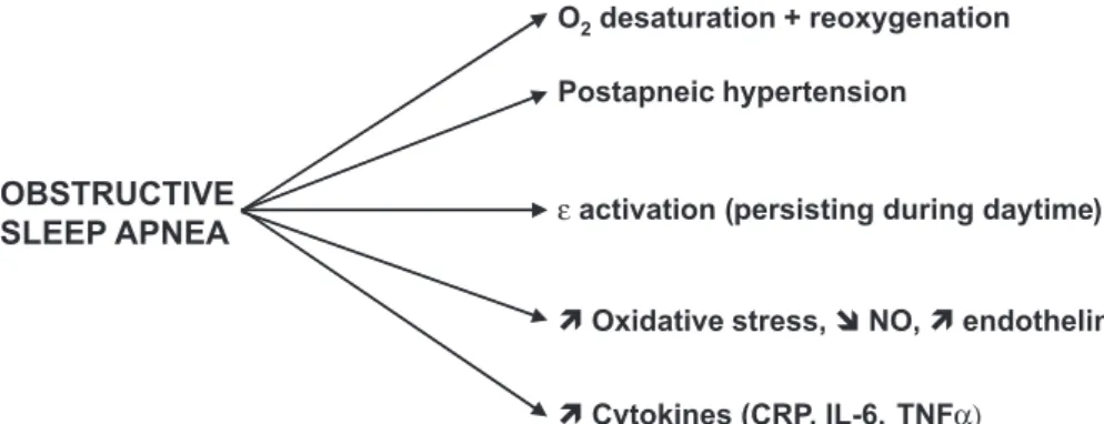
Figure 2 summarizes the main potential consequences of OSAs on metabolic and vascular parameters.
MECHANISMS OF OSA-INDUCED METABOLIC AND VASCULAR DISORDERS
The similarity of OSA-related pathologies with meta-bolic syndrome is striking: in addition to hypertension, risk factors for atherosclerosis, inflammation (cytokines), hemostatic disorders, oxidative stress, and defects in vas-cular reactivity are among the major common features. The following chapter will document the major recent findings in biochemical and cellular mechanisms underlying these complications.
Inflammation / Adhesion Molecules
As in metabolic syndrome, a mild inflammatory state characterizes OSA.1 Indeed, several studies have reported an increase in CRP, IL-6, IL-8, or TNFα in patients with
OSA.59–61 In adolescents free of CVD but with an AHI > 5, CRP levels were increased even after adjustment for the confounding obesity factor.62 Interestingly, partial sleep deprivation also increases CRP levels.63 TNFα levels are also elevated in serum and monocytes.60,61,64,65 Post apnea,
TNFα increases immediately after SaO
2 reaches a thresh-old of 85%.66 Moreover, the TNFα circadian rhythm is dis-turbed (less during night, more during day).67 These mol-ecules are known to be closely linked with IR and predic-tive of type 2 diabetes.68 Adhesion molecules such as ICAM-1, VCAM-1, or E-selectin are also increased in pa-tients suffering from OSA.1,69 A particular role for CD11c and CD15 has been recently proposed.70
Adipokines
Several studies have demonstrated an increase in cir-culating leptin levels in OSA patients, which correlates with SaO2.71 Indeed, it has been shown that leptin rises in order to compensate for IH.38 The leptin increase is seen even in otherwise healthy subjects suffering from OSA.47 High plasma soluble leptin receptor concentrations and reduced glucose uptake are correlated with OSA, and globally there seems to exist an inverse correlation between insulin sen-sitivity and leptin receptor concentration.33,72 Another sub-stance liberated by fat cells is adiponectin, the role of which is increasingly considered to be protective against IR and vascular disturbances. In OSA patients, in addition to in-creases in hsCRP and IL-6, adiponectin levels were re-duced36; however, another study reported the opposite.73 This should be taken into account, since leptin is consid-ered important in IR and promotes oxidative stress, another potential factor involved in prediabetes and diabetes (see below).74,75
HIF-1 Alpha
Intermittent hypoxia, even in vitro, increases levels of hypoxia-inducible factor 1 alpha (HIF-1 alpha).76 While HIF-1 might first increase as a protective, compensatory mechanism,77 its sustained liberation may have deleterious effects77: indeed, HIF-1 is able to reduce eNOS (the en-dothelial isoform of nitric oxide synthase) and increase iNOS (inducible nitric oxide synthase) and VEGF (vascu-lar endothelial growth factor), with possible consequences to vascular reactivity and permeability.77-80
Hemostasis
Blood platelets are activated and aggregate in OSA.1 The blood coagulation system is activated by acute hy-poxia.81 Type III procollagen, as seen in NASH, correlates with SaO2.71 Patients with reduced SaO
2 have elevated D-dimer concentrations, indicating defects in the fibrinolytic system.82
Lipid Metabolism
Intermittent hypoxia leads to increased levels of total cholesterol, phospholipids, and triglycerides in normal ani-mals. However, no further aggravation was seen if animals were already hyperlipidemic.83 In vitro, IH also leads to li-pid loading of macrophages, a process critical for the de-velopment of atherosclerosis.84
Vascular Reactivity, Endothelin, Nitric Oxide
Patients with moderate to severe AHI values show re-duced endothelial function and a strong correlation between both parameters.85 The endothelium-dependent vasodilata-tion in response to acetylcholine or bradykinin is reduced, while endothelium-independent vasodilatation is unaf-fected.86,87 Another study found this defect only in small resistance vessels but not in conduit vessels.88 Impaired en-dothelium-dependent vasodilatation may be due to a de-fect in nitric oxide (NO) production and/or an excess of vasoconstrictor molecules. In fact, both are seen in OSA: NO production is reduced88,90 (Fig. 3), and endothelin 1 (and possibly 2) is increased. In vitro, hypoxia reduces eNOS activity,91 and in vivo, basal NO release is decreased in arterioles of rats chronically submitted to IH.92 In addition, plasma levels of ADMA (plasma asymmetric dimethylarginine), an arginine metabolite that interferes with normal arginine uptake, are increased.93
As for endothelin, several groups have reported in-creases in endothelin-1 in sleep deprivation93 and OSA.94,95 Others have reported increases in big endothelin-1.96,97 In-terestingly, experimental IH-induced hypertension showed an oversensitivity in vascular constriction that was selec-tive towards endothelin.98 Endothelin is indeed a powerful vasoconstrictor98 that may be responsible for IH-induced
hypertension.99 It may also induce oxidative stress by gen-erating superoxide via NADPH oxidase activation.100,101 In contrast, one clinical investigation failed to find endothelin-1 modifications in patients with OSA.102 Thus, despite some remaining controversy, most data points towards an impor-tant implication of endothelin in peripheral vascular resist-ance and hypertension in OSA.
Oxidative Stress
The occurrence of multiple cycles of hypoxia/ reoxygenation occurring in OSA patients raises the ques-tion of oxidative stress as a logical, main cause of the com-plications found in this pathology. Indeed, both hypoxia per se and, more expectedly, the reoxygenation phase are prone to produce free radicals102; in particular superoxide pro-duced by mitochondria during hypoxia acts as an oxygen sensor for adaptive mechanisms to limit the harmfulness of low oxygen.103–109 Oxygen species released during hy-poxia may act as signalling molecules and have been pos-tulated to resemble a preconditioning stimulus.110 It is known from the impressive amount of literature on oxidative stress that this phenomenon is linked to all un-desirable side effects associated with OSA. For example, provoking oxidative stress in rats leads to vasoconstriction, to an increase in blood pressure, and a reduction in NO production.111 If animals are already insulin-resistant (Zucker rats), administration of a pro-oxidant for some days switches IR to frank diabetes.112
Surprisingly, however, the role of oxidative stress in OSA is controversial. Investigations about the occurrence and extent of oxidative stress in OSA have, paradoxically, generated conflicting results. In animals, sleep deprivation reduced glutathione levels in the thalamus and hypothalamus.113 Chronic IH led to left ventricular dilata-tion with increased lipid peroxides and lower superoxide dismutase levels.114 The so-called “ reductive stress” ob-served during hypoxia is mediated by NADPH oxidase, the gene and protein expression of which is increased in IH.115 Conversely, in mice with no or suppressed capacity to pro-duce NADPH oxidase, no lipid peroxidation or inflamma-tory reaction could be observed.115 However, in humans, data are more controversial; the production of superoxide was increased in neutrophils of OSA patients,116 and 8-isoprostane was elevated in the breath condensate in a man-ner correlating with AHI values.117 In contrast, several stud-ies failed to document oxidative stress; susceptibility of LDL (low-density lipoprotein) to oxidation, which is an in-direct way to detect oxidative stress, was not different from controls.118 Another study could detect neither differences in lipid peroxidation nor diminished superoxide dismutase
levels.119 Very recently, a clinical study dedicated to this topic completely failed to find changes in oxidized LDLs, thiobarbituric acid reactive substances, or isoprostanes, which are different substances that reflect various possible sources/targets of oxidative stress.120
Oxidative stress may precede endothelial dysfunction and IR,121 which makes it an attractive candidate. However, it seems that in humans, against expectations, there is pres-ently no firm evidence for oxidative stress, at least as a chronic pathological defect.122 Considering on one hand the complexity of this field and its numerous contradictions121 and, on the other hand, the possibility that local, repeti-tive, and transient peaks of oxidative stress are not neces-sarily translated into permanently measurable defects, a definitive conclusion should not be drawn here. Alterna-tive explanations may also exist. For example, early IR pri-marily occurs in the skeletal muscles, but we ignore whether the insult developing in OSA is sufficient to in-duce severe abnormalities in this tissue: a recent paper showed that, at least globally, oxygen levels in muscle tis-sue did not reach critical anaerobic levels during systemic hypoxia close to levels observed in OSA.123 Finally, because stress reactions are induced at the gene level, it could be that such mechanisms can compensate for the hypoxic stimulus over long periods of time.
OSA, IR, AND CVD: IS MICROCIRCULATION THE CLUE?
The similarities between metabolic syndrome (syn-drome X) and OSA are striking. If we do not consider the literature about OSA in IR or diabetes but take it from the point of view of OSA as the primary defect, we observe the same profile of disturbances as described for IR and its associated vascular pathologies.
Sympathetic Activation and Its Consequences
insu-lin and glycemia regulation.130,131 In addition, insulin, which increases in OSA, is itself able to stimulate the sympathetic system. Higher levels of heart rate and pulse pressure are responsible for a phenomenon called “hyperdynamic cir-culation,” which is linked to the metabolic syndrome131,132 and adverse cardiovascular risks.133,134 Interestingly, hyper-dynamic circulation occurring in parents predicts IR in their children.135
Left ventricular relative wall thickness is inversely cor-related with skeletal muscle glucose uptake, both in the presence and absence of hypertension.136 Despite normal hormonal signaling in muscles, insulin-stimulated glucose uptake is impaired in patients with chronic heart failure.137 Chronic OSA can lead to some degree of heart failure, and this complication, in turn, reduces flow to the hindquar-ters and limits the vascular flow capacity of mainly high oxidative muscle fibers, ie, those most sensitive to insu-lin.138 Therefore, heart failure reduces the proportion of cap-illaries supporting continuous whole blood flow.139 Inter-estingly, left ventricular dysfunction without concomitant heart failure may be sufficient to induce disturbances in skeletal muscle arteriolar dimensions and reactivity, sug-gesting that perturbations in muscle may start very early.140 Systemic blood pressure, on the other hand, is accom-panied by microvascular rarefaction in skeletal muscle and impaired vasodilatation.141 This, in addition to a decrease in type I oxidative muscle fibers and an increase in the di-ameter of fast-twitch fibers, would increase the diffusion distance for insulin to its sites of action, explaining at least in part how hypertension could be linked to peripheral IR.142–144 Indeed, serum cholesterol and glucose are in-versely related to the percent of highly insulin-sensitive type I fibers in hypertensive patients, whereas negative cor-relations were found between the degree of capillarization and glucose, cholesterol, and uric acid. Under conditions of stimulation, the reactivity of skeletal muscle arterioles is limited in hypertensive rats.145,146 Impaired endothelium-dependent vasodilatation of small arteries and arterioles is also seen in experimental IH.47,147To what extent these lim-ited reactions of the resistance vessels are responsible for IR in HT is still unclear, but they are clearly related.148,149
Microcirculation as a Key Player?
A somewhat provocative hypothesis should be discussed here: if we consider the group of nervous, chemical, and vascular factors found in OSA, all ingredients are present that favor malfunctioning of the microcirculation. It is in-teresting to note that the resistance vessels, and not the larger conduit vessels, are the site of impaired endothelium-dependent vasodilatation in OSA.88 The question then is
if—and how—impaired microcirculation can lead to—and aggravate—the cardiovascular disorders observed in OSA patients.
It has been proposed that microvascular (arterioles, cap-illaries) defects could precede or act in concert with IR.150,151 In skeletal muscle, each capillary supplies several muscle fibers; therefore, one might expect capillary rarefaction, as seen in hypertension or heart failure, to be one of these de-fects. Moreover, because several (10 to 15) capillaries are grouped into functional units and each unit is controlled by one terminal arteriole, the recruitment of capillaries upon demand depends on the reactivity of the feeding ar-teriole. Therefore, defects in arteriolar reactivity to nerv-ous inputs or local hemodynamic forces, such as shear stress, may impair the recruitment and/or perfusion of cap-illaries. Among substances involved in capillary recruit-ment, insulin itself has hemodynamic properties at the mi-crovascular level; low insulin concentrations that do not increase bulk blood flow dilate terminal arterioles and in-crease capillary flow.152–154 It has been suggested that small increases in insulin from meals, by opening additional mi-crocirculatory units, “open its own way” for reaching skel-etal muscle cells in order to store excess postprandial glu-cose.155 Other physiological regulatory processes that are specific for the microcirculation, such as precapillary ar-teriolar vasomotion, may also be defective and lead to a patchy and inadequate tissue perfusion.156 Any defect in these processes leads to reduced functional capillary den-sity and thereby to impaired muscle glucose delivery and uptake, which consequently favors the development of IR. Although this hypothesis still needs to be fully con-firmed,157–159 the profile of OSA fits largely with this theory. Today, OSAs can be reasonably well simulated in animals, new devices allow microcirculation to be measured noninvasively in humans, and clinical awareness of the fre-quency and importance of this pathology is sharply grow-ing. Therefore, targeted microcirculatory investigations should be implemented to evaluate the importance of this anatomical entity.
CONCLUSION / PERSPECTIVES
conse-quence of daytime sleepiness.
The development of continuous positive airway pres-sure (CPAP) as a therapy for OSA has generated mixed re-sults, even if one excludes the limited patient compliance due to discomfort. Thus, while surrogate endpoints such as AHI5, TNFα65, reactive oxygen species,87,93 or endothelial function were improved by CPAP, some global clinical out-comes have not necessarily shown the same benefit.5,160 Moreover, for various reasons, a good portion of the gen-eral population has mild OSA and, as such, is at risk for CVD but is not amenable to CPAP therapy. It is therefore important to know much more about this widespread and harmful pathology in order to possibly develop alternative therapeutic strategies.
As for most domains, available data in OSA are partly conflicting. This field of investigation is only beginning and
needs more experimental models, confirmations of prelimi-nary findings, and target-directed clinical protocols. In the present review, we have tried to give a broad and updated objective state of our actual knowledge to date. Neverthe-less, recent data indicates that OSA may not only be a con-sequence of IR or obesity but might also be causal, making these relationships bidirectional. If true, this would mean that not only could OSA aggravate existing IR and its related vas-cular abnormalities, but it could also directly induce an ar-ray of perturbations favoring—or directly generating—a state much resembling the well-known metabolic syndrome. We believe that, although the amount of sound data is still lim-ited, the convincing nature of this data supports this latter hypothesis. In view of the list of OSA-modified parameters, we also propose a hypothesis according to which microcir-culation might play a key role in this pathology.
RESUMO
Wiernsperger N, Nivoit P, Bouskela E. Apnéia obstrutiva do sono e resistência à insulina: qual o papel da microcirculação? Clinics. 2006;61(3):253-66.
A apnéia obstrutiva do sono é um problema médico cujo re-conhecimento tem aumentado. As últimas pesquisas mostran-do sua freqüência na população em geral e seu importante papel metabólico, vascular e comportamental aumentou o número e a natureza das investigações revelando, assim, no-vos aspectos que abrem caminhos para estudos. Embora a apnéia obstrutiva do sono seja um fenômeno bem
con-REFERENCES
1. Wieber SJ. The cardiac consequences of the obstructive sleep apnea-hypopnea syndrome. Mount Sinai J Med. 2005;72:10-12.
2. Wexler L, Javaheri S. Sleep apnea is linked to heart failure, but does treatment improve outcome? Cleveland Clin J Med. 2005; 72:929-36. 3. Peppard PE, Young T, Palta M, Skatrud J. Prospective study of the association between sleep-disordered breathing and hypertension. New Engl J Med. 2000;342:1378-84.
4. Patel SR. Shared genetic risk factors for obstructive sleep apnea and obesity. J Appl Physiol. 2005;99:1600-6.
5. Caples SM, Wolk R, Somers VK. Influence of cardiac function and failure on sleep-disordered breathing: evidence for a causative role. J Appl Physiol.2005;99:2433-9.
6. Vgontzas AN, Papanicolaou DA, Bixler EO, Hopper K, Lotsikas A, Lin HM, et al. Sleep apnea and daytime sleepiness and fatigue: relation to visceral obesity, insulin resistance, and hypercytokinemia. J Clin Endocrinol Metab. 2000;85:1151-8.
7. Chasens ER, Weaver TE, Umlauf MG. Insulin resistance and obstructive sleep apnea: is increased sympathetic stimulation the link? Biol Res Nurs. 2003;5:87-96.
8. Tasali E, Van Cauter E. Sleep-disordered breathing and the current epidemic of obesity. Consequence or contributing factor? Am J Respir Crit Care Med. 2002;165:562-3.
9. Vgontzas AN, Legro RS, Bixler EO, Grayev A, Kales A, Chrousos GP. Polycystic ovary syndrome is associated with obstructive sleep apnea and daytime sleepiness: role of insulin resistance. J Clin Endcrinol Metab. 2001;86:517-20.
10. Fatti LM, Scacchi M, Mincelli AI, Lavezzi E, Cavagnini F. Prevalence of sleep apnea and lung disease in acromegaly. Pituitary. 2001;4:259-62.
11. Blanco-Perez JJ, Blanco-Ramos MA, Zamarron Sanz C, Souto Ferna A, Mato Mato A, Lamela Lopez J. Acromegaly and sleep apnea. Arch Broncopneumol. 2004;40:355-9.
12. Tanne F, Gagnadoux F, Chazouilleres O, Fleury B, Wendum D, Lasnier E, et al. Chronic liver injury during obstructive sleep apnea. Hepatology. 2005;41:1290-6.
13. Wilcox I, McNamara SG, Collins FL, Grunstein RR, Sullivan CE. “Syndrome Z”: the interaction of sleep apnea, vascular risk factors and heart disease. Thorax. 1998;53(Suppl.3):S25-8.
14. Kaditis AG, Alexopoulos EI, Damani E, Karadonta I, Kostadima E, Tsolakidou A, et al. Obstructive sleep-disordered breathing and fasting insulin levels in nonobese children. Pediatr Pulmonol. 2005;40:515-23. 15. Lormeau B, Valensi P; Frenkel AL, Guedj F, Paries J, Cosson E et al. Obstructive sleep apnea in overweight subjects and type 2 diabetic patients. Link between oxygen desaturation, insulin resistance and abdominal adiposity. Diabetologia. 2003;46(Suppl.2):A1692. 16. Ip MS, Lam B, Ng MM, lam WK, Tsang KW, Lam KSL. Obstructive
sleep apnea is independently associated with insulin resistance. Am J Respir Crit Care Med. 2002;165:670-6.
17. Punjabi NM, Sorkin JD, Katzel LI, Goldberg AP, Schwartz AR, Smith PL. Sleep-disordered breathing and insulin resistance in middle-aged and overweight men. Am J Respir Crit Care Med. 2002;165:677-82. 18. de la Eva RC, Baur LA, Donaghue KC, Waters KA. Metabolic correlates with obstructive sleep apnea in obese subjects. J Pediatr. 2002;140:654-9.
19. Spiegel K, Leproult R, Van Cauter E. Impact of sleep debt on metabolic and endocrine function. Lancet. 1999;354:1435-9.
20. Spiegel K, Knutson K, Leproult R, Tasali E, van Cauter E. Sleep loss: a novel risk factor for insulin resistance and type 2 diabetes. J Appl Physiol. 2005 99:2008-19.
21. Punjabi NM, Shahar E, Redline S, Gottlieb DJ, Givelber R, Resnick HE. Sleep-disordered breathing, glucose intolerance, and insulin resistance: the Sleep Heart Health Study. Am J Epidemiol. 2004;160:521-30.
siderando que as mesmas são interligadas, propomos que a microcirculação, como ocorre nos casos de síndrome meta-bólica e diabetes, poderia representar o denominador comum que mediaria a progressão desta patologia. Esta hipótese é discutida em detalhe e deve ser verificada em estudos
pré-clínicos e pré-clínicos apropriados que são atualmente possíveis usando técnicas não-invasivas em humanos.
22. Meslier N, Gagnadoux F, Giraud P, Person C, Ouksel H, Urban T, et al. Impaired glucose-insulin metabolism in males with obstructive sleep apnea syndrome. Eur Respir J. 2003;22:156-60.
23. Ramadan W, Petitjean M, Loos N, Geloen A, Vardon G, Delanaud S, et al. Effect of high-fat diet and metformin treatment on ventilation and sleep apnea in nonobese rats. Resp Physiol Neurobiol. In press. 24. Sanders MH, Givelber R. Sleep disordered breathing may not be an independent risk factor for diabetes, but diabetes may contribute to the occurrence of periodic breathing in sleep. Sleep Med. 2003;4:349-50. 25. Yaggi HK, Concato J, Kernan WN, Lichtman JH, Brass LM, Mohsenin V. Obstructive sleep apnea as a risk factor for stroke and death. N Engl J Med. 2005;353:2070-3.
26. He J, Kryger MH, Zorick FJ, Conway W, Roth T. Mortality and apnea index in obstructive sleep apnea. Experience in 385 male patients. Chest. 1988;94:9-14.
27. Shahar E, Whitney CW, Redline S, Lee ET, Newman AB, Nieto FJ, et al. Sleep-disordered breathing and cardiovascular disease. Am J Respir Crit Care Med. 2001;163:19-25.
28. Peker Y, Hedner J, Norum J, Ktaiczi H, Carlson J. Increased incidence of cardiovascular disease in middle-aged men with obstructive sleep apnea: a 7-year follow-up. Am J Respir Crit Care Med. 2002;166:159-65. 29. Jelic S, Bartels MN, Mateika JH, Ngai P, DeMeersman RE, Basner
RC. Arterial stiffness increases during obstructive sleep apnea. Sleep. 2002;25:850-5.
30. Kasikcioglu HA, Karasulu L, Durgun E, Oflaz H, Kasikcioglu E, Cuhadaroglu C. Aortic elastic properties and left ventricular diastolic dysfunction in patients with obstructive sleep apnea. Heart Vessels. 2005;20:239-44.
31. Baguet JP, Hammer L, Levy P, Pierre H, Launois S, Mallion JM, et al. The severity of oxygen desaturation is predictive of carotid wall thickening and plaque occurrence. Chest. 2005;128:3407-12. 32. Yun AJ, Lee PY, Bazar KA. Autonomic dysregulation as a basis of
cardiovascular, endocrine, and inflammatory disturbances associated with obstructive sleep apnea and other conditions of chronic hypoxia, hypercapnia, and acidosis. Med Hypotheses. 2004;62:852-6. 33. Manzella D, Parillo M, Razzino T, Gnasso P, Buonanno S, Gargiulo
A, et al. Soluble leptin receptor and insulin resistance as determinant of sleep apnea. Int J Obesity. 202;26:370-5.
34. Strohl KP, Novak RD, Singer W, Cahan C, Boehm KD, Denko CW, et al. Insulin levels blood pressure and sleep apnea. Sleep. 1994;17:614-8.
35. Chin K, Shimizu K, Nakamura T et al. Changes in intraabdominal visceral fat and serum leptin levels in patients with obstructive sleep apnea syndrome following nasal continuous positive pressure therapy. Circulation. 1999; 100:706-712.
36. Teramoto S, Yamamoto H, Yamaguchi Y, Namba R, Ouchi Y. Obstructive sleep apnea causes systemic inflammation and metabolic syndrome. Chest. 2005;127:1074-5.
37. Abrams B. Add Alzheimer’s to the list of sleep apnea consequences. Med Hypotheses. 2005;65:1201-2.
38. Polotsky VY, Li J, Punjabi NM, Rubin AE, Smith PL, Schwartz AR, et al. Intermittent hypoxia increases insulin resistance in genetically obese mice. J Physiol. 2003;552:253-64.
39. Oltmanns KM, Gehring H, Rudolf S, Schultes B, Rook S, Schweiger U, et al. Hypoxia causes glucose intolerance in humans. Am J Respir Crit Care Med. 2004;169:1231-7.
40. Boethel CD. Sleep and the endocrine system: new associations to old diseases. Curr Opin Pulm Med. 2002;8:502-5.
41. Schlenker EH, Shi Y, Wipf J, Martin DS, Kost CK. Fructose feeding and intermittent hypoxia affect ventilatory responsiveness to hypoxia and hypercapnia in rats. J Appl Physiol. 2004;97:1387-94. 42. Djovkar A. Influence of intermittent hypoxia on intravenous glucose
tolerance and insulin sensitivity in anesthetized normal rats. Horm Metab Res. 1983;15:254-5.
43. Al-Delaimy WK, Manson JE, Willett WC, Stampfer MJ, Hu FB. Snoring as a risk factor for type II diabetes mellitus: a prospective study. Am J Epidemiol. 2002;155:387-93.
44. Elmasry A, Janson C, Lindberg E, Gislason T, Tageldin MA, Boman G. The role of habitual snoring and obesity in the development of diabetes: a 10-year follow-up study in a male population. J Int Med. 2000;248:13-20.
45. Reichmuth KJ, Austin D, Skatrud JB, Young T. Association of sleep apnea and type II diabetes: a population-based study. Am J Respir Crit Care Med. 2005;172:1590-5.
46. Sharabi Y, Rabin K, Grossman E. Sleep apnea-induced hypertension: mechanisms of vascular changes. Expert Rev Cardiovasc Ther. 2005;3:937-40.
47. Phillips BG, Kato M, Narkiewicz K, Choe I, Somers VK. Increases in leptin levels, sympathetic drive, and weight gain in obstructive sleep apnea. Am J Physiol. 2000;279:H234-7.
48. Meadows GE, O’Driscoll DM, Simonds AK, Morell MJ, Corfield DR. Cerebral blood flow response to isocapnic hypoxia during slow-wave sleep and wakefulness. J Appl Physiol. 2004;97:1343-8.
49. Campen MJ, Shimoda LA, O’Donnell CP. Acute and chronic cardiovascular effects of intermittent hypoxia in C57BL/6J mice. J Appl Physiol. 2005;99:2028-35.
50. Bradford A. Effects of chronic intermittent asphyxia on haematocrit, pulmonary arterial pressure and skeletal muscle structure in rats. Exp Physiol. 2004;89:44-52.
51. Gami AS, Somers VK. Obstructive sleep apnea, metabolic syndrome and cardiovascular outcomes. Eur Heart J. 2004;25:709-11. 52. Lefebvre B, Godin-Ribuot D, Joeux-Faure M, Caron F, Bessard G, Levy
P, et al. Functional assessment of vascular reactivity after chronic intermittent hypoxia in the rat. Respir Physiol Neurobiol. 2005; in press. 53. Prabhakar NR. Oxygen sensing during intermittent hypoxia: cellular
and molecular mechanisms. J Appl Physiol. 2001;90:1986-94. 54. Barcelo A, Barbe F, Llompart E, de la Pena M, Duran-Cantolla J,
Ladaria A, et al. Neuropeptide Y and leptin in patients with obstructive sleep apnea syndrome: role of obesity. Amer J Respir Crit Care Med. 2004;171:183-7.
55. Fletcher EC. Effect of episodic hypoxia on sympathetic activity and blood pressure. Respir Physiol. 2000;119:189-97.
57. Robinson GV, Stradling JR, Davies RJO. Obstructive apnoea/ hypopnoea syndrome and hypertension. Thorax. 2004;59:1089-94. 58. Lorenzi-Filho G, Genta PR, Figueiredo AC, Inoue D. Cheyne-stokes
respiration in patients with congestive heart failure: causes and consequences. Clinics. 2005;60:332-344.
59. Yokoe T, Minoguchi K, Matsuo H, Oda N, Minoguchi H, Yoshino G, et al. Elevated levels of C-reactive protein and interleukin-6 in patients with obstructive sleep apnea syndrome are decreased by nasal continuous positive airway pressure. Circulation. 2003;107:1129-34. 60. Ciftci TU, Kokturk O, Bukan N, Bilgihan A. The relationship between cytokine levels with obesity and obstructive sleep apnea syndrome. Cytokine. 2004;28:87-91.
61. Liu H, Liu J, Xiong S, Shen G, Zhang Z, Xu Y. The change of interleukin-6 and tumor necrosis factor in patients with obstructive sleep apnea syndrome. J Tongji Med Univ. 2000;20:200-2. 62. Larkin EK, Rosen CL, Kirchner HL, Storfer-Isser A, Emancipator JL,
Johnson N, et al. Variation of C-reactive protein levels in adolescents. Circulation. 2005;111:1978-84.
63. Meier-Ewert HK, Ridker PM, Rifai N, Regan MM, Price NJ, Dinges DF, et al. Effect of sleep loss on C-reactive protein, an inflammatory marker of cardiovascular risk. J Am Coll Cardiol. 2004;43:678-83. 64. Minoguchi K, Tazaki T, Yokoe T, Minoguchi H, Watanabe Y, Yamamoto
M, et al. Elevated production of tumor necrosis factor alpha by monocytes in patients with obstructive sleep apnea syndrome. Chest. 2004;126:1473-9.
65. Ryan S, Taylor CT, McNicholas WT. Selective activation of inflammatory pathways by intermittent hypoxia in obstructive sleep apnea syndrome. Circulation. 2005; 112:2660-7.
66. Alberti A, Sarchielli P, Gallinella E, Floridi A, Floridi A, Mazzotta G, et al. Plasma cytokine levels in patients with obstructive sleep apnea syndrome: a preliminary study. J Sleep Res. 2003;12:305-11. 67. Entzian P, Linneann K, Schlaak M, Zabel P. Obstructive sleep apnea
syndrome and circadian rythms of hormones and cytokines. Am J Respir Crit Care Med. 1996;153:1080-6.
68. Pradhan AD, Manson JE, Rifai N, Buring J, Ridker PM. C-reactive protein, interleukin-6 and risk of developing type 2 diabetes mellitus. JAMA. 2001;286:327-34.
69. Ohga E, Nagase T, Tomita T, Teramoto S, Matsuse T, Katayama H. Increased levels of circulating ICAM-1, VCAM-1, and L-selectin in obstructive sleep apnea syndrome. J Appl Physiol. 1999;87:10-14. 70. Dyugovskaya L, Lavie P, Lavie L. Increased adhesion molecules
expression and production of reactive oxygen species in leukocytes of sleep apnea patients. Am J Respir Crit Care Med. 2002;165:934-9. 71. Tatsumi K, Kasahara Y, Kurosu K, Tanabe N, Takiguchi Y, Kuryiama T. Sleep oxygen desaturation and circulating leptin in obstructive sleep apnea—hypopnea syndrome. Chest. 2005;127:716-21.
72. Ogawa T, Hirose H, Yamamoto Y, Nishikai K, Miyashita K, Nakamura H, et al. Relationships between serum soluble leptin receptor level and serum leptin and adiponectin levels, insulin resistance index, lipid profile, and leptin receptor gene polymorphisms in the Japanese population. Metabolism. 2004;53:879-85.
73. Wolk R, Svatikova A, Nelson CA, Gami AS, Govender K, Winnick M, et al. Plasma levels of adiponectin, a novel adipocyte-derived hormone, in sleep apnea. Obes Res. 2005;13:186-90.
74. Bouloumie A, Marumo T, Lafontan M, Busse R. Leptin induces oxidative stress in human endothelial cells. Faseb J. 1999;13:1231-8. 75. Yamagishi S, Edelstein D, Du L, Kaneda Y, Guzman M, Brownlee M. Leptin induces mitochondrial superoxide production and monocyte chemoattractant protein-1 expression in aortic endothelial cells by increasing fatty acid oxidation via protein kinase A. J Biol Chem. 2001;276:25096-100.
76. Yan G, nanduri J, Bhasker CR, Semenza GL, Prabhakar NR. Calcium/ calmodulin kinase-dependent activation of hypoxia inducible factor-1 transcriptional activity in cells subjected to intermittent hypoxia. J Biol Chem. 2005;280:4321-8.
77. Shao G, Gao CY, Lu GW. Alterations of hypoxi-inducible factor 1 alpha in the hippocampus of mice acutely and repeatedly exposed to hypoxia. Neurosignals. 2005;14:255-61.
78. Jurkovicova I, Celec P, Mueska I, Hodosy J. On the origin of cardiovascular complications of sleep apnea syndrome by the mans of molecular interactions. Bratisl Lek Listy. 2003;104:167-73. 79. Natarajan R, Jones DG, Fisher BJ, Wallace TJ, Ghosh S, Fowler J.
Hypoxia inducible factor-1: regulation by nitric oxide in posthypoxic microvascular endothelium. Biochem Cell Biol. 2005;83:597-607. 80. Kalaria RN, Spoors L, Laude EA, Emery CJ, Thwaites-Bee D, Fairlie
AE, et al. Hypoxia of sleep apnea: cardiopulmonary and cerebral changes after intermittent hypoxia in rats. Respir Physiol Neurobiol. 2004;140:53-62.
81. von Kanel R, Loredo JS, Powell FL, Adler KA, Dimsdale JE. Short-term isocapnic hypoxia and coagulation activation in patients with sleep apnea. Clin Hemorheol Microcirc. 2005;33:369-77.
82. Shitrit D, Peled N, Shitrit AB, Meidan S, Bendayan D, Sahar G. An association between oxygen desaturation and D-dimer in patients with obstructive sleep apnea syndrome. Thromb Haemost. 2005;94:544-7.
83. Li J, Thorne LN, Punjabi NM, Sun CK, Schwartz AR, Smith PL, et al. Intermittent hypoxia induces hyperlipidemia in lean mice. Circ Res. 2005;97:698-706.
84. Lattimore JD, Wilcox I, Nakhla S, Langenfeld M, Jessup W, Celermajer DS. Repetitive hypoxia increases lipid loading in human macrophage—a potentially atherogenic effect. Atherosclerosis. 2005;179:255-9.
85. Itzhaki S, Lavie L, Pillar G, Tal G, Lavie P. Endothelial dysfunction in obstructive sleep apnea measured by peripheral arterial tone response in the finger to reactive hyperemia. Sleep. 2005;28:594-600. 86. Ip MS, Tse HF, Lam BL, tsang KW, Lam WK. Endothelial function in
obstructive sleep apnea and response to treatment. Am J Respir Crit Care Med. 2004;169:348-53.
87. Duchna HW, Guillemainault C, Stoohs RA, Orth M, de Zeeuw J, Schultze-Werninghaus G, et al. Das obstructive Schlafapnoe-Syndrom: ein kardiovaskulärer Risikofaktor ? Z. Kardiol. 2001;90:568-75. 88. Kato M, Roberts-Thomson P, Phillips BG, Haynes WG, Winnicki M,
89. Ip MS, Lam B, Chan LY, Zheng L, Tsang KW, Fung PC, et al. Circulating nitric oxide is suppressed in obstructive sleep apnea and is reversed by nasal continuous positive airway pressure. Am J Respir Crit Care Med. 2000;162:2166-71.
90. Schulz R, Schmidt D, Blum A, Lopes-Ribeiro X, Luecke C, Mayer K, et al. Decreased plasma levels of nitric oxide derivatives in obstructive sleep apnea: response to CPAP therapy. Thorax. 2000;55:1046-51. 91. McQuillan LP, Leung GK, Marsden PA, Kostyk SK, Kourembanas S.
Hypoxia inhibits expression of eNOS via transcriptional and posttranscriptional mechanisms. Am J Physiol. 1994;267:H1921-7. 92. Tahawi Z, Orolinova N, Johsua IG, bader M, Fletcher EC. Altered
vascular reactivity in arterioles of chronic intermittent hypoxic rats. J Appl Physiol. 2001;90:2007-13.
93. Ohike Y, Kozaki K, Iijima K, Eto M, Kojima T, Ohga E, et al. Amelioration of vascular endothelial dysfunction in obstructive sleep apnea syndrome by nasal continuous positive airway pressure. Circ J. 2005;69:221-26.
94. Palma BD, gabirel A, Bignotto M, Tufik S. Paradoxical sleep deprivation increases plasma endothelin levels. Braz J Med Biol Res. 2002;35:75-9. 95. Saarelainen S, Seppala E, Laasonen K, Hasan J. Circulating endothelin-1 in obstructive sleep apnea. Endothelium. 1997;5:115-8. 96. Jordan W, Reinbacher A, Cohrs S, Grunewald RW, Mayer G, Ruether E, et al. Obstructive sleep apnea: plasma endothelin-1 precursor but not endothelin-1 levels are elevated and decline with nasal continuous positive airway pressure. Peptides. 2005;26:1654-60.
97. Kähler J, Mendel S, Weckmuller J, Orzechowski HD, Mittmann C, Köster R, et al. Oxidative stress increases biosynthesis of big endothelin-1 by activation of the endothelin-1 promoter. J Mol Cell cardiol. 2000;32:1429-37.
98. Allahdadi KJ, Walker BR, Kanagy NL. Augmented endothelin vasoconstriction in intermittent hypoxia-induced hypertension. Hypertension. 2005;45:705-9.
99. Hergenroder S, Munter K, Kirchengast M. Effects of endothelin and endothelin receptor antagonism in arteriolar and venular microcirculation. Vasa. 1998;27:216-9.
100. Kanagy NL, Walker BR, Nelin LD. Role of endothelin in intermittent hypoxia-induced hypertension. Hypertension. 2001;37:511-15. 101. Pollock DM, Pollock JS. Endothelin and oxidative stress in the vascular
system. Curr Vasc Pharmacol. 2005;3:365-7.
102. Grimpen F, Kanne P, Schulz E, Hagenah G, Hasenfuss G, Andreas S. Endothlein-1 plasma levels are not elevated in patients with obstructive sleep apnea. Eur Respir J. 2000; 15: 320-25.
103. Lavie L. Obstructive sleep apnea syndrome—an oxidative stress disorder. Sleep Med Rev. 2003;7:35-51.
104. Hitka P; Vizek M, Wilhelm J. Hypoxia and reoxygenation increase H2O2 production in rats. Exp Lung Res. 2003;29:585-92.
105. Pyner S, Coney A, Marshall JM. The role of free radicals in the muscle vasodilatation of systemic hypoxia in the rat. Exp Physiol. 2003;88:733-40.
106. Duranteau J, Chandel NS, Kulisz A, Shao Z, Schumaker PT. Intracellular signalling by reactive oxygen species during hypoxia in cardiomyocytes. J Biol Chem. 1998;273:11619-24.
107. Zuo L, Clanton TL. Reactive oxygen species formation in the transition to hypoxia in skeletal muscle. Am J Physiol Cell Physiol. 2005;289:C207-16.
108. Abe J, Herk BC. Reactive oxygen species as mediators of signal transduction in cardiovascular disease. Trends Cardiovasc Med. 1998;8:59-64.
109. Wolin MS, Ahmad M, Gupte A. Oxidant and redox signalling in vascular oxygen sensing mechanisms: basic concepts, current controversies, and potential importance of cytosolic NADPH. Am J Physiol, Lung Cell Mol Physiol. 2005;289:L159-73.
110. Clanton TL, Klawitter PF. Adaptive responses of skeletal muscle to intermittent hypoxia: the known and the unknown. J Appl Physiol. 2001;90:2476-87.
111. Ganafa AA, Socci RR, Eatman D, Silvestrov K, Abukhalaf IK, Bayorh MA. Acute inhibition of glutathione biosynthesis alters endothelial function and blood pressure in rats. Eur J Pharmacol. 2002;454:217-23. 112. Laight DW, Desai KM, Gopaul NK, Anggard EE, Carrier MJ. Pro-oxidant challenge in vivo provokes the onset of NIDDM in the insulin resistant obese Zucker rat. Br J Pharmacol. 1999;128:269-71. 113. D’Almeida V, Lobo LL, Hipolide DC, de Oliveira AC, Nobrega JN,
Tufik S. Sleep deprivation induces brain region-specific decreases in glutathione levels. Neuroreport. 1998;9:2853-6.
114. Chen L, Einbinder E, Zhang Q, Hasday J, Balke CW, Scharf SM. Oxidative stress and left ventricular function with chronic intermittent hypoxia in rats. Am J Respir Crit Care Med. 2005;172:915-20. 115. Zhan G, Serrano F, Fenik P, Hsu R, Kong L, Pratico D et al. NADPH
oxidase mediates hypersomnolence and brain oxidative injury in a murine model of sleep apnea. Am J Respir Crit Care Med. 2005;172:921-9.
116. Schulz R, Mahmoudi S, Hattar K, Sibelius U, Olschewski H, Mayer K, et al. Enhanced release of superoxide from polymorphonuclear neutrophils in obstructive sleep apnea. Am J Respir Crit Care Med. 2000;162:566-70.
117. Carpagnano GE, Khavitonov SA, Resta O, Foschino-Barbaro MP, Gramicioni E, Barnes PJ. Increased 8-isoprostane and interleukin-6 in breath condensate of obstructive sleep apnea patients. Chest. 2002;122:1162-7.
118. Wali SO, Bahammam AS, Massaeli,H, Pierce GN, Iliskovic N, Singal P, et al. Susceptibility of LDL to oxidative stress in obstructive sleep apnea. Sleep. 1998;21:290-6.
119. Alzoghaibi MA, Bahammam AS. Lipid peroxides, superoxide dismutase and circulating IL-8 and GCP-2 in patients with severe obstructive sleep apnea: a pilot study. Sleep breath. 2005;9:119-26. 120. Svatikova A, Wolk R, Lerman LO, Juncos LA, Greene EL, McConnell
JP, et al. Oxidative stress in obstructive sleep apnea. Eur Heart J. 2005;24:2435-9.
121. Gopaul NK, Manraj MD, Hebe A, Yan S, Johnston A, Carrier MJ, et al. Oxidative stress could precede endothelial dysfunction and insulin resistance in Indian Mauritians with impaired glucose metabolism. Diabetologia. 2001;44:706-12.
123. Johnson PC, Vandegriff K, Tsai AG, Intaglietta M. Effect of acute hypoxia on microcirculatory and tissue oxygen levels in rat cremaster muscle. J Appl Physiol. 2005;98:1177-84.
124. Morgan BJ, Crabtree DC, Palta M, Skatrud JB. Combined hypoxia and hypercapnia evokes long-lasting sympathetic activation in humans. J Appl Physiol. 1995;79:205-13.
125. Smith MJ, Muenter NE. Effects of hypoxia on sympathetic neural control in humans. Resp Physiol. 2000;121:163-71.
126. Spaak J, Egri ZJ, Kubo T, Yu E, Ando S, Kaneko Y, et al. Muscle sympathetic nerve acitvity during wakefulness in heart failure patients with and without sleep apnea. Hypertension. 2005;46:1327-32. 127. Cutler MJ, Swift NM, Keller DM, Wasmund WL, Burk JR, Smith ML.
Periods of intermittent hypoxic apnea can alter chemoreflex control of sympathetic nerve activity in humans. Am J Physiol, Heart Circ Physiol. 2004;287:H2054-60.
128. Buckley TM, Schatzberg AF. On the interactions of the hypothalamic-pituitary-adrenal (HPA) axis and sleep: normal HPA axis activity and circadian rhythm, exemplary sleep disorders. J Clin Endocrinol Metab.2005;90:3106-14.
129. Björntorp P, Holm G, Rösmond R. Hypothalamic arousal, insulin resistance and type 2 diabetes mellitus. Diab Med. 1999;16:373-83. 130. Bruce DG, Chisholm DJ, Storlien LH, Kraegegn EW, Smythe GA. The effects of sympathetic nervous system activation and psychological stress on glucose metabolism and blood pressure in subjects with type 2 (non-insulin-dependent) diabetes mellitus. Diabetologia. 1992;35:835-43.
131. Jarhult J, Falck B, Ingemansson S, Nobin A. The functional importance of sympathetic nerves to the liver and endocrine pancreas. Ann Surg. 1979;189:96-100.
132. Stern MP, Morales PA, Haffner SM, Valdez RA. Hyperdynamic circulation and the insulin resistance syndrome (“syndrome “). Hypertension. 1992;20:802-8.
133. Jiang X, Srinivasan SR, Urbina E, Berenson GS. Hyperdynamic circulation and cardiovascular risk in children and adolescents. The Bogalusa Heart Study. Circulation. 1995;91:1101-6.
134. Merritt SL. Sleep-disordered breathing and the association with cardiovascular risk. Prog Cardiovasc Nurs. 2004;19:19-27. 135. Palatini P, Vriz O, Nesbitt S, Amerena J, Majahalme S, Valentini M et
al. Parental hyperdynamic circulation predicts insulin resistance in offspring: the Tecumseh Offspring Study. Hypertension. 1999;33:769-74.
136. Sundstrom J, Lind L, Valind S, Holmang A, Bjorntorp P, Andren B, et al. Myocardial insulin-mediated glucose uptake and left ventricular geometry. Blood Press. 2001;10:27-32.
137. Kemppainen J, Tsuchida H, Stolen K, Karlsson H, Bjornholm M, Heinonen OJ, et al. Insulin signalling and resistance in patients with chronic heart failure. J Physiol. 2003;550:305-15.
138. McAllister RM, Laughlin MH, Musch TL. Effects of chronic heart failure on skeletal muscle vascular transport capacity of rats. Am J Physiol. 1993;264:H689-91.
139. Richardson TE, Kindig CA, Musch TI, Poole DC. Effects of chronic heart failure on skeletal muscle capillary hemodynamics at rest and during contractions. J Appl Physiol. 2003;95:1055-62.
140. Thomas DP, Hudlicka O, Brown MD, Deveci D. Alterations in small arterioles precede changes in limb skeletal muscle after myocardial infarction. Am J Phsyiol. 1998;275:H1032-9.
141. Salvetti A, Brogi G, DiLegge V, Bernini GP. The inter-relationship between insulin resistance and hypertension. Drugs. 1993;46(Suppl.2):149-59.
142. Egan BM. Neurohumoral, hemodynamic and microvascular changes as mechanisms of insulin resistance in hypertension: a provocative but partial picture. Int J Obes. 1991;15 (Suppl.2):133-9.
143. Saad MF, Rewers M, Selby J, Howard G, Jinagouda S, Fahmi S. Insulin resistance and hypertension: the Insulin Resistance Atherosclerosis Study. Hypertension. 2004;43:1324-31.
144. Sauleda J, Garcia-Palmer FJ, Tarraga S, Maimo A, Palou A, Agusti AG. Skeletal muscle changes in patients with obstructive sleep apnea syndrome. Respir Med. 2003;97:804-10.
145. Koller A, Huang A. Shear stress-induced dilation is attenuated in skeletal muscle arterioles of hypertensive rats. Hypertension. 1995;25:758-63.
146. Losada L, Torres SH, Hernandez N, Lippo M, Sosa A. Muscle arteriolar and venular reactivity in two models of hypertensive rats. Microvasc Res. 2005;69:142-8.
147. Tahawi Z, Orolinova N, Joshua IG, bader M, Fletcher EC. Altered vascular reactivity in arterioles of chronic intermittent hypoxic rats. J Appl Physiol. 2001;90:2007-13.
148. Serne EH, Stehouwer CD, ter Maaten JC, ter Wee PM, Rauwerda JA, Donker AJ, et al. Microvascular fucntion relates to insulin sensitivity and blood pressure in normal subjects. Circulation. 1999;99:896-902. 149. Olsen MH, Andersen UB, Wachtell K, Ibsen H, Dige-Petersen H. A possible link between endothelial dysfunction and insulin resistance in hypertension. A LIFE substudy. Losartan Intervention For Endpoint-Reduction in Hypertension. Blood Press. 2000;9:132-9.
150. Clark MG, Barrett EJ, Wallis MG, Vincent MA, Rattigan S. The microvasculature in insulin resistance and type 2 diabetes. Semin Vasc Med. 2002;2:21-31.
151. Wiernsperger NF, Bouskela E. Microcirculation in insulin resistance and diabetes: more than just a complication ? Diabetes Metab. 2003;295(suppl.4):S77-S88.
152. Bouskela E, Cyrino FZ, Wiernsperger N. Effects of insulin and the combination of insulin plus metformin (Glucophage) on microvascular reactivity in control and diabetic hamsters. Angiology. 1997;48:503-14. 153. 153. Coggins M, Lindner J, Rattigan S, Jahn L, Fasy E, Kaul S et al. Physiologic hyperinsulinemia enhances human skeletal muscle perfusion by capillary recruitment. Diabetes. 2001;50:2682-90. 154. Vincent MA, Clerk LH, Lindner JR, Klibanov AL, Clark MG et al.
Microvascular recruitment is an early insulin effect that regulates skeletal muscle glucose uptake in vivo. Diabetes. 2004;53:1418-23. 155. Wiernsperger N. Vascular defects in the etiology of peripheral insulin resistance in diabetes. A critical review of hypotheses and facts. Diabetes Metab Rev. 1994;10:287-307.
157. Wiernsperger N. Defects in microvascular haemodynamics during prediabetes contributor or epiphenomenon? Diabetologia. 2000;43:1439-48.
158. Wassermannn DH. Interaction of physiological mechanisms in control of muscle glucose uptake. Clin exp Pharmacol Physiol. 2005;32:319-23.
159. Matsumoto Y, Ohno H, Noguchi I, Kikuchi Y, Kurihara T. Disturbance of microcirculation due to unhealthy lifestyle: cause of type 2 diabetes. Med hypotheses. 2005; Epub