○ ○ ○ ○ ○ ○ ○ ○ ○ ○ ○ ○ ○ABST RAC T○ ○ ○ ○ ○ ○ ○ ○ ○ ○ ○ ○ ○ ○ ○ ○IN T RO D U C T IO N○ ○ ○ ○ ○ ○ ○ ○ ○ ○
Pheochromocytomas are tumors that arise in the adrenal medulla or in other foci of chro-maffin cells and most of them are benign. Some 10% of pheochromocytomas are extra-adrenal, the majority of them being found throughout the length of the sympathetic cell chain, which contains chromaffin cells, whether in the head, the neck, the thorax or the abdomen. T he most common site of extra-adrenal pheochromocy-toma is Zuckerkandl’s organ.1 T he excessive se-cretion of catecholamines by the tumor is re-sponsible for most of the adrenergic signs and symptoms such as hypertension, headache and tachycardia, among others.
Although pheochromocytomas are rarely a cause of hypertension (less than 1% of the hy-pertensive population), their diagnosis is im-portant by virtue of the potential cure of the hypertension which it represents, and also be-cause of the oncological character of the lesion.2 T he available literature reports only 16 cases of this tumor with invasion of the inferior vena cava.3-18 When two or more tumors are found in a patient, the possibility of a family disease as well as multiple endocrine neoplasia should be investigated. T he family pheochromocytoma may also be associated with Von Hippel Lindau and Von Recklinghausen’s disease.19
Our purpose is to report a case of malig-nant right adrenal pheochromocytoma with vena cava invasion and its surgical approach.
○ ○ ○ ○ ○ ○ ○ ○ ○ ○ ○ ○CA SE R EPO RT○ ○ ○ ○ ○ ○ ○ ○
A 43-year-old man was admitted for in-vestigation of recent hypertension onset
asso-ciated with headache and tremors. Urinary vanillimandelic acid (VMA) was 16 mg/24h (normal range 2 to 12 mg/24h) and the uri-nary methanephrines were 2.1 mcg/mg of cre-atinine (normal ranges 0.05 to 1.2). Abdomi-nal ultrasonography showed a solid lesion measuring 6.0 x 6.5 cm in the topography of the right adrenal. A computerized tomogra-phy (CT ) scan confirmed the ultrasonogratomogra-phy findings and revealed its extension into the inferior vena cava above the diaphragm with-out reaching the right atrium.
A right thoracophrenic laparotomy al-lowed good access to the tumor and inferior vena cava. First, the supra-diaphragmatic in-ferior vena cava was dissected and repaired. T he same procedure was used to control the left renal vein and inferior vena cava below the renal vein. T he tumor and the right kidney were isolated and removal en bloc was per-formed, preserving the right adrenal vein with its tumoral thrombi inside. Vascular clamps were then placed on the infra-renal vena cava, left renal vein, hepatic pedicle (Pringle’s maneuver) and intra-thoracic inferior vena cava. T he anterior wall of the abdominal vena cava was opened and the thrombi were re-moved using Randall’s clamps. Flushing with 1:200 heparin solution was carried out. At this time, the bleeding from the lumbar veins was not serious and did not jeopardize the removal of the thrombi with good visibility. The vessels were closed with a running prolene suture. Dur-ing the period of clampDur-ing, which lasted for 12 minutes, there was light arterial hypotension, controlled by the anesthesiologist. The clamps were removed in reverse order (Figure 1).
T he patient became free of symptoms for • Antonio M armo Lucon
• Renato Falci Júnior
• José N ery Praxedes
• M arcel Cerqueira Cesar M achado
• Luis Balthazar Saldanha
• M arcelo M arcondes M achado
• Sami Arap
Multicentric pheochromocytoma
and involvement of
the inferior vena cava
Division of Urology, Hospital das Clínicas, Faculty of Medicine,
Universidade de São Paulo, São Paulo, Brazil
CO N TEX T: Extensio n o f pheo chro mo cyto mas to the infe-rio r vena cava is rare. Multicentric tumo rs are rare as well, being present in up to 1 0 % o f cases. Surg ery is the treatment o f cho ice because o f the lo nSurg -term survival free o f disease.
DESIGN : Case repo rt.
CASE REPO RT: W e repo rt o n a case o f rig ht adrenal pheo c hro mo c yto ma with extensio n to the supra -diaphrag matic vena cava, which underwent surg i-c a l exi-c isio n thro ug h tho ra i-c o phrenii-c la pa ro to my witho ut the need fo r cardio pulmo nary bypass. In a 6 -year fo llo w-up, ano ther pheo chro mo cyto ma was fo und in the infra-renal Zuckerkandl’s o rg an. Co m-plete surg ical excisio n o f the tumo r was perfo rmed by a median laparo to my and co mplete retro perito -neal dissectio n. In bo th cases, the to tal remo val o f the pheo chro mo cyto ma has been g uaranteed by having marg ins free o f tumo r and a no rmal po st-o perative level st-o f catechst-o lamines. The pathst-o lst-o g ical study revealed a malig nant pheo chro mo cyto ma with marg ins free o f neo plasia in bo th specimens.
KEY W O RDS: Pheo chro mo cyto ma. Retro perito neal neo -plasia. Adrenal tumo r. Adrenal surg ery. Vena cava.
C
a
se
R
ep
o
rt
São Paulo M edical Journal - Revista Paulista de M edicina
87
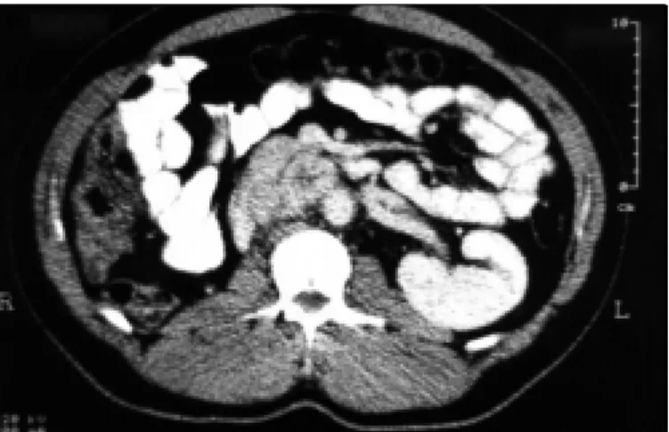
six years, with normal catecholamine serum levels. After this period, a rise in arterial blood pressure level was found during a medical fol-low-up assessment. T he norepinephrine serum level was found to be 3347 (normal 40 to 268 pg/ml) and urinary norepinephrine 741 (nor-mal 65 to 400 mcg/24h). T he epinephrine se-rum level was normal. T he CT scan of the ab-domen revealed a pre-aortic mass of 3 x 4 cm situated immediately below the renal artery (Figure 2). T he existence of the mass was con-firmed by magnetic resonance imaging (MRI), appearing with hypersignal in T 2. T he radio-isotope study with meta-iodobenzyl-guanidine (MIBG) demonstrated considerable enhance-ment of the tumor.
T he surgical removal of the mass described was undertaken by median laparotomy with retroperitoneal dissection. A frozen biopsy of the specimen showed free margins. T he nor-mal postoperative catecholamine levels con-firmed the total removal of the tumor.
Alpha and beta-blockers (prazosin and propranolol, respectively) were used as preoperative treatment in both operations. T he adrenergic blockage was begun 15 days before the surgery with gradual adjustment of dos-age, reaching 6 mg of prazosin and 80 mg of propranolol per day. T he pathological study of the specimen showed malign pheochromo-cytoma with free margins. T he patient is well after a 7-year follow-up.
○ ○ ○ ○ ○ ○ ○ ○ ○ ○ ○ ○ ○D ISC U SSIO N○ ○ ○ ○ ○ ○ ○
T he invasion of the inferior vena cava has already been described in relation to many
ab-dominal tumors. T he retroperitoneal tumor most commonly associated with extension into this vessel is renal cell carcinoma12 but it rarely occurs in pheochromocytomas. T he extension may occur by direct invasion of the vessel wall or by luminal progression within the vein. T he direct invasion is more frequently observed in malignant neoplasia.17 T he technique of choice employed by many authors for the removal of tumors that present a thrombus inside the in-fra or supra-hepatic vena cava without affect-ing the right atrium utilizes extra corporeal cir-culation with hypothermia and total cardiac arrest for their safe removal.12 However, in such cases, radical surgery is also practicable with-out the use of extra corporeal circulation,20 thus avoiding the morbidity related to this proce-dure as well as the increase in hospital costs, as demonstrated in our case.
Others prefer to use the piggyback style of mobilization of the liver to access the infe-rior vena cava, which is the technique em-ployed for orthotopic liver transplantation.21 T he extension of the tumor into the inferior vena cava with no invasion of the vessel wall does not necessarily mean malignancy,22 which occurs when there is direct invasion of the vessel wall.23 T he presence of distant metastases is the unquestionable criterion for the classification of malignant pheochromo-cytoma.24 Although malignant pheochromo-cytomas tend to reappear or present metastases during long-term follow-up, sur-gical removal of these tumors is the best op-tion for treatment,25 considering that the pa-tients have a ten year survival rate with no evidence of disease.
T he incidence of extra adrenal pheochro-mocytomas or multicentric tumors varies from 3.8 to 10%,26,27 the most common site being Zuckerkandl’s organ. In these cases, where the concentration of norepinephrine is consider-ably greater than that of epinephrine, the ex-istence of extra adrenal pheochromocytoma may be suspected, as the methylation occurs mainly in the adrenal gland. T his was observed in the case presented. In rare cases, measure-ment of plasma catecholamine values in blood samples taken from different levels of the in-ferior vena cava may also be useful in the search for possible sites of metastases.26
At present, MRI is tending to become the method of choice for the assessment of thrombi in the large vessels, visualized mainly in T 1 and T 2 sequences.17 In the past, laparotomy was the approach of choice for pheochromo-cytomas because of the frequency of bilateral disease and the possibility of the existence of extra adrenal tumors misdiagnosed prior to surgery. Nevertheless, with the improvement in methods of localization such as CT scan, MRI and MIBG scintigraphy, the surgeon enjoys the possibility of choosing the best sur-gical approach beforehand.
T he use of alpha and beta blocking agents preoperatively ensures a fully expanded vascu-lar system and also minimizes intraoperative blood pressure fluctuation. Recently, the use of other drugs such as calcium channel blockers, instead of alpha and beta adrenergic antagonists has been demonstrated.28 However, due to the effectiveness and low costs of alpha and beta blocking agents, they are still being used as drugs of choice in our experience.
Figure 2. CT scan of the abdomen showing a 3 x 4 cm pre-aortic mass. Figure 1. Hematoxylin-eosin staining showing vascular invasion of the malignant
pheochro-mocytoma.
São Paulo M edical Journal - Revista Paulista de M edicina
88
1. Korobkin M, Francis IR, Kloos RT, Dunnick NR. T he incidental
adrenal mass. Radiol Clin North Am 1996;34(5):1037-54.
2. Bravo EL, Gifford RW Jr. Current concepts –
pheochromocy-toma: diagnosis, localization and management. N Engl J Med 1984;311(20):1298-303.
3. Young JD Jr, Qureshi AS, Connor T B, Wiswell JG. Problem
le-sion in adrenal surgery. J Urol 1969;101:233-40.
4. Rote AR, Flint LD, Ellis FH Jr. Intracaval recurrence of
pheochro-mocytoma extending into right atrium. Surgical management us-ing extra corporeal circulation. N Engl J Med 1977;296:1269-71.
5. Costello P, Clouse ME, Kane RA, Paris A. Problems in the
diag-nosis of adrenal tumors. Radiology 1977;125:335-41.
6. Scott HW, Reynolds V, Green N, Page D, Oates JA, Robertson
D. Clinical experience with malignant pheochromocytoma. Surg Gynecol Obstet 1982;154:801-18.
7. Russinovich NAE, Recio MG, Tishler JM, Zornes SL, Luna RF.
Intracaval extension of pheochromocytoma: ultrasonographic dem-onstration. Can Assoc Radiol J 1982;33:53-5.
8. Hoffman JC, Weiner SN, Koenigsberg M, Morehouse H, Smith
T. Pheochromocytoma invasion of the inferior vena cava: sonographic evaluation. Radiology 1983;149:793-5.
9. Smith EJ, McPherson GAD, Lynn J. Inferior vena cava
involve-ment by a pheochromocytoma. Br J Surg 1987;74:597. 10. Dicke T E, Henry ML, Minton JP. Intracaval extension of
pheo-chromocytoma simulating pulmonary embolism. J Surg O ncol 1987;34:160-4.
○ ○ ○ ○ ○ ○ ○ ○ ○ ○ ○ ○ ○ ○ ○ ○ ○ ○ ○ ○ ○ ○ ○ ○ ○ ○ ○ ○ ○ ○ ○ ○ ○ ○ ○ ○ ○ ○ ○ ○ ○ ○ ○ ○ ○ ○ ○ ○ ○ ○ ○ ○ ○ ○ ○ ○ ○ ○ R EFER EN C ES○ ○ ○ ○ ○ ○ ○ ○
11. Levine E, de Vries P, Wetzel LH. MR imaging of inferior vena cava recurrence of extra-adrenal pheochromocytoma: a case re-port. J Comput Assist Tomogr 1987;11:717-8.
12. Novick AC, Kaye MC, Cosgrove DM, et al. Experience with car-diopulmonary bypass and deep hypothermic circulatory arrest in the management of retroperitoneal tumors with large vena cava thrombi. Ann Surg 1990;212(4):472-6.
13. Dumm CW, Snyder WH, Ring WS, Latson T W. Pheochromocy-toma with extension into the inferior vena cava: a case report. Surgery 1992;111(4):472-4.
14. Boneschi M, Miani S, Erba M, Giuffrida GF, Giordanengo F. Malignant neoplasms invading into the inferior vena cava. Minerv Cardioangiol 1995;43(3):91-5.
15. Raghavan R, Ince PG, Walls T J, Gholkar A, Dark JH, Foster JB. Malignant cerebrovascular thromboembolization by pheochromo-cytoma. Clin Neuropathol 1995;14(2):69-71.
16. Rotker J, Oberpenning F, Scheld HH, Hertle L, Knichwitz G, Hammel D. Pheochromocytomas with extension into central vas-cular structures. Ann T horac Surg 1996;61(1):222-4. 17. Lau T N, Goddard P, Vaidya M, Calloway M, Bullimore J.
In-volvement of the inferior vena cava by adrenal pheochromocy-toma: MRI findings. Br J Radiol 1997;70:303-5.
18. Melicow MM. One hundred cases of pheochromocytoma (107 tumors) at the Columbia Presbyterian Medical Center, 1926-1976. Cancer 1997;40:1987-2004.
19. Arroj a J M , G udinchet F, M aeder P, Fournier D .
Phéochromocytome multiple familial: démonstration échographique de multiples localisations surrénaliennes, coeliaques et vésicales chez un enfant. Schweiz Rundsch Med Prax 1995;84(43):1231-4.
20. Hedican SP, Marshall FF. Adrenocortical carcinoma with intracaval extension. J Urol 1997;158(6):2056-61.
21. Ciancio G, Hawke C, Soloway M. T he use of liver transplanta-tion technique to aid in the surgical management of urological tumors. J Urol 2000;164:665-72.
22. Dunnick NR, Dompman JL, Geelhoed GW. Intravenous exten-sion of endocrine tumors. A J R 1980;135:471-6.
23. Meyers MA, King MC. Unusual radiological features of pheo-chromocytoma. Clin Radiol 1969;20:52-6.
24. D avis P. Malignant pheochromocytoma with functioning metastases. Lancet 1955;269:274-5.
25. Mahoney EM, Harrison JH. Malignant pheochromocytoma: clini-cal course and treatment. J Urol 1977;18(2):225-9. 26. Delarue NC, Morrow JD, Kerr JH, Colapinto RF.
Pheochromo-cytoma in the modern context. Can J Surg 1978;21(5):387-94. 27. van Heerden JA, Roland CF, Carney JA, Sheps SG, Grant CS.
Long-term evaluation following resection of apparently benign pheochromocytoma(s)/ paraganglioma(s). World J Surg 1990;14(3):325-9.
28. Ulchaker JC, Goldfarb DA, Bravo EL, Novick AC. Successful outcomes in pheochromocytoma surgery in the modern era. J Urol 1999;161(3):764-7.
CONTEXTO: Feocromocitoma com invasão de veia cava inferior é raro. Tumores multicêntricos são igualmente raros, estando presentes em até 10% dos casos. A cirurgia é o tratamento de escolha, uma vez que a sobrevida livre da doença é longa.
TIPO DE ESTUDO: Relato de caso.
RELATO DE CASO: Relatamos um caso de feocromocitoma de adrenal direita com extensão para veia cava inferior supra-diafragmática, retirado cirúrgicamente através de tóraco-freno-laparotomia, sem a necessidade de circulação extra-corpórea. Após seis anos de
○ ○ ○ ○ ○ ○ ○ ○ ○ ○ ○ ○ ○ ○ ○ ○ ○ ○ ○ ○ ○ ○ ○ ○ ○ ○ ○ ○ ○ ○ ○ ○ ○ ○ ○ ○ ○R ESU M O○ ○ ○ ○ ○ ○
Antonio M a rmo Lucon, M D, PhD. Asso ciate Pro fesso r, Divisio n o f Uro lo g y, Department o f Surg ery, Faculty o f Medi-cine, University o f São Paulo , São Paulo , Braz il.
Rena to Fa lci Júnior, M D. Resident in Uro lo g y, Divisio n o f Uro lo g y, Department o f Surg ery, Faculty o f Medicine, Uni-versity o f São Paulo , São Paulo , Braz il.
José N ery Pra x edes, M D, PhD. Asso ciate Pro fesso r, De-partment o f N ephro lo g y, Faculty o f Medicine, University o f São Paulo , São Paulo , Braz il.
M a rcel Cerqueira Cesa r M a cha do, M D, PhD. Asso ci-ate Pro fesso r, Divisio n o f G astro entero lo g ical Surg ery, De-partment o f Surg ery, Faculty o f Medicine, University o f São Paulo , São Paulo , Braz il.
Luis Ba ltha za r Sa lda nha , M D, PhD. Asso ciate Pro fesso r, Department o f Patho lo g y, Faculty o f Medicine, University o f São Paulo , São Paulo , Braz il.
M a rcelo M a rcondes M a cha do, M D, PhD. Full Pro fesso r and Head, Department o f N ephro lo g y, Faculty o f Medicine, University o f São Paulo , São Paulo , Braz il.
Sa mi Ara p, M D, PhD. Full Pro fesso r and Head, Divisio n o f Uro lo g y, Department o f Surg ery, Faculty o f Medicine, Uni-versity o f São Paulo , São Paulo , Braz il.
Sources of funding: N o t declared
Conflict of interest: N o t declared
La st received: 2 2 September 2 0 0 0
Accepted: 3 0 O cto ber 2 0 0 0
Address for correspondence:
Anto nio Marmo Luco n
Divisão de Clínica Uro ló g ica, Ho spital das Clínicas da Faculdade de Medicina da Universidade de São Paulo Av. Dr. Eneas de Carvalho Ag uiar, 2 5 5
7o andar - sala 7 1 0 F
São Paulo / SP – Brazil - CEP 0 5 4 0 3 -9 0 0 E-mail: webmaster@ uro lo g ia.hcnet.usp.br
CO PYRIG HT© 2 0 0 1 , Asso ciação Paulista de Medicina
○ ○ ○Pu b l i sh i n g i n f o r m a t i o n○ ○ ○ ○ ○ ○ ○ ○ ○ ○ ○ ○ ○ ○ ○ ○ ○
seguimento, outro feocromocitoma foi achado no órgão de Zuckerkandl. A excisão cirúrgica completa do tumor foi realizada através de laparotomia mediana e dissecção retroperito-neal. Em ambos os casos, margens cirúrgicas livres do tumor e níveis pós-operatórios normais de catecolaminas garantiram remoção total do feocromocitoma. O estudo anatomopatológico revelou feocromocitoma maligno com margens livres de neoplasia em ambos os espécimes.
PALAVRAS-CHAVE: Feocromocitoma. Neoplasia retroperitoneal. Tumor adrenal. Veia cava.