C
ASER
EPORT| R
ELATO DEC
ASO477
Recovery of renal function after bilateral renal vein
thrombo-sis episode as complication of membranous
glomerulopa-thy: case report
Recuperação da função renal após episódio de trombose de veia
renal bilateral como complicação da glomerulopatia membranosa:
relato de caso
Authors
Ana Larissa Pedrosa Ximenes 1
Elizabeth De Francesco Daher 2,3
Pedro Duarte Barreto Castillo 4
Francisco Eduardo Siqueira da Rocha 5
Camila Freire Salem de Miranda 2
Flavio Bezerra de Araujo 2
1 Hospital Geral de
Fortaleza, Departamento Medicina Interna, Fortaleza - CE, Brazil.
2 Hospital Geral de
Fortaleza, Departamento de Nefrologia, Fortaleza - CE, Brazil.
3 Universidade Federal
do Ceará, Departamento Medicina Interna, Fortaleza - CE, Brazil.
4 Hospital Geral de
Fortaleza, Fortaleza - CE, Brazil.
5 Hospital Geral de
Fortaleza, Departamento Cirurgia Vascular, Fortaleza - CE, Brazil.
Submitted on: 01/17/2017. Approved on: 03/20/2017.
Correspondence to:
Ana Larissa Pedrosa Ximenes. E-mail: [email protected]
C
ASEREPORTA 33-year-old male patient, previously healthy and without known comorbidi-ties, was admitted to the Nephrology Service of the General Hospital of Fortaleza (HGF), complaining of gastric fullness, lower limb edema, unproductive cough and frothy urine for three months. He also reported dyspnea on average ef-forts progressing to great efef-forts 15 days from admission. He had a weight loss of 10 kg in this period.
Good general condition, eupneic, alert and oriented; small, palpable, mobile, fi-broelastic lymph node with approxima-tely one centimeter in the left posterior
DOI: 10.5935/0101-2800.20170085
Renal vein thrombosis (RVT) is a com-plication often associated with nephrotic syndrome. It occurs due to a state of hy-percoagulability common in the diseases that attend to this syndromic diagnosis. It should be suspected whenever there is ne-phrotic syndrome associated with sudden flank pain, hematuria and worsening of proteinuria. Bilateral RVT also presents with frequently oliguric renal dysfunction. This case reports a 33-year-old patient hospitalized for a nephrotic syndrome, with etiologic investigation suggestive of primary membranous glomerulopathy, which evolved with bilateral RVT associ-ated with deterioration of renal function and need for renal replacement therapy. He promptly performed angiography with thrombectomy and thrombolysis, evolving with recovery of renal function in two weeks.
A
BSTRACTKeywords: glomerulonephritis,
membra-nous; proteinuria; venous thrombosis.
A trombose de veia renal (TVR) é uma complicação muitas vezes associada à sín-drome nefrótica. Ocorre devido a um es-tado de hipercoagulabilidade comum nas enfermidades que cursam com esse diag-nóstico sindrômico. Deve ser suspeitada sempre que houver síndrome nefrótica associada à dor súbita em flanco, hema-túria e piora da proteinúria. TVR bilate-ral cursa, ainda, com disfunção renal fre-quentemente oligúrica. Esse caso reporta um paciente de 33 anos internado por um quadro de síndrome nefrótica, com inves-tigação etiológica sugestiva de glomerulo-patia membranosa primária, que evoluiu com TVR bilateral associada à deterio-ração da função renal e necessidade de terapia substitutiva renal. Realizou, pron-tamente, angiografia com trombectomia e trombólise, evoluindo com recuperação da função renal em duas semanas.
R
ESUMOPalavras-chave: glomerulonefrite mem-branosa; trombose venosa; proteinúria.
cervical chain. Heart auscultation wi-thout changes. Respiratory auscultation with universal vesicular murmur present, reduced in the left base. Flat abdomen, flaccid, painless to palpation, without visceromegaly, Traube free. Palpable peri-pheral pulses with lower limb edema (+/4 +), absence of cyanosis and well perfused extremities.
Braz. J. Nephrol. (J. Bras. Nefrol.) 2017;39(4):477-480 Bilateral renal vein thrombosis
478
absence of monoclonal peak. The remaining labora-tory tests are described in Table 1.
Urinary tract ultrasonography (US) evidenced slightly increased kidneys (RD: 13.8 x 6.8 x 5.8cm Parenchyma 1.5cm - RE: 13 x 7.1 x 6.1cm - Parenchyma: 1,5cm) and increased cortical echo-genicity, suggestive of parenchymal nephropathy with no stones. She undertook investigation of secondary causes of nephrotic syndrome that were all negative, and a renal biopsy was performed, which was sugges-tive of membranous glomerulopathy, according to the light microscopy illustrated in Figure 1.
After 1 week of admission, he was submitted to another complete abdomen US, due to an ill-defined abdominal pain, which showed signs suggestive of thrombosis of the right renal vein. After that, full anticoagulation with continuous infusion of heparin was initiated; on the following day, the patient de-veloped anuria for more than 12 hours, nausea, two emetic episodes, two febrile episodes (37.8ºC and 38.1ºC) and worsening of nitrogenous slags (cre-atinine 5.6 and urea 60), with suspicion of bilateral renal vein thrombosis. The patient was submitted to renal angiography (arteriography and phlebography), which confirmed the hypothesis of bilateral renal vein thrombosis (Figure 2). Bilateral thrombectomy and thrombolysis were performed on the left and the pa-tient was maintained in anticoagulation (initially with heparin and subsequently with warfarin).
The patient remained on hemodialysis for two weeks, evolving with progressive improvement in di-uresis and renal function. He was discharged with re-nal function recovery, creatinine of 1.66 mg/dl. First outpatient visit after discharge the patient had creati-nine of 0.77mg/dl.
D
ISCUSSIONRenal vein thrombosis (RVT) was described by Rayer in 1840 and its association with nephrotic syndrome (NS) was first reported in 1939 by Doroe, Schlesinger and Savitz.1
Initially, there were conflicting reports about the cause and effect relationship of the RVT in the NS, but in the last years RVT was better described as a consequence of NS.2
RVT is seen more frequently in membranous glo-merulopathies and membranoproliferative than in other types, such as minimal lesion and FSGS.3
Advanced age, membranous nephropathy, severe proteinuria and hypoalbuminemia are recognized as increased risk factors for the development of
thromboembolism.4
The RVT pathogenic mechanism in the NS is not fully understood, but it is established that the NS is associated with a state of hypercoagulability, and it is further reinforced by urinary loss and, consequently, reduced serum antithrombin level III.5
The clinical condition results from the balance be-tween acute occlusion, extension of thrombosis and development of collateral circulation. The acute pre-sentation of renal vein thrombosis is infrequent and is mainly characterized by acute flank pain and hematu-ria. The laboratory findings that may suggest RVT are proteinuria (significant increase after event), increase in serum creatinine, hematuria, glycosuria, pyuria, hyperchloremic acidosis.6,7 In most cases, the patients are asymptomatic, making the RVT underdiagnosed.8
Early diagnosis is essential because it is a revers-ible condition. The gold standard diagnostic test is renal phlebography, but USG with renal vein Doppler and contrast abdominal CT have been fast and safe noninvasive measurements for the direct visualization of the thrombus.9,10
The recommended treatment is full anticoagula-tion, which should be started immediately. The cur-rent recommendation is to begin with heparinization and after combining warfarin, and the total time for anticoagulation for a first episode of venous throm-boembolism is at least 3-6 months, and until the cause of NS has been resolved or is in remission.11,12
In relation to the new oral anticoagulants (direct factor Xa inhibitors and direct thrombin inhibitor), warfarin anticoagulation is already recommended as an option in the treatment of general deep venous thromboembolism and pulmonary embolism.13,14 The
great limitation in the use of these medications is the impossibility of using them in patients with creatinine clearance lower than 15ml/min.15
Braz. J. Nephrol. (J. Bras. Nefrol.) 2017;39(4):477-480 Bilateral renal vein thrombosis
479
Figure 1. Light microscopy of the renal biopsy fragments - silver staining - evidencing thickening of glomerular basement membrane with spicules.
Figura 2. Bilateral renal angiography (arterial and venous).
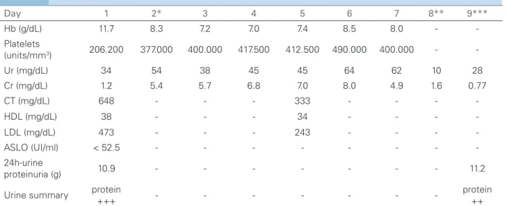
Day 1 2* 3 4 5 6 7 8** 9***
Hb (g/dL) 11.7 8.3 7.2 7.0 7.4 8.5 8.0 -
-Platelets
(units/mm3) 206.200 377.000 400.000 417.500 412.500 490.000 400.000 -
-Ur (mg/dL) 34 54 38 45 45 64 62 10 28
Cr (mg/dL) 1.2 5.4 5.7 6.8 7.0 8.0 4.9 1.6 0.77
CT (mg/dL) 648 - - - 333 - - -
-HDL (mg/dL) 38 - - - 34 - - -
-LDL (mg/dL) 473 - - - 243 - - -
-ASLO (UI/ml) < 52.5 - - -
-24h-urine
proteinuria (g) 10.9 - - - 11.2
Urine summary protein
+++ - - -
-protein ++
Hb - hemoglobin, PTA - prothrombin time of activity, Ur - urea, Cr - creatinine, CT - total cholesterol, HDL - high density lipoprotein, LDL - low density lipoprotein. * Initiated hemodialysis. ** Renal function at the time of the patient discharge. *** Renal function upon the patient’s first outpatient return visit.
TABLE 1 LABORATORYTESTSPERFORMEDDURINGHOSPITALIZATION
in a patient with NS. The patient did not present the classical clinical signs. Bilateral RVT was suspected due to anuria and sudden worsening of renal func-tion. The venogram, gold standard, was performed to obtain the diagnosis, as well as the therapeutic inter-vention, with bilateral thrombectomy and thromboly-sis located in the left renal vein. There was modest improvement in renal flow immediately and complete recovery of renal function after two weeks of the event.
R
EFERENCES1. Chugh KS, Malik N, Uberoi HS, Gupta VK, Aggarwal ML, Sin-ghal PC, et al. Renal vein thrombosis in nephrotic syndrome-a prospective study and review. Postgrad Med J 1981;57:566-70. PMID: 7329894 DOI: http://dx.doi.org/10.1136/ pgmj.57.671.566
Braz. J. Nephrol. (J. Bras. Nefrol.) 2017;39(4):477-480 Bilateral renal vein thrombosis
480
3. Llach F, Arieff AI, Massry SG. Renal vein thrombosis and nephrotic syndrome. A prospective study of 36 adult pa-tients. Ann Intern Med 1975;83:8-14. DOI: http://dx.doi. org/10.7326/0003-4819-83-1-8
4. Llach F, Koffler A, Finck E, Massry SG. On the incidence of renal vein thrombosis in the nephrotic syndrome. Arch Intern Med 1977;137:333-6. PMID: 843151 DOI: http://dx.doi. org/10.1001/archinte.1977.03630150039012
5. Janda SP. Bilateral renal vein thrombosis and pulmonary embo-lism secondary to membranous glomerulonephritis treated with percutaneous catheter thrombectomy and localized throm-bolytic therapy. Indian J Nephrol 2010;20:152-5. DOI: http:// dx.doi.org/10.4103/0971-4065.70848
6. Qian Q, Saucier NA, King BF. Acute bilateral renal vein throm-bosis. Am J Kidney Dis 2009;54:975-8. PMID: 19748714 DOI: http://dx.doi.org/10.1053/j.ajkd.2009.06.035
7. Shumei S, Ling X, Yanxia W, Lei Z, Yuanyuan S. Acute kidney injury as the first sign of spontaneous renal vein thrombosis: re-port of 2 cases. J Thromb Thrombolysis 2012;33:129-32. DOI: http://dx.doi.org/10.1007/s11239-011-0633-2
8. Laville M, Aguilera D, Maillet PJ, Labeeuw M, Madonna O, Zech P. The prognosis of renal vein thrombosis: a re-evaluation of 27 cases. Nephrol Dial Transplant 1988;3:247-56.
9. Sandhu G, Bansal A, Ranade A, Jones J, Cortell S. Bilateral renal vein thrombosis can cause nephrotic range proteinuria. QJM 2014;107:763-5. PMID: 22279146 DOI: http://dx.doi. org/10.1016/j.ejvs.2007.02.017
10. Asghar M, Ahmed K, Shah SS, Siddique MK, Dasgupta P, Khan MS. Renal vein thrombosis. Eur J Vasc Endovasc Surg 2007;34:217-23.
11. Wu CH, Ko SF, Lee CH, Cheng BC, Hsu KT, Chen JB, et al. Successful outpatient treatment of renal vein thrombosis by low-molecular weight heparins in 3 patients with nephrotic syndrome. Clin Nephrol 2006;65:433-40.
12. Singhal R, Brimble KS. Thromboembolic complications in the nephrotic syndrome: pathophysiology and clini-cal management. Thromb Res 2006;118:397-407. PMID: 15990160
13. Madan S, Shah S, Dale P, Partovi S, Parikh SA. Use of novel oral anticoagulant agents in venous thromboembolism. Cardiovasc Diagn Ther 2016;6:570-81.
14. Kearon C, Akl EA, Ornelas J, Blaivas A, Jimenez D, Bou-nameaux H, et al. Antithrombotic Therapy for VTE Di-sease: CHEST Guideline and Expert Panel Report. Chest 2016;149:315-52.
15. Belmar Vega L, de Francisco ALM, Bada da Silva J, Galván Espinoza L, Fernández Fresnedo G. Nuevos anticoagulantes orales en pacientes con enfermedad renal crónica. Nefrologia 2017;37:244-52.