Acoustic radiation force impulse (ARFI) elastography
compared with biopsy for evaluating hepatic ibrosis after
liver transplantation: a cross-sectional diagnostic study
Elastograia ARFI (
acoustic radiation force impulse
) comparada com biópsia na
avaliação de ibrose hepática após transplante: estudo transversal diagnóstico
Joel Schmillevitch
I, Maria Cristina Chammas
II, Vincenzo Pugliese
III, Edson Abdala
IV, Adriana Cortez Rizzon
V, Venâncio Alves
VI,
Luiz Augusto Carneiro
VII, Giovanni Cerri
VIIIInstituto de Radiologia (InRAD), Hospital das Clínicas (HC), Faculdade de Medicina da Universidade de São Paulo (FMUSP),
São Paulo (SP), Brazil
ABSTRACT
CONTEXT AND OBJECTIVE: Biopsies are used after liver transplantation to evaluate ibrosis. This study aimed to evaluate the elasticity of transplanted livers by means of a non-invasive examination, acoustic radiation force imaging (ARFI) elastography, correlating the results with the histological analysis.
DESIGN AND SETTING: Cross-sectional study in a public university hospital.
METHODS: All patients consecutively operated between 2002 and 2010 with an indication for biopsy were evaluated by means of elastography. The radiologist evaluating ARFI and the pathologist doing anatomopathological examinations were blinded to each other’s evaluations.
RESULTS: During the study period, 33 patients were included. The indication for transplantation was cir-rhosis due to hepatitis C in 21 cases (63%). Liver biopsies showed absence of ibrosis (F0) in 10 patients, F1 in 11, F2 in 8 and F3 in 4. There were no cases of F4 (cirrhosis). The diference in ARFI values (degree of ibrosis) was 0.26 (95% conidence interval, CI: 0.07-0.52) between the groups F0-F1 and F2-F4 (P = 0.04). An area under the curve of 0.74 (CI: 0.55-0.94) and a cutof of 1.29 m/s between the groups resulted in the best relationship between sensitivity and speciicity. Sensitivity (0.66; CI: 0.50-0.83) was lower than speciic-ity (0.85; CI: 0.72-0.97). There was no signiicant diference in ARFI between patients with hepatitis C and those with other diseases.
CONCLUSIONS: The values obtained from elastography were not afected by inlammatory reaction or anatomical alterations. A cutof point of 1.29 m/s separating patients with or without signiicant ibrosis was identiied.
RESUMO
CONTEXTO E OBJETIVO: Biópsias são utilizadas para avaliar ibrose após transplante de fígado. O estudo objetivou avaliar a elasticidade hepática após transplante por meio de um exame não invasivo, a elastogra-ia ARFI (acoustic radelastogra-iation force imaging), correlacionando-a com a análise histológica.
TIPO DE ESTUDO E LOCAL: Estudo transversal em hospital público universitário.
MÉTODOS: Todos os pacientes consecutivamente operados entre 2002 e 2010, com indicação para bióp-sia, foram avaliados por elastograia. O radiologista avaliando ARFI e o patologista fazendo exames anato-mopatológicos estavam cegos para as avaliações um do outro.
RESULTADOS: No período do estudo, 33 pacientes foram incluídos. A indicação para o transplante foi cirrose por hepatite C em 21 (63%). As biópsias mostraram ausência de ibrose (F0) em 10 pacientes, F1 em 11, F2 em 8, F3 em 4 e nenhum caso de F4 (cirrose). A diferença nos valores de ARFI (grau de ibrose) foi de 0,26 (intervalo de coniança, IC, de 95%: 0,07-0,52) entre os grupos F0-F1 e F2-F4 (P = 0,04). A área sob a curva de 0,74 (IC: 0,55-0,94) e o valor de corte de 1,29 m/s entre os grupos resultaram na melhor relação entre sensibilidade e especiicidade, de 0,57. A sensibilidade (0,66; IC: 0,50-0,83) foi menor que a especi-icidade (0,85; IC: 0,72-0,97). Não houve diferença signiicativa em ARFI entre pacientes com hepatite C e aqueles com outras doenças.
CONCLUSÕES: Os valores obtidos com a elastograia não foram afetados por reação inlamatória ou alte-rações anatômicas. Foi identiicado ponto de corte de 1,29 m/s que separa pacientes com ou sem ibro-se signiicativa.
IMD. Researcher, Instituto de Radiologia (InRad), Hos pi tal das Clínicas (HC), Faculdade de Medicina da Universidade de São Paulo (FMUSP), São Paulo, SP, Brazil.
IIMD, PhD. Director, Ultrasonography Service, Instituto de Radiologia (InRad), Hospital das Clínicas (HC), Faculdade de Medicina da Universidade de São Paulo (FMUSP), São Paulo (SP), Brazil. IIIMD, PhD. Attending Physician, Liver and Digestive System Organ Transplantation Division, Hospital das Clínicas (HC), Faculdade de Medicina da Universidade de São Paulo (FMUSP), São Paulo (SP), Brazil. IVMD, PhD. Professor, Department of Infectious and Parasitic Diseases, Hospital das Clínicas (HC), Faculdade de Medicina da Universidade de São Paulo (FMUSP), São Paulo (SP), Brazil. VNurse, Coordinator of the Liver and Digestive System Organ Transplantation Division, Hospital das Clínicas, Faculdade de Medicina da Universidade de São Paulo (FMUSP), São Paulo (SP), Brazil. VIMD, PhD. Titular Professor, Department of Pathology, Faculdade de Medicina da Universidade de São Paulo (FMUSP), São Paulo (SP), Brazil.
VIIMD, PhD. Titular Professor and Director, Liver and Digestive System Organ Transplantation Division, Hospital das Clínicas (HC), Faculdade de Medicina da Universidade de São Paulo (FMUSP), São Paulo (SP), Brazil.
VIIIMD, PhD. Titular Professor, Department of Radiology, Universidade de São Paulo (FMUSP), São Paulo (SP), Brazil.
KEY WORDS:
Elasticity imaging techniques. Liver transplantation. Fibrosis.
Liver cirrhosis. Ultrasonography.
PALAVRAS-CHAVE:
Técnicas de imagem por elasticidade. Transplante de fígado.
INTRODUCTION
Liver transplantation is an established form of therapy for treat-ing acute and chronic end-stage hepatic diseases and for some hepatic tumors, with ten-year grat survival greater than 70% at most transplantation centers.1-4 Nonetheless, histological changes
in transplanted livers can occur subclinically and progressively even in asymptomatic patients and without signiicant laboratory alterations.3-6
Liver biopsy is currently the gold standard for histological evaluation of the liver.7-10 However, it is invasive and poses a risk
of complications with morbidity around 0.3 to 0.6% and mor-tality of 0.05%, besides hospitalization of at least 6-18 hours.3,4
In order to diagnose these alterations, some transplantation centers recommend routine biopsies at ixed intervals (annu-ally) ater liver transplantation, thus called “protocol biopsies”.11
Other centers do not follow this protocol, taking the view that liver biopsy is both risky and invasive.3,4
he main histological alterations observed in anatomopatho-logical evaluations on biopsies on transplanted livers involve inlammation and ibrosis.6 However, the degree of ibrosis may
be underestimated in situations of inadequate sampling, given that the volume of the sample evaluated in the biopsy is only 1/50,000 of the organ. here have been reports of discrepancies in both inter and intraobserver histological analyses, at estimated rates of 10 to 30% of the cases.12
New noninvasive diagnostic methods, such as serum tests, magnetic resonance imaging and elastography, which have shown signiicant correlation with the histological test,13 can be introduced
with the objective of reducing the number of biopsies. his has already been happening in relation to patients with chronic hepa-topathy. Several noninvasive methods have been proposed for stag-ing hepatic ibrosis. hese include indices based on biochemical tests and imaging examinations.14 Among the biochemical tests,
the aspartate aminotransferase (AST) to platelet ratio index (APRI) stands out due to its simplicity and low cost. he APRI was pro-posed in 2003 by Wai et al. and promising results were reported.15,16
Transient hepatic elastography (Fibroscan, Echosens, France) was the irst noninvasive diagnostic method for quantifying the degree of hepatic ibrosis, and the irst scientiic paper on this method was published in 2003.17 Its use on patients with liver
disease is based on the relationship between stifness and hepatic ibrosis. It measures the elasticity of a volume of hepatic tissue corresponding to a cylinder with a thickness of 1 cm and depth of 2 cm, with results expressed in kilopascals. Diferent studies have assessed its diagnostic accuracy. Using optimized cutof points, Castéra et al.7 found a positive predictive value (PPV) of 95% and
a negative predictive value (NPV) of 48% for the presence of sig-niicant ibrosis, and PPV of 77% and NPV of 95% for the pres-ence of cirrhosis. he same group also reported concordance of
83% with liver biopsy regarding the diagnosis of signiicant ibro-sis and 90% regarding the diagnoibro-sis of cirrhoibro-sis.7
Another method for quantifying the degree of hepatic ibro-sis was presented more recently, in 2009: acoustic radiation force imaging (ARFI) (S2000, Siemens, Germany). In the ARFI elas-tography method, short-duration pulses are emitted in the region of interest (ROI), thus generating waves with velocities measured in meters per second.18,19 Fierbinteanu-Braticevici et al.
mea-sured the area under the ROC curve of ARFI elastography and found accuracy of 90.2% in predicting ibrosis.20 Some
advan-tages of ARFI elastography merit special emphasis, including the facts that manual compression is unnecessary and that this elas-tography is coupled to conventional ultrasound equipment, thus also enabling analysis of other organs.
Elastography and other noninvasive methods have been used over the short term (one to ive years) on patients undergoing liver transplantation, in order to determine the degrees of
ibro-sis.8,9,11,21 Determination of the degrees of hepatic ibrosis in liver
transplant patients is crucial for early detection of ibrosis and for making decisions on the therapeutic approach to be adopted.1
OBJECTIVE
he current study aimed to evaluate liver elasticity in patients who had undergone liver transplantation at least one year ear-lier, by means of ARFI elastography, correlating the results with the histological analysis and determining a cutof value for ARFI results by comparing patients presenting ibrosis grades 0 and 1 with those presenting grades 2 to 4.
METHODS
Study design, location and ethics
In this cross-sectional observational study, we analyzed patients operated on for liver transplantation in the Department of Gastroenterology, Liver Transplantation and Surgery of the School of Medicine of the University of São Paulo. During the period between April 2012 and March 2015, all patients with an indication for liver biopsy were recruited for ARFI evaluation. he research project was approved by the Ethics Committee for Analysis of Research Projects (CAPPESQ), and the patients signed informed consent forms.
Participants
elastography. Patients who presented post-transplantation vascu-lar and biliary complications were excluded.
Procedures: biopsy, elastography and laboratory tests he biopsy, elastography and laboratory tests were carried out on the same day, or with not more than 15 days between the pro-cedures. All the ultrasound scans were performed by the same radiologist (JS), and all the anatomopathological exams by the same pathologist (VA). he radiologist and the pathologist were blinded to each other’s evaluations and to the patients’ labora-tory results.
Blood samples were also taken from the patients (not more than 15 days before or ater the biopsy and the ARFI examina-tion), to evaluate the following variables: aspartate aminotrans-ferase (AST), alanine aminotransaminotrans-ferase (ALT), platelets, albumin, direct and indirect total bilirubin, blood glucose, insulin, Homa index, total cholesterol and fractions and triglycerides. he bio-chemical reference values were deined in accordance with the ranges of the institution’s biochemistry laboratory.
he liver biopsies were collected subcutaneously,3,4 guided
by ultrasonography, using needles of 1.2-1.4 mm in diameter. he length of the liver biopsy tissue sample was at least 1.5 mm, with a minimum of 10 port spaces in all cases. he biopsy frag-ments were immediately ixed in formalin.
he inlammatory activity was graded and the ibrosis was staged in accordance with the criteria of the METAVIR Cooperative Study Group12 classiication and other
histologi-cal parameters (Chart 1), for each liver biopsy. According to the METAVIR classiication, patients staged as F0 or F1 were considered to have no ibrosis or minimal ibrosis. Signiicant ibrosis was deined as stages F2, F3 and F4. Patients classi-ied as F4 were considered to have hepatic cirrhosis. For this study, the patients were divided in two groups: the F0-F1 group and the F2-F3-F4 group, given that patients with ibro-sis grade 2 and above may require treatment, depending on the disease etiology.
he patients fasted for six hours prior to the elastography exam-ination. he equipment used on all the patients was the Siemens
S2000 with CH41 transducer (Siemens Healthcare, Ultrasound Business Unit, Mountain View, CA, USA). Twenty hepatic elas-ticity acquisitions were performed, consisting of 10 in segment V and 10 in segment VIII, with the patient in dorsal decubitus or in let lateral decubitus. he patients held their breath for 5 seconds for each acquisition. he region of interest (ROI) was established in segments 5 and 8, with distances of 3 to 5 cm from the skin in all patients. he results from the measurement acquisitions were expressed in meters per second.
Statistical analysis
he MS-Excel electronic worksheet (Microsot Oice 2010 ver-sion) was used to organize the data, and IBM SPSS (Statistical Package for the Social Sciences), version 22.0, was used for the analysis. A signiicance level of 5% (P < 0.05) was used.
For inferential statistics, the measurements from ARFI elas-tography were compared between the ibrosis groups using the Mann-Whitney test. he Kruskal-Wallis and Mann-Whitney tests were used on the ARFI elastography measurements and groups were formed according to the ibrosis scores from the dif-ferent hepatic regions. he ARFI elastography was analyzed via the median and the mean of the replicates. Cutof values for elas-tography were investigated using receiver operating characteris-tic (ROC) curves.
RESULTS
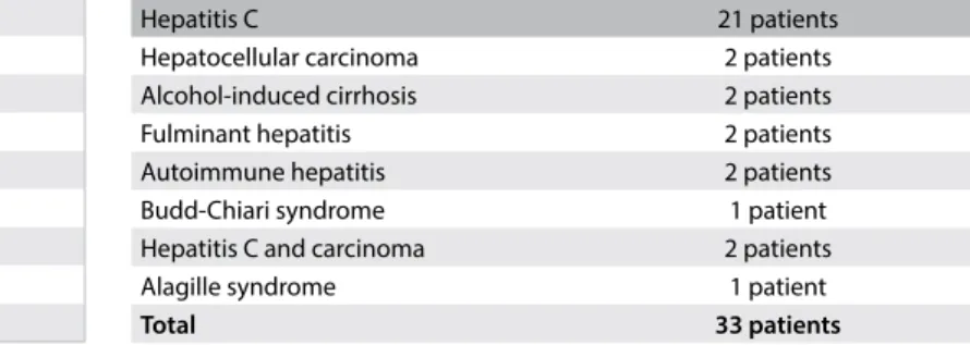
During the study period, 33 patients who had undergone a liver transplant more than one year previously, between 2002 and 2010, were included and subjected to liver biopsy and elastog-raphy examinations. he original indication for transplantation had been cirrhosis due to the hepatitis C virus in 21 cases (63%) and other causes in 37% (Table 1). Among these 21 patients, 8 were alcohol users. he ARFI elastography was performed successfully in all cases. No complications from the liver biopsy were registered.
Twenty-two of these patients were men. he patients’ average age was 55 ± 11 years (mean 49.7; minimum 21; maximum 74). he mean body mass index was 26.2 kg/m2 (range: 16.3-32.7).
Fibrosis Portal ibrosis Septa
Perisinusoidal ibrosis Portal and periportal iniltrate Lobular iniltrate
Steatosis Balloonization Sinusoidal dilation
Chart 1. Histological variables analyzed by means of liver biopsy
Hepatitis C 21 patients
Hepatocellular carcinoma 2 patients
Alcohol-induced cirrhosis 2 patients
Fulminant hepatitis 2 patients
Autoimmune hepatitis 2 patients
Budd-Chiari syndrome 1 patient
Hepatitis C and carcinoma 2 patients
Alagille syndrome 1 patient
Total 33 patients
he anatomopathological results from the liver biopsies showed absence of ibrosis (stage F0) in 10 patients (out of 33, 30.3%), stage 1 ibrosis (F1) in 11 patients (33.3%), stage 2 ibrosis (F2) in eight patients (24.2%), stage 3 ibrosis (F3) in four patients (12.1%) and no cases of stage 4 ibrosis (F4; cirrhosis). he mean ARFI result was 1.11 ± 0.14 for patients staged as F0; 1.21 + 0.28 for those who were F1; 1.36 + 0.35 for patients in the F2 group; and 1.57 + 0.47 for F3 patients. here was no signiicant diference between groups for ARFI values (P = 0.10). here were signiicant associations between the ibrosis classiication and the number of septa, portal ibrosis, perisinusoidal ibrosis, centrilobular ibrosis and periportal and portal iniltrate (P < 0.05). However, steatosis observed in the histological examination did not show any correla-tion with ibrosis stage according to ARFI (P > 0.05).
he ibrosis classiication groups were then regrouped: FO-F1 (with 21 of the 33 cases, 63%) and F2-F3-F4 (with 12 cases, 37%). he ARFI value was signiicantly diferent between these two subgroups: 1.16 + 0.22 (median 1.12, minimum of 0.91 and maximum of 1.99) versus 1.43 + 0.38 (median of 1.38, minimum 0.84 and maximum 2.22), respectively.
he best cutof value for ARFI that was able to diferentiate F0-F1 from F2-F3-F4 was 1.29 m/s, which presented sensitivity of 0.66 (95% conidence interval, CI: 0.50 to 0.83) and speciicity of 0.85 (95% CI: 0.72 to 0.97). he area under the ROC curve was 0.74 (95% CI: 0.55 to 0.94). Table 2shows the accuracy of ARFI at the diferent stages of ibrosis.
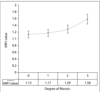
he Kruskal-Wallis test demonstrated statistical diferences between the two groups, with P = 0.02 for the general degree of ibrosis (Figure 1), P = 0.031 for septa and P = 0.027 for centri-lobular ibrosis. he cutof point between the groups F0-F1 and F2-F3 was 1.29 m/s (Figure 2).
here was no statistical diference between the values from the ARFI elastography between the patients with hepatitis C and those with other hepatic diseases.
DISCUSSION
he prospect of a reduction in the number of biopsy proce-dures performed on operated patients is very important, both for patients and for hospitals, because of the risk of the invasive examination and the cost. herefore, this study strove to contrib-ute through presenting an alternative to liver biopsy for evaluat-ing post-transplantation liver elasticity.
New noninvasive diagnostic methods have arisen for evaluat-ing the degree of liver ibrosis over recent years: serological tests, elastography and magnetic resonance imaging. Hepatic elastog-raphy has been highlighted due to its harmlessness, speed and technical ease of implementation.13
Figure 1. Boxplot of the results from acoustic radiation force impulse (ARFI) elastography regarding the general degree of ibrosis in the two groups of patients, with or without ibrosis.
Mean ARFI value
2.25
2.00
1.75
1.50
1.25
1.00
F0 or F1 F2 or F3
Figure 2. Analysis on the acoustic radiation force impulse (ARFI) values to detect a cutof point that separates patients with and without ibrosis.
2
1.8
1.6
1.4
1.2
1.1
0.8
0.6
0.4
0.2
ARFI value
ARFI value
Degree of fibrosis
1.13 1.17 1.29 1.58
3 2
1 0
0
Degreee of ibrosis > F0 > F1 > F2
Cutof ARFI value 1.26 1.29 1.42
AU-ROC [95% CI] 0.67
[0.50 to 0.87]
0.74 [0.55 to 0.94]
0.77 [0.5 to 1.00]
Sensitivity [95% CI] 0.43
[0.31 to 0.66]
0.68 [0.50 to 0.83]
0.75 [0.60 to 0.90]
Speciicity [95% CI] 0.9
[0.81 to 1]
0.86 [0.72 to 0.97]
0.86 [0.76 to 0.99]
PPV [95% CI] 0.91
[0.76 to 10
0.91
[0.76 to 10] 1
NPV [95% CI] 0.43
[0.21 to 0.64]
0.41 [0.20 to 0.62]
0.38 [0.19 to 0.57]
Table 2. Cutof values for acoustic radiation force impulse (ARFI)
Transient elastography was the irst widely disseminated method used for evaluating chronic hepatopathy.17 Piscaglia et al.8
demonstrated that transient elastography produced better results than did biochemical tests for quantifying the degree of ibrosis in patients undergoing liver transplantation because of the hepa-titis C virus. In 56 patients, accuracy rates greater than 85% were obtained among those with signiicant ibrosis.22 he study by
Piscaglia et al.8 contained a description of the importance of
future investigations evaluating factors that interfere with hepatic ibrosis, such as hepatic steatosis, perisinusoidal ibrosis and other histological information.
here are few studies on transient elastography performed on transplanted livers, and a meta-analysis only gathered 470 patients.22 he studies reviewed showed excellent results
regarding identiication of cirrhosis and good results in major ibrosis cases. However, indings of low levels of ibrosis from transient elastography may not rule out cirrhosis, and in these cases, biopsy is still needed. he major limitation of transient elastography and other noninvasive methods lies in the inter-pretation of results from intermediate stages of liver ibrosis. Additionally, transient elastography has the disadvantages of only using A and M modes, and the absence of a B mode pre-cludes evaluation of the liver.
ARFI elastography, the subject of the present study, was the irst technique to be coupled with conventional ultra-sound equipment, and it has the advantage of viewing both the liver and the measurement site. A meta-analysis on more than 3,000 patients demonstrated that it had high accuracy for quanti-fying the degree of hepatic ibrosis,23 and that its results
were sim-ilar to those from transient elastography. However, few studies have used ARFI elastography on transplanted livers.24
Crespo et al.25 examined 87 patients and found that the
sen-sitivity and speciicity of ARFI elastography were 76% for F2 and 85% for F4. Wildner et al.26 used ARFI elastography on 58 patients
who underwent transplantation and showed that the velocities were signiicantly higher in the patients with advanced ibrosis. In the present prospective study, despite the limited number of patients, two primary results can be noted: irstly, ARFI elastog-raphy was able to distinguish between livers transplanted in the group with F0-F1 and those in the group with F2-F3, with a sta-tistically signiicant diference. here was signiicant correlation between the velocities obtained and the presence of septa and centrilobular ibrosis.
he current case study was on patients who received trans-plants due to hepatitis C virus or other forms of chronic hepa-topathy. In the analysis on the two ibrosis groups, the underly-ing disease was not taken into consideration. his was in fact one limitation of this study: its analysis of patients with hepatitis C and other liver diseases together. Another limitation was the low
number of patients, which serves as a stimulus for further inves-tigations on this subject. he velocities obtained in the various degrees of ibrosis following transplantation were similar to those described in populations of chronic hepatopathy cases that did not undergo transplantation.10,13
hese preliminary results indicated that the values obtained through ARFI elastography were not afected by conditions that could change these values, such as inlammatory reactions or ana-tomical alterations. Absence or a low number of patients with F4 was expected, as also found by other authors, due to the good results from antiviral therapies. he diferences between biopsy and ARFI results may be due to histological analyses performed in diferent regions of the liver, heterogeneous liver tissue samples and obese patients. We believe that the following factors could pos-sibly lead to wrong diagnostic conclusions: biopsy samples smaller than 1.5 cm with less than 10 portal spaces; heterogeneous liver tis-sue, with variable ibrosis density; or obese patients, in which the elastography results are false negatives or false positives.
he second conclusion is that a cutof value of 1.29 meters per second that separates patients with or without signiicant ibrosis was identiied. his may inluence antiviral therapy over the short term, because higher velocities indicated by ARFI mean higher degrees of ibrosis, which require antiviral treatment. he follow-up on ibrosis progression could include this noninvasive method in future post-transplantation protocols.
he goal of ARFI elastography is not to replace liver biopsy in all transplanted livers. Other alterations, such as rejections and vascular or biliary abnormalities, usually require a liver biopsy for diagnosis and follow-up.
his study has demonstrated promising results with regard to diferentiation of patients with ibrosis grades 0 and 1 from those with grades 2 to 4, through ARFI elastography. Further studies with larger samples of patients are necessary in order to conirm these results and possibly include ARFI in the protocol for evalu-ating transplantation patients.
REFERENCES
1. Hübscher SG. What is the long-term outcome of the liver allograft? J Hepatol. 2011;55(3):702-17.
2. Sebagh M, Rifai K, Féray C, et al. All liver recipients beneit from the protocol 10-year liver biopsies. Hepatology. 2003;37(6):1293-301. 3. Shiroma RK, Chaib E, Amed-Filho AM, et al. Transplante de fígado de
acordo com os critérios de Milão: revisão dos últimos 10 anos. [Liver transplantation according to Milan criteria: an overview of the past ten years]. Rev Med (São Paulo). 2012;91(2):120-4.
5. Evans HM, Kelly DA, McKiernan PJ, Hübscher S. Progressive histological damage in liver allografts following pediatric liver transplantation. Hepatology. 2006;43(5):1109-17.
6. Ekong UD, Melin-Aldana H, Seshadri R, et al. Graft histology characteristics in long-term survivors of pediatric liver transplantation. Liver Transpl. 2008;14(11):1582-7.
7. Castéra L, Vergniol J, Foucher J, et al. Prospective comparison of transient elastography, Fibrotest, APRI, and liver biopsy for the assessment of ibrosis in chronic hepatitis C. Gastroenterology. 2005;128(2):343-50.
8. Piscaglia F, Cucchetti A, Terzi E, Gianstefani A. Validation of noninvasive methods for the assessment of liver ibrosis in patients with recurrent hepatitis C after transplantation. Liver Transpl. 2010;16(8):1006-7. 9. J S Cross T, Jothimani D, Heneghan MA, Harrison PM. Non-invasive
assessment of ibrosis in liver grafts due to hepatitis C virus recurrence. Clin Transplant. 2011;25(3):345-51.
10. Friedrich-Rust M, Nierhof J, Lupsor M, et al. Performance of Acoustic Radiation Force Impulse imaging for the staging of liver ibrosis: a pooled meta-analysis. J Viral Hepat. 2012;19(2):e212-9.
11. Cholongitas E, Tsochatzis E, Goulis J, Burroughs AK. Noninvasive tests for evaluation of ibrosis in HCV recurrence after liver transplantation: a systematic review. Transpl Int. 2010;23(9):861-70.
12. Intraobserver and interobserver variations in liver biopsy interpretation in patients with chronic hepatitis C. The French METAVIR Cooperative Study Group. Hepatology. 1994;20(1 Pt 1):15-20.
13. Silva Junior RG, Schmillevitch J, Nascimento Mde F, et al. Acoustic radiation force impulse elastography and serum ibrosis markers in chronic hepatitis C. Scand J Gastroenterol. 2014;49(8):986-92. 14. Huwart L, Peeters F, Sinkus R, et al. Liver ibrosis: non-invasive
assessment with MR elastography. NMR Biomed. 2006;19(2):173-9. 15. Loaeza-del-Castillo A, Paz-Pineda F, Oviedo-Cárdenas E, et al. AST to
platelet ratio index (APRI) for the noninvasive evaluation of liver ibrosis. Ann Hepatol. 2008;7(4):350-7.
16. Wai CT, Greenson JK, Fontana RJ, et al. A simple noninvasive index can predict both signiicant ibrosis and cirrhosis in patients with chronic hepatitis C. Hepatology. 2003;38(2):518-26.
17. Sandrin L, Fourquet B, Hasquenoph JM, et al. Transient elastography: a new noninvasive method for assessment of hepatic ibrosis. Ultrasound Med Biol. 2003;29(12):1705-13.
18. Rifai K, Bahr MJ, Mederacke I, et al. Acoustic radiation force imaging (ARFI) as a new method of ultrasonographic elastography allows accurate and lexible assessment of liver stifness. Journal of Hepatology. 2009;50(Supplement 1):S88 [abstract 217]. Available from: http://www.journal-of-hepatology.eu/article/S0168-8278 % 2809 % 2960219-1/pdf. Accessed in 2016 (Sep 1).
19. Lupsor M, Badea R, Stefanescu H, et al. Performance of a new elastographic method (ARFI technology) compared to unidimensional transient elastography in the noninvasive assessment of chronic hepatitis C. Preliminary results. J Gastrointestin Liver Dis. 2009;18(3):303- 10.
20. Fierbinteanu-Braticevici C, Andronescu D, Usvat R, et al. Acoustic radiation force imaging sonoelastography for noninvasive staging of liver ibrosis. World J Gastroenterol. 2009;15(44):5525-32.
21. Donato MF, Rigamonti C, Colombo M. Can protocol liver biopsy be avoided to evaluate post-transplant hepatitis C recurrence? Transient elastography makes it possible. Dig Liver Dis. 2010;42(4):307.
22. Adebajo CO, Talwalkar JA, Poterucha JJ, Kim WR, Charlton MR. Ultrasound-based transient elastography for the detection of hepatic ibrosis in patients with recurrent hepatitis C virus after liver transplantation: a systematic review and meta-analysis. Liver Transpl. 2012;18(3):323-31.
23. Nierhof J, Chávez Ortiz AA, Herrmann E, Zeuzem S, Friedrich-Rust M. The eiciency of acoustic radiation force impulse imaging for the staging of liver ibrosis: a meta-analysis. Eur Radiol. 2013;23(11):3040- 53.
24. Pinto J, Matos H, Nobre S, et al. Comparison of acoustic radiation force impulse/serum noninvasive markers for ibrosis prediction in liver transplant. J Pediatr Gastroenterol Nutr. 2014;58(3):382-6. 25. Crespo G, Fernández-Varo G, Mariño Z, et al. ARFI, FibroScan,
ELF, and their combinations in the assessment of liver ibrosis: a prospective study. J Hepatol. 2012;57(2):281-7.
26. Wildner D, Strobel D, Konturek PC, et al. Impact of acoustic radiation force impulse imaging in clinical practice of patients after orthotopic liver transplantation. Med Sci Monit. 2014;20:2027-35.
Sources of funding: None
Conlict of Interest: None
Date of irst submission: June 24, 2016
Last received: August 15, 2016
Accepted: August 17, 2016
Address for correspondence:
Joel Schmillevitch
Hospital das Clínicas, Universidade de São Paulo, Instituto de Radiologia
Rua Dr. Ovídio Pires de Campos, 75 Cerqueira César – São Paulo (SP) – Brasil CEP 05403-010