Evaluation of glial fibrillar acidic protein as a marker of hepatic
Evaluation of glial fibrillar acidic protein as a marker of hepatic
Evaluation of glial fibrillar acidic protein as a marker of hepatic
Evaluation of glial fibrillar acidic protein as a marker of hepatic
Evaluation of glial fibrillar acidic protein as a marker of hepatic
ischemia-reperfusion
ischemia-reperfusion
ischemia-reperfusion
ischemia-reperfusion
ischemia-reperfusion
Avaliação da proteína acídica fibrilar glial como marcador da injúria por
Avaliação da proteína acídica fibrilar glial como marcador da injúria por
Avaliação da proteína acídica fibrilar glial como marcador da injúria por
Avaliação da proteína acídica fibrilar glial como marcador da injúria por
Avaliação da proteína acídica fibrilar glial como marcador da injúria por
isquemia-reperfusão hepática
isquemia-reperfusão hepática
isquemia-reperfusão hepática
isquemia-reperfusão hepática
isquemia-reperfusão hepática
GIULIANO ANCELMO BENTO, ACBC-RJ1; VIVIANI REISDA CUNHA2; RODRIGO MARTINEZ, TCBC-RJ3; FLÁVIA CARVALHO ALCANTARA GOMES4;
ALBERTO SCHANAIDER, TCBC-RJ5
A B S T R A C T A B S T R A C T A B S T R A C T A B S T R A C T A B S T R A C T
Objective Objective Objective Objective
Objective: To evaluate the expression of Glial Fibrillary Acidic Protein (GFAP) after ischemia-reperfusion injury. MethodsMethodsMethodsMethodsMethods: twenty four rats were divided into four groups: Control, submitted to anesthesia and liver biopsy; Sham, receiving injection of heparin through the vena cava and hepatic pedicle dissection, with liver biopsy after 24 hours; Ischemia-30, the same as Sham group, plus hepatic pedicle clamping for 30 minutes; and Ischemia-90, the same procedure of Ischemia-30 group, but with clamping period of 90 minutes. After 24 hours of observation, the animals underwent laparotomy and we evaluated their livers macroscopically, microscopically by hematoxylin-eosin (HE) and analyzed the expression of GFAP by Western Blotting. ResultsResultsResultsResultsResults: There was no difference in the gross appearance of the livers between the different experimental groups, all having demonstrated normal morphology. HE analysis showed no significant differences with respect to lobule morphology. On the other hand, in the ischemia groups we observed neutrophilic infiltrates and small areas of necrosis. GFAP expression was similar in all groups, either qualitatively and quantitatively. ConclusionConclusionConclusionConclusionConclusion: The expression of Glial Fibrillary Acidic Protein did not change in our model of ischemia-reperfusion.
Key words Key words Key words Key words
Key words: Ischemia. Reperfusion injury. Liver. Glial fibrillary acidic protein. Immunohistochemistry.
Work performed at the Center for Experimental Surgery, Department of Surgery, Faculty of Medicine, and at the Institute of Biomedical Sciences, Federal University of Rio de Janeiro – UFRJ, city of Rio de Janeiro, Rio de Janeiro State – RJ, Brazil.
1. General Surgeon, MSc, Postgraduation Program in Surgical Sciences, Department of Surgery, Faculty of Medicine, UFRJ; 2. Medical School Graduate, Scientific Initiation Program, Faculty of Medicine, UFRJ; 3. Associate Professor, Department of Surgery, Faculty of Medicine, UFRJ; 4. Professor, Institute of Biomedical Sciences, UFRJ; 5. Professor, Department of Surgery, Faculty of Medicine, UFRJ.
INTRODUCTION
INTRODUCTION
INTRODUCTION
INTRODUCTION
INTRODUCTION
H
istological evaluation of liver grafts used in liver transplantation is a challenging problem in medicine due to the lack of malfunction prognostic markers 1. Currently, the only histological marker that can be used is hepatic steatosis, whose accuracy is low to predict the quality of the organ, though 2. For this reason, the evaluation of these grafts is mainly based on clinical data from donors, such as age and hemodynamic parameters, these too of little accuracy 3. One of the changes proposed in this field is the characterization of other markers, which could assist in this evaluation. Surely, the candidate molecules would be those that have their expression modified according to different types of liver injury.The Glial Fibrillary Acidic Protein (GFAP) is a cytoskeletal molecule highly expressed in the central nervous system, which has its expression increased in
ischemic lesions, such as post-infarction brain scar 4-6. In the liver parenchyma, the GFAP is expressed in perisinusoidal cells 7. After various forms of liver injury, the expression of this marker is increased, which is related to the phenotype modification of perisinusoidal cells which, after a period of persistent injury (eg weeks after exposure to the hepatotoxic agent carbon tetrachloride or after ligation of the common bile duct) are finally differentiated into myofibroblastic cells 8-10. The expression of this marker is also altered in other types of liver disease, as has been described in recurrence of infection by hepatitis C after liver transplantation 11.
METHODS
METHODS
METHODS
METHODS
METHODS
All experiments followed the guidelines for the care and use of laboratory animals. Our experimental protocol was approved by the Ethics Committee for Use of Laboratory Animals in Research, Education and Extension (CEUA), registered under the number 79/09.
Twenty-four Wistar rats weighing about 220g from the rat vivarium of the Center for Experimental Surgery, Department of Surgery, Faculty of Medicine, UFRJ were divided into four groups, each consisting of six rats.
The control animals were anesthetized with ketamine (100mg/kg) and xylasina (10mg/kg) and submitted to laparotomy. After removal of specimens of the cerebellum and the liver, corresponding, in the latter, to the same segments studied in the other groups, the animals were killed. The fragments were subjected to histological analysis by hematoxylin-eosin, and GFAP expression analysis by Western Blotting technique.
In the sham group, the animals were anesthetized and underwent laparotomy with isolation of their liver pedicles with a ribbon (Figure 1A); we proceeded to the injection of heparin (1000U/kg) in the vena cava (Figure 1B). Soon after, the abdominal wall was sutured and the animals kept under regular analgesia regimen for 24 hours. After this period, the animals were killed and liver fragments representative of the segments were collected for analysis by Western blotting. Perfusion was performed with 10 ml of saline using an infusion pump at 5ml/minute followed by perfusion with 4% paraformaldehyde at 4°C, diluted in sodium phosphate buffer, pH 7.4, for 15 minutes.
In rats of groups “Inchemia-30” and “Ischemia-90,” the same surgical protocol of the sham group was applied, but a selective clamping of the superior hepatic pedicle (Figure 1C), comprising the left lateral and median lobes, was applied for 30 or 90 minutes (Figures 1D and E), respectively, followed by reperfusion (Figure 1F) and synthesis of the abdominal wall. The animals were kept under regular analgesia and after 24 hours they were killed and liver tissue fragments representative of the segments obtained for performing Western blotting for GFAP. The animals were then perfused similarly to the sham group, and fragments of liver tissue segments were obtained for representative histological analysis.
Liver biopsies were preserved in 4% paraformaldehyde for 24 hours and then subjected to graded dehydration in sucrose 10, 20 and 30% for cryoprotection. After this tissue blocks were immersed in TissueTek® and frozen at -20° C. Frozen sections of 12 ìm were obtained and mounted on glass slides, which were then dried in formaldehyde vapor for one hour.
For staining with hematoxylin-eosin, the sections were dehydrated in absolute alcohol and rehydrated in water. They were then immersed in hematoxylin dye for four minutes, washed with water, differentiated in 70% alcohol and stained in 0.01% eosin for five seconds,
immersed in 95% ethanol, dehydrated with absolute ethanol and subjected to washing in xylene for five minutes.
The protein expression analysis was performed according to the protocol of Sampaio et al. 13. The samples of liver tissue were lysed in loading buffer (100 mM Tris-Cl [pH 6.8], 4% SDS, 0.2% bromophenol blue, 20% glycerol, 200 mM dithiothreitol DTT) and boiled for five minutes before loading in gel. Protein concentration was determined with the BCA kit. Ten micrograms of protein were applied in each analyzed case and subjected to electrophoresis on a mini SDS-PAGE 12% gel, followed by electric transfer to a PVDF membrane for one hour. The membranes were blocked in 5%PBS-milk and primary antibodies were added for 24 hours at 4° C, followed by incubation for one hour at room temperature with peroxidase-conjugated secondary antibodies. The proteins were revealed using the chemiluminescence detection system. The following antibodies were used: anti-GFAP made in rabbit and IgG anti-rabbit made in goat.
Densitometries of the bands were measured with the aid of the program Image J (National Institutes of Health, USA). The values were computed on a GraphPad Prism 4.0 table. The single factor ANOVA test was applied. We used a confidence interval of 95% and p-value less than 0.05 was considered statistically significant.
RESULTS
RESULTS
RESULTS
RESULTS
RESULTS
As shown in Figure 1, livers subjected to ischemic insults did not show any changes in their microscopic structure, with sharp edges and smooth surfaces, just like the control and sham groups. These results suggest that the macroscopic evaluation of these grafts, considered one of the most important parameters used during the organ collection 12, is not able to accurately identify livers previously submitted to an ischemia-reperfusion injury.
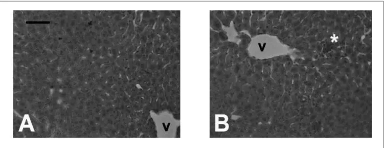
Histological sections stained with hematoxylin-eosin representative of the different experimental conditions were evaluated. All cuts demonstrated the typical lobular pattern described in mammalian species14 (Figure 2A). More specifically, the portal areas located along the central venules were clearly identified and showed the typical elements of the portal triad. Infiltrates of neutrophils and small areas of necrosis were found in the groups subjected to ischemia, as previously described 15 (Figure 2B). On the other hand, hepatic steatosis, either micro or macro, was not detected in any of the sections examined.
expression was also detected (Figure 3B). Densitometry analysis detected no significant differences in the expression of this protein between the different experimental conditions (Figure 3C).
DISCUSSION
DISCUSSION
DISCUSSION
DISCUSSION
DISCUSSION
Hepatic ischemia is one of the most common forms of injury found in cadaveric liver donors 12. It is most commonly a consequence of the use of high doses of vasopressors or episodes of cardiac arrest. Under these conditions, the decision about whether or not to use grafts is always seen as a dilemma, since there is no safe limit set for the doses of vasopressors. Thus, many clinical and laboratory parameters end up being considered in the final analysis 3. Many surgeons consider the macroscopic appearance of the liver as one of the most important parameters to be evaluated 12.
In our experiment, the hepatic pedicle was clamped over two different time periods, with the goal of creating an ischemia that would mimic the different forms of injury previously described. This maneuver is commonly performed during liver surgery as a way to control bleeding during resection of hepatic parenchyma, and is held at the cost of causing some degree of liver dysfunction. The period of 30 minutes is considered “safe”, since it rarely causes severe hepatic dysfunction, except when performed in livers with cirrhosis or steatosis. The period of 90 minutes, on its turn, is considered much less safe and is hardly applied in the clinical setting, since it often results in severe hepatic dysfunction 16. The option of performing the selective clamping was taken because, in rats, total clamping of the pedicle is poorly tolerated due to rapid installation of mesenteric congestion followed by infarction 17. This approach allowed further to compare the parameters
Figure 1 Figure 1 Figure 1 Figure 1
Figure 1 - Surgical procedure. The animals were anesthetized and their superior pedicles isolated (A). Heparin (1000U/Kg) injection in the vena cava (B). Clamping of the superior hepatic pedicle (C). Ischemia of median lobes for 30 minutes (D) and 90 minutes (E). Homogeneous reperfusion verified after releasing the clamps (F).
Figure 2 Figure 2 Figure 2 Figure 2
analyzed not only with the control groups, but also with the segments not subjected to ischemia of the same animals. The gross appearance of the livers analyzed 24 hours after ischemic injury did not change. We observed aspects commonly used by liver collection surgeons such as color, edge and regularity of the organ’s surface 12. No differences were found between the groups, whereas all livers analyzed had aspects compatible with healthy organs, and therefore grafts said to be ideal, such as sharp edges, normal color and smooth surface. These results speak against the empirical argument that the macroscopic appearance of the liver is a very accurate parameter to take into account in deciding whether or not to use the organ, since it is known that organs previously submitted to prolonged ischemic injury bring risk high of dysfunction 3.
A second form of analysis was done by histological evaluation of liver tissues, using the most common approach in medical practice, staining with hematoxylin-eosin 12. Under these conditions, neutrophilic infiltrates and small areas of necrosis were found in the Ischemia groups. Our results are in agreement with the work of Jaeschke et al., who described the presence of
neutrophilic infiltration and necrosis of hepatocytes after ischemia-reperfusion injury 17.
In general clinical practice, hepatic steatosis is certainly the most widely used clinical parameter for the histological evaluation of liver grafts for transplantation, and most authors agree that grafts showing levels of steatosis greater than 30% are considered at risk 2. Steatosis may be a consequence of various forms of liver injury such as obesity, prolonged parenteral nutrition, alcohol abuse and treatment with chemotherapeutic drugs18. There are no previous reports describing the development of hepatic steatosis after injury by ischemia-reperfusion. Our results support this knowledge, since steatosis was not detected in any of the experimental conditions, and thus we conclude that other parameters predictive of a graft high risk must also be evaluated.
Experiments were performed to evaluate whether the expression of GFAP could be altered after ischemic injury. This filament was chosen as a candidate molecule mainly due to the fact that its expression was altered in many models of liver injury, such as ligation of the common bile duct, injection of carbon tetrachloride and infection by hepatitis C virus, and it is related to the phenotypic modification of perisinusoidal cells during their transformation into myofibroblastic cells 8,19,20. Therefore, we screened representative samples of liver tissue of different experimental conditions through the Western Blotting technique. No differences were found in the pattern of expression of this protein after hepatic ischemia. The Western blotting technique was chosen because our primary goal was to develop an alternative method that would provide additional data to histological exams and faster results, thus having an application in clinical practice, where the short period of ischemia tolerated by these grafts does not allow more time consuming methods, such as immunohistochemistry, to be applied. Our results, nonehteless, showed no differences in the expression of this protein after ischemic insult. Likewise, no differences were found in the pattern of protein degradation. These findings, however, do not preclude the occurrence of later changes in the expression of this molecule after injury by ischemia-reperfusion. For example, a change in the expression of this filament is only detectable in three to eight weeks after the injection of carbon tetrachloride and in four weeks after ligation of the common bile duct 19,20. Considering these data, a modification of the expression of this marker in our model could only be deleted after the completion of other tests, involving, for example, a more prolonged ischemic insult. From a practical standpoint, the establishment of such an injury would be technically complex and involve approaches such as induction of arterial and portal stenosis or keeping animals at high doses of vasoactive drugs. Another option would be to employ the analysis of gene expression of GFAP, through techniques like Reverse Transcription Polymerase Chain Reaction (RT-PCR), in situ hybridization or the use of transgenic
Figure 3 Figure 3Figure 3 Figure 3
animals 21. Nevertheless, the practical applicability of such experiments would probably be much lower.
Our results, although negative, bring great importance with them, since they represent some of the few efforts already described in order to detect molecular markers of high-risk grafts 22,23. Our extensive search of the literature found few studies with this objective. It is worth noting that some authors have employed more sophisticated techniques for this purpose. Ray et al. employed analysis by microsatellite of pre-reperfusion biopsies from lung transplantation grafts and detected an altered expression of 24 genes in organs that have come to be dysfunctional after transplantation 22. Most of these genes were linked to
signaling pathways and are thus translated into proteins that have a much shorter half-life, which makes the practical use of these markers as diagnostic tools in clinical practice unlikely. We expect that the results obtained by Ray et al., like ours, encourage other researchers to direct their efforts to a so obscure and little explored area of the science of transplantation, since the discovery of a reliable predictor of organ dysfunction and, more optimistically, the development of a method for accurate diagnosis, would surely revolutionize the practice in this field. We conclude that GFAP showed no expression changes in response to the liver injury produced by ischemia-reperfusion in our experimental model.
R E S U M O R E S U M O R E S U M O R E S U M O R E S U M O
Objetivo: Objetivo: Objetivo: Objetivo:
Objetivo: Avaliar a expressão da Proteína Acídica Fibrilar Glial após a injúria por isquemia-reperfusão. Métodos:Métodos:Métodos:Métodos: vinte e quatroMétodos: ratos foram distribuídos em quatro grupos: Controle, submetidos à anestesia e biópsia hepática; Simulação, injeção de heparina através da veia cava e dissecção do pedículo hepático superior, biópsia após 24 horas; Isquemia 30 minutos, mesmo procedimento do grupo Simulação, acrescido de clampeamento do pedículo hepático superior por 30 minutos; Isquemia 90 minutos, mesmo procedimento do grupo Isquemia 30 minutos, porém com período de clampeamento de 90 minutos. Após 24 horas de observação, os animais foram submetidos à laparotomia e seus fígados avaliados macroscopicamente, microscopicamente, por coloração de Hematoxilina-Eosina (HE) e submetidos à análise da expressão da GFAP por Western Blotting. Resultados:Resultados:Resultados:Resultados:Resultados: Não se observou diferença no aspecto macroscópico dos fígados entre os diferentes grupos experimentais, tendo todos evidenciado morfologia normal. A análise por HE não evidenciou diferenças significativas, no que diz respeito à morfologia lobular. Por outro lado, nos grupos isquemia, foram encontrados infiltrados neutrofílicos e pequenas áreas de necrose. A expressão de GFAP foi semelhante em todos os grupos, seja qualitativamente quanto quantitativamente. Conclusão:Conclusão:Conclusão:Conclusão:Conclusão: A expressão da Proteína Acídica Fibrilar Glial não se alterou em nosso modelo de isquemia-reperfusão.
Descritores: Descritores: Descritores: Descritores:
Descritores: Isquemia. Traumatismo por reperfusão. Fígado. Proteína glial fibrilar ácida. Imunoistoquímica.
REFERENCES
REFERENCES
REFERENCES
REFERENCES
REFERENCES
1. Uemura T, Randall HB, Sanchez EQ, Ikegami T, Narasimhan G, McKenna GJ, et al. Liver retransplantation for primary nonfuntion: analysis of a 20-year single-center experience. Liver Transpl. 2007;13(2):227-33.
2. Doyle MB, Vachharajani N, Wellen JR, Anderson CD, Lowell JA, Shenoy S, et al. Short-and long-term outcomes after steatotic liver transplantation. Arch Surgery. 2010;145(7):653-60. 3. Gastaca M. Extended criteria donors in liver transplantation:
adapting donor and quality and recipient. Transplant Proc. 2009;41(3):975-9.
4. Bignami A, Eng LF, Dahl D, Uyeda CT. Localization of the glial fibrillary acidic protein in astrocytes by immunofluorescence. Brain Res. 1972;43(2):429-35.
5. Petito CK, Morgello S, Felix JC, Lesser ML. The two patterns of reactive astrocytosis in postischemic rat brain. J Cereb Blood Flow Metab. 1990;10(6):850-9.
6. Yasuda Y, Tateishi N, Shimoda T, Satoh S, Ogitani E, Fujita S. Relationship between S100beta and GFAP expression in astrocytes during infarction and glial scar formation after mild transient ischemia. Brain Res. 2004;1021(1):20-31.
7. Baratta JL, Ngo A, Lopez B, Kasabwalla N, Longmuir KJ, Robertson RT. Cellular organization of normal mouse liver: a histological, quantitative immunocytochemical, and fine structural analysis. Histochem Cell Biol. 2009;131(6):713-26.
8. Parsons CJ, Takashima M, Rippe RA. Molecular mechanisms of hepatic fibrogenesis. J Gastroenterol Hepatol. 2007;22 Suppl 1:S79-84.
9. Gressner AM. The cell biology of liver fibrogenesis – an imbalance of proliferation, growth arrest and apoptosis of myofibroblasts. Cell Tissue Res. 1998;292(3):447-52.
10. Pinzani M, Gentilini P. Biology of hepatic stellate cells and their possible relevance in the patogenesis of portal hypertension in cirrhosis. Semin Liver Dis. 1999;19(4):397-410.
11. Carotti S, Morini S, Corradini SG, Burza MA, Molinaro A, Carpino G, et al. Glial fibrillary acidic protein as an early marker of hepatic stellate cell activation in chronic and posttransplant recurrent hepatitis C. Liver Transpl. 2008;14(6):806-14.
12. Busuttil RW, Tanaka K. The utility of marginal donors in liver transplantation. Liver Transpl. 2003;9(7):651-63.
13. de Sampaio e Spohr TC, Martinez R, da Silva EF, Neto VM, Gomes FC. Neuro-glia interaction effects on GFAP gene: a novel role for transforming growth factor-beta1. Eur J Neurosci. 2002;16(11):2059-69.
14. Loud AV. A quantitative sterological description of the ultrastructure of normal rat liver parenchymal cells. J Cell Biol. 1968;37(1):27-46. 15. Jaeschke H. Vascular oxidant stress and hepatic ischemia/reperfusion
injury. Free Radic Res Commun. 1991;12-13 Pt 2:737-43. 16. Belghiti J, Noun R, Malafosse R, Jagot P, Sauvenet A, Pierangeli F,
17. Jaeschke H, Farhood A, Smith CW. Neutrophils contribute to ischemia/reperfusion injury in rat liver in vivo. FASEB J. 1990;4(15):3355-9.
18. Angele MK, Rentsch M, Hartl WH, Wittmann B, Graeb C, Jauch KW, et al. Effect of graft steatosis in liver function and organ survival after liver transplantation. Am J Surg. 2008;195(2):214-20.
19. Niki T, De Bleser PJ, Xu G, Van Den Berg K, Wisse E, Geerts A. Comparison of glial fibrillary acidic protein and desmin staining in normal and CCl4-induced fibrotic rat livers. Hepatology. 1996;23(6):1538-45.
20. Cassiman D, Libberecht L, Desmet V, Denef C, Roskams T. Hepatic stellate cell/myofibrobast subpopulations in fibrotic human and rat livers. J Hepatol. 2002;36(2):200-9.
21. Gomes FC, Maia CG, de Menezes JR, Neto VM. Cerebellar astrocytes treated by thyroid hormone modulate neuronal proliferation. Glia. 1999;25(3):247-55.
22. Ray M, Dharmarajan S, Freudenberg J, Zhang W, Patterson GA. Expression profiling of human donor lungs to understand primary graft dysfunction after lung transplantion. Am J Transplant. 2007;7(10):2396-405.
23. Boutros T, Nantel A, Emadali A, Tzimas G, Conzen S, Chevet E, et al. The MAP Kinase phosphatase-1 MKP-1/DUSP1 is a regulator of human liver response to transplantation. Am J Transplant. 2008;8(12):2558-68.
Received on 05/07/2012
Accepted for publication 19/08/2012 Conflict of interest: none
Source of funding: Foundation for Research Support of the State of Rio de Janeiro, FAPERJ.
How to cite this article: How to cite this article:How to cite this article: How to cite this article:How to cite this article:
Bento GA, Cunha VR, Martinez R, Gomes FCA, Schanaider A. Evaluation of glial fibrillar acidic protein as a marker of hepatic ischemia-reperfusion injury. Rev Col Bras Cir. [periódico na Internet] 2013;40(3). Disponível em URL: http://www.scielo.br/rcbc
Address correspondence to: Address correspondence to:Address correspondence to: Address correspondence to:Address correspondence to: Rodrigo Martinez