ESCOLA UNIVERSITÁRIA VASCO DA GAMA
MASTER’S DEGREE
UTERINE ADENOCARCINOMA IN PET RABBITS: A REVIEW
ESCOLA UNIVERSITÁRIA VASCO DA GAMA
MASTER’S DEGREE
UTERINE ADENOCARCINOMA IN PET RABBITS: A REVIEW Coimbra, April 2015
Author
Carolina Salgueiro Costa e Silva
Supervisors
Prof.ª Dr.ª Anália do Carmo
Abstract
The uterine adenocarcinoma is a slow growing malignant tumour that affects 50-80% of pet rabbits over 4 years old, having age as its major risk factor.
The mechanisms underlying the development of this tumour are not completely understood and some of the existing studies presented contradictory results. In fact, in rabbits there is not an adequate classification of these tumours and until now the cellular and molecular features were not fully investigated.
Regarding treatment and until today, ovariohysterectomy is the only effective treatment. Without it, this neoplasia leads to the female rabbit’s death within 1 to 2 years after the appearance of symptoms and metastasis. In spite of the fact that these tumours could in other species be treated by chemotherapy, radiotherapy and hormonal therapy, in rabbits these treatments were mainly used in laboratory animals and therefore, there is no data regarding the adequate protocol. The limited studies available and the existing doubts around the development of the uterine adenocarcinoma in rabbits show that further investigation is necessary to provide other ways to make an accurate diagnosis, to establish an adequate treatment protocol and to determine the prognosis.
Resumo
O adenocarcinoma uterino é um tumor maligno de crescimento lento que afecta 50-80% dos coelhos com mais de 4 anos, sendo a idade o principal factor de risco para esta neoplasia. Os mecanismos que envolvem o desenvolvimento deste tumor não estão totalmente clarificados e alguns estudos apresentam resultados contraditórios. Actualmente, a classificação bem como a caracterização celular e molecular do adenocarcinoma uterino nesta espécie permanece pouco aprofundada.
Até à data, a ovariohisterectomia é o único tratamento eficaz e, na sua ausência, o adenocarcinoma provoca a morte num período de 1 a 2 anos após o aparecimento de sintomas e metástases. Em diferentes espécies, a quimioterapia, a radioterapia e a terapia hormonal são outras opções de tratamento, no entanto nos coelhos a sua aplicação é apenas experimental e associada a animais de biotério, não existindo informação suficiente para o estabelecimento de protocolos adequados à espécie.
O número limitado de estudos e as permanentes dúvidas associadas ao desenvolvimento desta patologia em coelhos evidencia a necessidade de investimento na investigação, a qual poderá revelar novas formas de diagnóstico e tratamento e, consequentemente, alterar o prognóstico.
Index
1. Introduction 1
2. Anatomy and physiology of the reproductive system 1
3. Uterine adenocarcinoma
3.1 Epidemiology and risk factors 3.1.1 Age
3.1.2 Breed 3.1.3 Hormones
3.2 Macroscopic characteristics 3.3 Microscopic characteristics
3.3.1 Histological characteristics
3.3.2 Immunohistochemical characteristics
4 4 4 5 6 7 9 9 10
4. Clinical signs 13
5. Diagnosis
5.1 Differential diagnosis 5.2 Diagnostic procedures
14
14
15
6. Treatment
6.1 Surgical treatment
6.1.1 Anaesthesia and Analgesia
6.1.2 Surgical technique – Ovariohysterectomy 6.1.3 Postoperative considerations
6.2 Oncological treatment
16 16 16 17 18 19
7. Prognosis 21
8. Prevention 21
9. Future perspectives 22
10. Acknowledgments 22
Figure Index
Figure 1. The female rabbit reproductive organs. 2
Figure 2. Detail of the two cervices of a rabbit that underwent ovariohysterectomy. 3
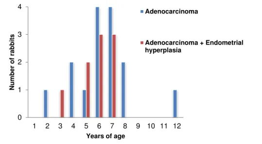
Figure 3. Age distribution of rabbits with adenocarcinoma (n=17) or concurrent adenocarcinoma and endometrial hyperplasia (n=9).
5
Figure 4. Incidence of uterine adenocarcinoma in different breeds according to a study by Green et al.
6
Figure 5. Multiple nodules in both uterine horns of a rabbit. 8
Figure 6. Metastatic disease in the lung of a rabbit with uterine adenocarcinoma. 8
Figure 7. Papillary adenocarcinoma of a female rabbit. HE. 10
Figure 8. Tubular adenocarcinoma of a female rabbit. HE. 10
Figure 9. Differential diagnosis for a palpable mass in the ventral abdomen of a female rabbit.
15
Figure 10. Pulmonary metastases in a laterolateral thoracic radiograph of a rabbit with uterine adenocarcinoma.
19
Table Index
Table 1. Physiological and reproductive data regarding the rabbit 3
Table 2. Oestrogen and progesterone receptor expression in 42 uterine adenocarcinomas of rabbits
12
Table 3. Percentage of several clinical signs of 31 pet rabbits diagnosed with uterine disorders
14
Abbreviations
FSH – Follicle Stimulating Hormone LH – Luteinizing Hormone
Bpm – Beats per minute Rpm – Respirations per minute CSCs – Cancer Stem Cells HE – Haematoxylin Eosin CK – Cytokeratin
P63 – Protein 63
ER-α– Oestrogen Receptor-α PR – Progesterone Receptor CT – Computerized Tomography SC – Subcutaneous
IM – Intramuscular
PO –Per os
1. Introduction
The domestic rabbit descends from the European rabbit, Oryctolagus cuniculus, which belongs to the Lagomorpha order.1 Since a few years, the rabbit that once was considered just a domestic animal for food purposes, is nowadays one of the modern day exotic pets, being the third most popular after dogs and cats.2–4
There are more than 50 different breeds of domestic rabbits that vary from small to large and giant breeds. Although many of the pet rabbits belong to smaller breeds such as Dwarf lops, Dutch or English,1,3 the majority of the pet rabbits are mixed breeds.3
A well cared house pet rabbit, fed a suitable diet and kept in a healthy environment has a lifespan of 8 to 12 years. An increased longevity is associated to the development of several tumours such as lymphoma, skin, bone and uterine tumours being the uterine adenocarcinoma one of the most frequent.5 In fact, previous studies reported that in females over the age of 4 years, the endometrium undergoes a decrease in cellularity and an increase in the production of collagen, that seems to be associated with the development of uterine tumours.6,7 According to a study performed in 47 rabbits presenting uterine disorders, 21.3% of the rabbits presented uterine adenocarcinoma, while 8.5% of the rabbits presented leiomyoma.8 Moreover, in a recent study performed in 50 pet rabbits, uterine adenocarcinoma was detected in 27 out of the 50 rabbits (54%) and 18% of the rabbits presented simultaneously two or three types of uterine disorders.9 Adenoma, leiomyosarcoma, carcinosarcoma, choriocarcinoma, malignant mixed müllerian tumour and hemangioma are other types of tumours that have been described in pet rabbits.9,10 Altogether, the published results indicated that uterine adenocarcinoma is the most frequently diagnosed malignant tumour of the female rabbit with an incidence that may reach 79.1% in rabbits with 5 to 6 years old.11 Considering that rabbits are one of the most popular pets, that uterine adenocarcinoma is the most prevalent tumour among female rabbits and the survival rate after diagnosis is reduced, the present review aims to provide the current state in the diagnosis and treatment of the rabbit uterine adenocarcinoma.
2. Anatomy and physiology of the reproductive system
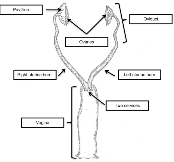
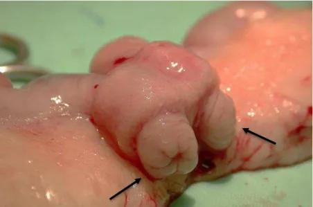
Anatomically, lagomorphs present two uterine horns and two cervices, one for each horn, which open separately into an elongated vagina (Figure 1. and 2.).3,12,13 The uterine body is inexistent or not distinct in this species.3,12 The ovaries are usually surrounded by fat and are not in a true ovarian bursa.13 The uterus can be palpated in the ventral abdomen, caudally to the caecum1 and the bladder is positioned ventrally to the uterine horns.13
in oestrus almost permanently (Table 1.).14 Nonetheless, a cyclic rhythm exists and variety in sexual receptivity can occur among females.1
The female rabbit is receptive to the male when the follicle stimulating hormone (FSH) stimulates the mature ovarian follicles to produce oestrogens, which may take 12 to 14 days.1 During this period, if mating does not occur, the follicles will degenerate and a decrease in oestrogen level and sexual receptivity to the male rabbit will happen, followed by maturation of new ovarian follicles. Therefore, the female rabbit presents a period of receptivity that lasts 12-14 days, succeeded by 1-2 days of non-receptivity to the male.1,14 Full sexual receptivity happens every 18 days.1 This cyclic rhythm may be influenced by different factors such as light, temperature, nutrition, sexual stimulation and individual variation.1 When mating does occur, ovulation is induced by the mounting 10 to 12 hours post-coitus1,2,14, causing the release of luteinizing hormone (LH) from the pituitary gland.14 Besides mounting, ovulation can also be induced by mechanical stimulation of the vagina1,14, mounting by other females and proximity of an entire male.1
Figure 1. The female rabbit reproductive organs. Original scheme
Right uterine horn Left uterine horn
Ovaries
Oviduct Pavillion
Two cervices
Figure 2. Detail of the two cervices of a rabbit that underwent ovariohysterectomy. Courtesy of Dr. Neus Morera, Clinica Veterinaria Exótics, Barcelona, Spain.
Table 1. Physiological and reproductive data regarding the rabbit.
Rabbit physiological data
Life Span 6-10+ years
Adult Body Weight
Female: 2-6 kg (depending on breed)
Male: 2-5 kg (depending on breed)
Rectal Temperature 38,5ºC – 40ºC
Heart Rate 130-325 bpm
Respiratory Rate 32-60 rpm
Blood Volume 55-70 ml/kg
Dental Formula 2-0-3-3 / 1-0-2-3
Rabbit reproductive data
Puberty Small breeds: 4-5 months
Large breeds: 5-8 months
Oestrus Cycle Induced ovulatory
Gestation 30-32 days
Litter Size 5-8 (average)
Birth Weight 40-100 g
Weaning 4-6 weeks
Breeding Duration 1-3 years
3. Uterine adenocarcinoma
The adenocarcinoma of the uterus is a slow growing malignant tumour having its origin in the epithelial cells of the deep glandular portion of the endometrium.15,16 Recently, several studies pointed that in addition to mature epithelial cells there is a small subpopulation of malignant tumour cells with a high proliferative potential and with the capacity to self-renew and differentiate termed cancer stem cells (CSCs), that may initiate and maintain the uterine adenocarcinoma17– 19. The CSCs have a high proliferative potential, an increased resistance to death and an increased ability to migrate, properties that confer to the tumour a bigger potential to metastasize and to resist to chemotherapy and radiation therapy. In spite of the fact that CSCs are mainly studied in humans, recent studies pointed that CSCs are a common feature across solid tumours from different tissues and species and therefore should also be considered in future studies of the rabbit uterine adenocarcinoma.17,20
Regarding the ability to metastasize, the uterine adenocarcinoma starts to invade the myometrium, extending to the uterine wall and to the surround structures of the peritoneal cavity.6,21 Due to the uterus rich blood supply, haematogenous metastatic spread to distant locations such as the lungs and liver is frequent and in a late phase, within 1 to 2 years, the metastatic spread may reach the brain, bones and skin.3,5,6,9,21,22 The rabbit’s life span between the appearance of symptoms and death by metastasis is approximately6,22 1 to 2 years without treatment.22
3.1 Epidemiology and risk factors
In female rabbits, the uterine adenocarcinoma is the most common tumour being diagnosed with an incidence of 50-80% in animals over 4 years old.3,6,7 Tumours in production and laboratory rabbits are practically inexistent due to these animals short period of life.23 There is no difference in the incidence of adenocarcinoma in breeding vs. nonbreeding rabbits.22
3.1.1 Age
Figure 3. Age distribution of rabbits with adenocarcinoma (n=17) or concurrent adenocarcinoma and endometrial hyperplasia (n=9). Adapted from Walter et al. (2010).
3.1.2 Breed
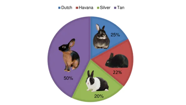
Although most races may present adenocarcinomas, there are breeds that have a higher incidence which seems to indicate an increased susceptibility to the development of this tumour.25 An extensive study performed by Greene et al. showed that the Tan breed was the one which presented a higher incidence of uterine adenocarcinoma (50% of rabbits were affected).15 The Silver breed had an incidence of 25% followed by the Havana breed with an incidence of 22% and by the Dutch breed with an incidence of 20% (Figure 4.). In other breeds, such as Polish, Himalayan, Sable, Beveren, Chinchilla, English and Marten, the incidence is low, varying between 8% and 17%.15,24 In breeds such as Rex and Belgian this neoplasia has not been reported.23,24 This data was collected from a colony of 849 rabbits, observed for 9 years, with a tumour incidence of 79% in 5 year-old animals.15
0 1 2 3 4
1 2 3 4 5 6 7 8 9 10 11 12
Num
be
r
of
rab
bi
ts
Years of age
Adenocarcinoma
Figure 4. Incidence of uterine adenocarcinoma in different breeds according to a study by Green et al. Adapted from Baba et al. (1972).
3.1.3 Hormones
Hormonal related risk factors such as pseudopregnancy, pregnancy toxaemia and obesity remain controversial.23 Several studies pointed that oestrogens may contribute to the development of uterine adenocarcinoma.8,24,25 Nevertheless, Baba et al. also pointed that the administration of oestrogens to aged female Dutch laboratory rabbits with a transplanted tumour reduced the incidence of endometrial carcinoma from 17% to 3%.15 In addition, a recent study suggested that the hormone progesterone may not be directly involved in neoplastic transformation, but may be involved in the modulation of the proliferative activity of the endometrial tissue.25 Therefore, more studies are needed in order to clarify the contribution of sexual hormones to the development of uterine adenocarcinoma.
a. Pseudopregnancy
In a mature female, pseudopregnancy is common25 and is characterized by the maintenance of the corpora lutea for a prolonged period of time, leading to secretion of progesterone in the absence of fetuses.25 This condition mimics a true pregnancy, even though it lasts 16-18 days rather than 31-32 days of a true pregnancy.1 In case of pseudopregnancy, the endometrium undergoes a continuous hormonal stimulation that can predispose to the development of uterine tumours.23,25
b. Pregnancy toxemia
According to a study24, all rabbits that experienced pregnancy toxaemia have also developed uterine tumours later. Pregnancy toxaemia is characterized by a development of ketosis in
Dutch Havana Silver Tan
22%
50%
20%
late pregnancy or during early lactation, when glucose is needed the most.5 It is most frequently seen in overweight females.2 This condition leads to a drop in blood glucose which stimulates the mobilization of fatty acids from the adipose tissue to the liver, developing hepatic lipidosis and sometimes liver failure.26 It was hypothesised that due to the liver failure, there is a significant increase in the oestrogens levels in circulation which may contribute to the development of uterine adenocarcinoma.24
c. Obesity
Although in rabbits, there is not much information regarding the direct association of obesity and the development of uterine adenocarcinoma, in humans, obesity is a well-known risk factor.
Obesity in rabbits is common and according to the Rabbit Clinic of the University of Edinburgh, 1 in 4 rabbits visiting the clinic is obese 27.
Rabbits presenting this condition are more predisposed to health problems, showing an increased risk of cardiovascular diseases, hyperglycaemia, high cholesterol and consequently hepatic lipidosis, amongst other complications 27,28. Also, overweight rabbits are poor surgical candidates28, leading to a more difficult treatment in case of uterine adenocarcinoma.
In humans, the excess of body weight is associated with an increased risk of endometrial tumours in postmenopausal women since the adipocytes have an endocrine function in addition to fat storage.29 Adipocytes secrete cytokines and growth factors and play a significant role in the modulation of the oestrogens levels through aromatase.29,30 Aromatase is an enzyme present in the adipose tissue that converts testosterone into oestrogen, inducing cell proliferation in oestrogen receptor positive tissues such as endometrial and breast tumour tissue.30 In rabbits the association between adipose tissue and the expression of aromatase was not evaluated. However, considering the genotypic homologies it is possible to hypothesise that a similar cellular and molecular mechanism may occur in obese rabbits and also that these rabbits are probably under an increased risk to develop uterine adenocarcinoma.
3.2 Macroscopic characteristics
Macroscopically, it is also possible to put in evidence the non-encapsulated and infiltrative characteristics of this tumour.25,33,34 Due to this infiltrative ability, metastasis in the mediastinum, diaphragm and lung lobes have been described as “numerous firm, smooth, white, glistening, poorly demarcated nodules with a white, smooth and flat cut surface”, by Zadravec et al. (Figure 6.).10
Figure 5. Multiple nodules in both uterine horns of a rabbit.
Courtesy of Dr. Neus Morera, Clinica Veterinaria Exótics, Barcelona, Spain.
Figure 6. Metastatic disease in the lungs of a rabbit with uterine adenocarcinoma.
3.3 Microscopic characteristics 3.3.1 Histological characteristics
Histologically, the epithelial cells of the rabbit uterine adenocarcinoma have been characterized as cuboidal, columnar or polygonal presenting indistinct cell borders and an eosinophilic or basophilic cytoplasm.25,31 The nuclei can be round to oval with a variable dimension and nuclear anisokaryosis and pleomorphism have been described.6,35,36The mitotic index was variable amongst studies.8,11,25,33,34
Infiltrations of eosinophils, areas of necrosis, cellular debris and haemorrhage were also described in association with the adenocarcinoma.6,25,33 Aggregates of lymphocytes and plasma cells at the periphery of the tumour have also been identified.33
Regarding tumour growth architecture and tumour cellular organization, several patterns have been described. In fact, much like canine tumour classification37, growth patterns such as tubular, solid, cystic, papillary and comedo-type were observed in the rabbit uterine adenocarcinoma. Although different growth patterns can coexist in the same tumour, according to several studies, a tubulopapillary growth pattern is usually dominant and characterized by neoplastic epithelial cells arranged irregularly in multilayers supported by a fibrous stroma.26,39,36,37 In effect, in a study presenting 42 cases of uterine adenocarcinomas, 26 had a papillary growth pattern (Figure 7.) and 16 had a tubular growth pattern (Figure 8.).11
Figure 7. Papillary adenocarcinoma of a female rabbit. HE. Courtesy of M. G. Asakawa from Asakawa et al. (2008)
Figure 8. Tubular adenocarcinoma of a female rabbit. HE. Courtesy of M. G. Asakawa from Asakawa et al. (2008)
3.3.2 Immunohistochemical characteristics
Nowadays, the macroscopic evaluation and the microscopic study of tumours is not complete without the study of the expression of molecular markers by immunohistochemistry. This study allows a better characterization of the tumour cells and consequently to set an adequate treatment and in some cases the establishment of a prognosis.
a. Protein Ki-67
Ki-67 is a nuclear protein strictly associated with cell proliferation. This protein is present in all the active phases of the cell cycle but it is absent from the resting cells. This characteristic makes it an excellent marker for determining the so-called growth fraction of a given cell population. Although the Ki-67 protein is well characterized on the molecular level and frequently used as a proliferation marker, its functional significance still remains unclear. 44
b. Calponin
Calponin is an Actin- and Tropomyosin-binding protein that acts as an inhibitory factor of smooth muscle actomyosin activity. It regulates smooth muscle cell contraction and it is a marker of smooth muscle differentiation and therefore it is useful to distinguish adenocarcinoma from smooth muscle tumours.45
c. Vimentin
Vimentin is a constituent of the protein intermediate filament family of proteins, contributes to the cellular integrity and provides resistance against stress. It is frequently used as a myoepithelial and mesenchymal cells marker. More recently, vimentin has also been associated to undifferentiated tumour cells.43 Several studies reported that the expression of vimentin is increased in several epithelial tumours and also that its expression is associated to more aggressive tumours, to an higher ability to invade tissues and to a poor prognosis.46
d. Protein 63 (P63)
P63 is considered a marker of myoepithelial and of basal cells from the stratified epithelia and seems to contribute to the epithelial development in several organs. Previous studies pointed that loss of the p63 expression frequently occurs in endometrial carcinoma and also that this loss is associated to an increased agressivity of the tumours cells. 47,48
e. Cytokeratin 19 (CK19)
Cytokeratins constitute a subgroup of intermediate filament proteins and are divided into two types: acidic type I (CK9-CK20) and basic type II (CK1-CK8) keratins. These proteins form the cytoskeleton of epithelial cells and their main function is to maintain the epithelial cell integrity. Therefore, epithelial cells usually express at least a type I and a type II keratin. CK19 is one of the main keratins that has been used as marker of myometrial invasion and subtle foci of lympho-vascular space invasion.49
f. Cytokeratin AE1/AE3
epithelial tumour cells evidenced positivity to the two CK, suggesting that they possibly originate from the endometrial surface epithelium.43
In previous studies that evaluated 6 rabbits’ uterine adenocarcinomas, 83.3% of the adenocarcinomas were positive for CK19, whereas uterus with endometrial hyperplasia and normal endometrium presented a much lower expression of this marker. Also, positive labelling for Ki-67 was detected for both benign and malign tumours, including the adenocarcinoma, for which proliferation indices were 19.41±7.9%.25
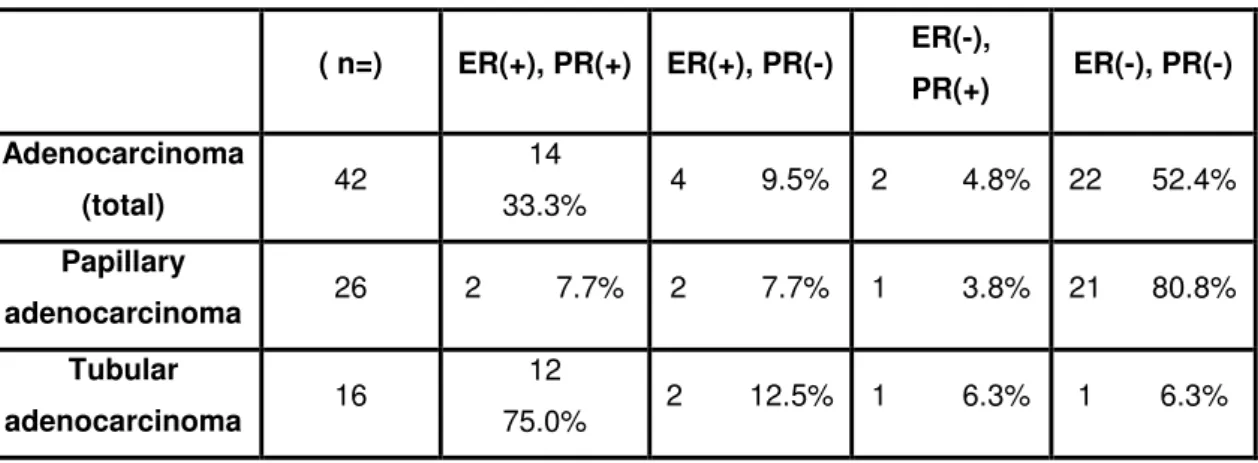
In addition to the evaluation of these markers, the study of the oestrogen receptor-α (ER-α) and progesterone receptors (PR) expression may also contribute to better understand the characteristics of the uterine adenocarcinoma. Considering the immune-reactive scores of ER-α and PR and the mode of myometrium invasion, a previous study proposed 2 different development patterns for uterine adenocarcinoma in the rabbit: a papillary growth pattern, which is negative for both ER-α and PR and a tubular growth pattern, which is positive to ER-α, PR or both (Table 2.). Regarding tumour invasion, this study also pointed that papillary adenocarcinomas infiltrated the myometrium later in the disease, whereas tubular adenocarcinomas invaded into the deep myometrium early in the disease.11 This classification could be compared to the one applied to humans, even though there are no studies supporting this idea. In spite of the scarce information regarding the expression of tumour markers, previous publications emphasize that the accurate evaluation of ER-α and PR and the establishment of a growth pattern may contribute to help determine the survival rate and to establish a selective therapy in the future.
These and other tumour markers contribute to characterize tumours, to evaluate the proliferative potential of the tumour cells, to establish a more accurate treatment, to establish a prognosis and confirm the origin of metastases. Therefore, an effort should be made in order to perform a more consistent evaluation of the rabbit uterine adenocarcinoma.
Table 2. Oestrogen and progesterone receptor expression in 42 uterine adenocarcinomas of rabbits.
( n=) ER(+), PR(+) ER(+), PR(-) ER(-),
PR(+) ER(-), PR(-) Adenocarcinoma
(total) 42
14
33.3% 4 9.5% 2 4.8% 22 52.4% Papillary
adenocarcinoma 26 2 7.7% 2 7.7% 1 3.8% 21 80.8% Tubular
adenocarcinoma 16
12
75.0% 2 12.5% 1 6.3% 1 6.3%
4. Clinical signs
Owners of female rabbits bearing uterine diseases frequently present complains about the presence of blood in the urine50, although in this cases blood originates in the uterus and is released when the rabbit urinates.9,10,22,50,51 Haematuria is intermittent and occurs more frequently at the end of the urination.22 In a recent study regarding uterine disorders in 50 pet rabbits, in 31 out of the 50 rabbits, 71% presented a serosanguineous vaginal discharge or haematuria and/or an enlarged uterus during abdominal palpation. In the same study, uterine adenocarcinoma was detected in 27 out of the 50 rabbits studied.9 In another study, 68.1% of the rabbits also presented haematuria and the bleeding complaints in 8 of 10 cases corresponded to uterine adenocarcinoma.51 Together, the results from these studies pointed that bleeding, usually detected by the rabbit owner as haematuria, could be one of the cardinal symptoms in rabbit’s uterine disorders9. However, as it was demonstrated by Walter et al., the absence of bleeding does not exclude the existence of a uterine disorder. In fact, in a study with 59 rabbits that presented uterine disorders, only 10 presented bleeding.52
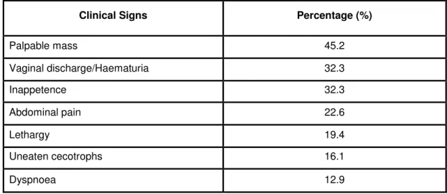
Enlargement of the abdomen and abdominal pain are some of the unspecific predominant clinical signs.9 During abdominal palpation it is possible to palpate masses consistent with uterine adenocarcinoma in the caudal portion of the abdomen.9,50 In a study performed by Kunzel et al. in 31 pets with uterine disorders, 45.2% of the rabbits presented a palpable mass in the caudoventral portion of the abdomen and 22.6% of the rabbits presented abdominal pain during palpation. In the same study, clinical signs such as innapetence, lethargy, uneaten cecotrophs and dyspnea in patients with lung metastases were also found (Table 3.).9
In another study with 47 rabbits, 15 (31.9%) of the rabbits presented mammary gland disorders in addition to uterine disorders.51 The association between uterine disorders and the occurrence of mammary gland disorders was confirmed in a recent study performed by Kreso et al. who reported that 30% of the rabbits with uterine tumours may also present mastitis and mammary gland tumours.22 These studies seem to emphasize the role of hormones in the development of both uterine and mammary tumours, since it has been proven in other species such as rats, dogs and humans that there is a direct correlation between oestrogen and progesterone receptors concentration in both uterine and mammary tissues53.
Table 3. Percentage of several clinical signs of 31 pet rabbits diagnosed with uterine disorders.
Clinical Signs Percentage (%)
Palpable mass 45.2
Vaginal discharge/Haematuria 32.3
Inappetence 32.3
Abdominal pain 22.6
Lethargy 19.4
Uneaten cecotrophs 16.1
Dyspnoea 12.9
Adapted from Kunzel et al. (2015).
5. Diagnosis
Because of their prey instinct, rabbits have a tendency to mask their illnesses which requires an exhaustive and patient examination performed by the veterinarian in order to achieve a successful diagnosis.3
The rabbit uterus lies caudally to the caecum in the ventral abdomen and when enlarged has a tendency to fall forward into the abdomen.28 In an initial phase, due to its small size, the uterine adenocarcinoma can be unnoticeable during general examination.50 When detected, the tumour may have up to 1 cm in diameter and if left without treatment can reach 5 cm in diameter within 6 months, being easily palpable through the abdominal and uterine walls.23
5.1 Differential Diagnosis
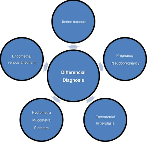
Figure 9. Differential diagnosis for a palpable mass in the ventral abdomen of a female rabbit.
5.2 Diagnostic Procedures
In rabbits with a clinically suspected uterine adenocarcinoma, a diagnostic workup is required in order to achieve a correct diagnosis. One of the main complementary diagnostic exams is imaging.9,50 Abdominal radiographs could be used to assess the reproductive system in case of a mass in the caudalventral abdomen. It is important to take at least two radiographs one in right lateral and the other ventrodorsal to achieve an adequate image of the mass 3. In rabbits presenting dyspnoea, thoracic radiographs can detect and diagnose metastatic neoplasia3,9 being this technique recommended to detect early metastases.22
Another imaging technique proven to be useful in the diagnosis of uterine adenocarcinoma is the ultrasound. If a 7.5 to 10 MHz transducer is used it is possible to obtain quality images of the abdomen and thorax.3,9 Using this technique, alterations such as enlargement of the uterus’ diameter, fluid within the lumen, masses and alterations in echogenicity were found in all rabbits.9 Although it has not been reported in rabbits, in humans, the use of computerized tomography (CT) is routinely used in the management of uterine tumours.54
In a recent study, both radiography and ultrasonography were used. Abdominal radiography was performed in 11 out of the 50 rabbits and uterine alterations were detected in 8 out of the 11 rabbits. The confirmation of the diagnosis was given by abdominal ultrasonography in 28 rabbits with possible uterine disorders. The authors,9 based on this results, consider abdominal
Differencial Diagnosis Uterine tumours
Pregnancy
Pseudopregnancy
Endometrial
hyperplasia Hydrometra
Mucometra
Piometra Endometrial
ultrasonography to be the most accurate method to diagnose uterine disorders such as the uterine adenocarcinoma.9
In ambiguous cases of females presenting haematuria or serosanguineous vaginal discharge, an urine dipstick can be useful to differentiate blood from porphyrin-pigmented urine caused by plant pigments9 and can detect the presence of other cells like leucocytes.10 Ultrasound-guided cystocentesis can be used to differentiate haematuria from uterine blood.22 Also, an hematologic test or at least an haematocrit determination should be done due to the fact that, in more advanced cases, the rabbit can be anaemic due to blood loss.50
The definite diagnosis is only confirmed by histopathology after ovariohysterectomy and removal of the masses. 6,8
6. Treatment
6.1 Surgical Treatment
The treatment of choice in clinically stable rabbits with uterine adenocarcinoma is ovariohysterectomy.7,9,22,24,50,52 This surgical technique involves the removal of the reproductive tract of the animal.55
Prior to the surgery, several unique features regarding the reproductive system of the female rabbit must be taken in consideration, such as the presence of two uterine horns and two cervices, one for each horn. The uterine body is not identified in the rabbit.55 Also, the mesometrium stores a great amount of fat, which can make it difficult to identify.12,55 Due to the fact that the uterus is localised in the abdominal cavity, care must be taken to avoid the caecum and bladder when making the abdominal incision.1 Some authors12,55 recommend expressing the bladder prior to surgery to avoid filling of the vagina with urine and possible contamination.
In general, the rabbit uterus is fragile in comparison with other species such as dogs and cats, so it must be handled with care.12,55
6.1.1 Anaesthesia and Analgesia
For routine surgical procedures such as an ovariohysterectomy56, a combination of medetomidine (0,25 mg/kg SC IM)55, ketamine (15 mg/kg SC IM)55 and butorphanol (0,5 mg/kg SC IM)57,58 is recommended for the induction of anaesthesia and analgesia.55,58 At the end of the surgery, medetomidine should be reversed with its antagonist atipamezole55 (same volume SC or IM as medetomidine)57.
Monitoring of the patient must be constant.2 Respiration rate, depth and pattern are the best indicators of anaesthetic depth.58 Also, in a rabbit, the absence of an ear pinch and hind-leg withdrawal reflexes and loss of jaw tone are reliable indicators of surgical anaesthesia.2,58 Heart rate, colour of the mucous membranes and rectal temperature are other important aspects to take in consideration during the monitoring of an anaesthetised rabbit.58
6.1.2 Surgical Technique – Ovariohysterectomy
In female rabbits, the ovariohysterectomy is performed in dorsal recumbency via a ventral midline incision12,55,60, starting half way between the umbilicus and the pubic symphysis.12,56 Once the
linea alba is exposed, before making the incision and in order to prevent perforation of abdominal organs, elevation of the body wall from the abdominal structures is recommended due to the fact that the caecum and bladder may be directly under the incision site12,55,56 Within the peritoneal cavity, identification of the uterus must be done60 and the cervices may be seen immediately after the incision.61 Elevate one of the uterine horns from the incision12 and follow it cranially to the ovary.61 The ovarian ligament can be detached manually for further exteriorization of the ovary.55,56 To tie off the ovarian blood vessels it is necessary to dissect through the fat and to pass a ligature around the portion containing the vessels supplying the ovary.12 Ligation of large blood vessels in the broad ligament of the uterus is frequently required.12,55 After the procedure is repeated in the contralateral uterine horn, the uterus should then be ligate caudally to the cervices with a transfix ligature around the vagina side, in order to remove the cervices completely.12,60,61 This technique will carry a smaller risk of leaving uterine tissue and will consequently reduce the possibility of a tumour recurrence.56,61 Risks like incorporating the ureters and the bladder blood vessels in a misplaced ligature must be taken in consideration.56 Also, due to the fact that the rabbit’s proximal aspect of the vagina fills with urine during micturition, leakage of urine through the vaginal stump can result in abdominal cavity contamination when the ligature is not securely performed.56,60,61
During this procedure, inspection of the abdominal organs for metastasis should be done and, in case of any suspected metastasis, biopsy is recommended.3 If there are neoplastic mammary glands associated with the adenocarcinoma its removal should be done at the same time. Cystic breast changes such as cystic mastitis will reverse after the ovariohysterectomy.21,22
Closure of the abdominal wall in three layers is routine12,60, with muscular wall, subcutaneous tissue and skin being closed as separated layers12, using an intradermal suture pattern with a 3-0 absorbable suture is recommended.55,61
6.1.3 Postoperative Considerations
When recovering from surgery, hypothermia, stress, anorexia and reduced gut motility are some of the possible symptoms that a small mammal like the rabbit can experience.58 Recovery time in a quiet, warm and comfortable environment is the key to achieve an adequate recovery2,55,58 To reduce hypothermia, frequently associated with clipping of the fur, asepsis and prolonged recovery time, towels or heating pads can be used to elevate the body temperature58, which should be carefully monitored.62 Stressful environments (noises, odour and sight of potential predators)62 have a negative impact on rabbits recovery due to the stimulation of the sympathetic nervous system and consequently decreasing of gastrointestinal motility.58 Physiological parameters such as respiratory and heart are also affected and should be monitorized.58 Although it is a rare complication, a recent study presented 3 cases of colonic obstruction after ovariohysterectomy, in which clinical signs such as decreased appetite, anorexia, abdominal pain and decreased faecal output were present.63 To avoid a decreased gastrointestinal motility, a gastrointestinal kinetic stimulant such as metoclopramide (0,2-0,5 mg/kg PO, SC, every 6 to 8 hours)57 should be administrated if the rabbit is anorexic or does not defecate within 24-48h after the surgery.55,58
Rabbits and rodents present very subtle signs of pain making it difficult to assess.55,58 Signs like absence of grooming, crouched position and tooth grinding are suggestive of postoperative pain.55,58 The presence of such signs should determinate whether further analgesics should be given.55 To guarantee an adequate analgesia, association of opioids such as butorphanol (0,1-0,5 mg/kg SC, IM, IV, every 4 hours)57 or buprenorphine (0,03 mg/kg IM every 12 hours)57 and non-steroidal analgesics such as carprofen (1-2,2 mg/kg PO every 12 hours)57 or meloxicam (0,2 mg/kg SC, IM, every 24 hours)57 can be done without adverse effects on rabbits.58
Palatable food (Critical Care®) and water should be available right after the rabbit is recovered2,55,58,62 to ensure gut motility is restored as soon as possible.55
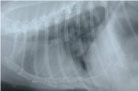
Figure 10. Pulmonary metastasis detected in a laterolateral thoracic radiograph of a rabbit with uterine
adenocarcinoma.
Courtesy of Dr. Neus Morera, Clinica Veterinaria Exótics, Barcelona, Spain
6.2 Oncological treatment
In addition to surgery, the use of chemotherapy, radiotherapy and hormonal therapy could be hypothesised in the treatment of the rabbit uterine adenocarcinoma. However, effective treatment using these therapies has not yet been reported.21,22
Admitting that there are no official approved chemotherapy agents for use in exotic animals, the few reports of chemotherapy in rabbits result from the use of canine and feline protocols adapted to the rabbit.64,65 As described in Table 4., several chemotherapy agents have been experimented in rabbits, mainly in a laboratory setting but also in pet rabbits.66,64 One of the main limitations of the chemotherapy use in rabbits is the toxicity.64,65 In fact, moderate to severe myelosupression with associated neutropenia and gastrointestinal toxicity are two of the main effects described mainly in laboratory animals with induced tumours. 64,66
Regarding radiotherapy, there is also very little literature regarding its utilization alone and in combination with surgery and/or with chemotherapy.65 In women, the treatment of endometrial tumours with radiation therapy involves two techniques: external beam radiation, which consists of a machine that focuses a beam of radiation at the tumour, meaning that the treatment is delivered from a source outside the body, and brachytherapy or internal radiation therapy, a technique in which radioactive materials are placed near the tumour, inside the body.67 Information regarding the association of these techniques and the treatment of uterine adenocarcinoma in veterinary medicine articles has not been found.
aromatase inhibitors and tamoxifen is one of the main therapeutic methods in the treatment of hormone-dependent tumours. Therefore, in rabbits, to determine the role of hormonal therapy it is important to make an exhaustive analysis of the contribution of hormones for the development of this tumour.
a. Progestins
Progestins such as medroxyprogesterone acetate and megestrol acetate are drugs that present similarity to progesterone. Their function is to slow the growth of the endometrial tumour.68
b. Luteinizing hormone-releasing hormone agonists
In women with functioning ovaries this agonist is given to inhibit the production of oestrogens and consequently to lower the oestrogen level in the body. Goserelin and leuprolide are some of the drugs with this effect.68
c. Aromatase inhibitors
Aromatase inhibitors are used to block the enzyme aromatase which converts testosterone to oestrogens, leading to a decrease in oestrogen production in the tumour tissues. An in vitro
treatment of endometrial tumour tissue with the aromatase inhibitors showed that in situ
depletion of oestrogen leads to a lower proliferation of tumour cells.69 Examples of aromatase inhibitors are letrozole, anastrozole and exemestane. They are most frequently used in the treatment of mammary neoplasia and its therapy success towards more advanced endometrial tumours is still unclear.68,69
d. Tamoxifen
Tamoxifen is an anti-oestrogen nonsteroidal drug used commonly in the treatment of mammary tumours from different species including the rabbit. It binds to the oestrogen receptors present in the body, including the ones in the tumour tissue, blocking their activity.3
Table 4. Chemotherapy agents and dosage recommendations for rabbits.
Chemotherapy Agent Dosage
Carboplatin 150-180 mg/m² IV, every 21-28 days
Lomustine (CCNU) 50 mg/m² PO, every 3-6 weeks
Cyclophosphamide 5-6 mg/m² IV, every 21 days
Doxorubicin 1 mg/kg IV, every 14-21 days
L-asparaginase 400 U/kg SC, IV
Prednisone 0.5-2 mg/kg PO
Vincristine 0.5-0.7 mg/m² IV, every 7-14 days
Adapted from Mayer et al. (2013).
7. Prognosis
If surgical treatment is performed prior to metastatic disease or associated mammary neoplasia, the prognosis of the uterine adenocarcinoma is considered excellent and frequently curative in clinically stable patients.7,9,22,50 In case of metastatic spread, the prognosis is poor, since there is no oncological treatment considered successful. In these cases the survival rate is approximately 12 to 24 months upon the diagnosis.7,22 Moreover, to reach an accurate prognosis, evaluation of the physical condition of the patient is essential51, since a poor clinical condition is associated with a worse prognosis9. Euthanasia should only be considered in the referred cases.9,52
According to Saito et al., the mortality rate immediately after ovariohysterectomy was 21.3%.51 However, according Kunzel et al., when rabbits survive to surgery the survival rate at 6 months was 80% 9, and, in a similar study performed by Walter et al. 52, the survival rate at two years was 28%.
8. Prevention
Prevention is the answer when it comes to the management of uterine adenocarcinoma.7,21,50 In spite of the fact that the contribution of hormones to tumour development is not yet well investigated, female rabbit owners that are not considering breeding should be advised to neuter their pet as soon as the procedure can be done and before they reach 2 years of age.7,50 It is preferable to spay the rabbit between 6 and 12 months since at an early age ovariohysterectomy tends to be less complicated due to less fat content of the abdomen, and healing time is minimal in comparison with older rabbits. 7,21,22 Although the procedure can be done after 4 to 5 months of age, neutering of a pre-pubescent female is not recommended on account that it can be difficult to clearly identify the uterus and ovaries since they are not fully developed.28
When it comes to breeding rabbits, it is recommended to stop breeding and perform ovariohysterectomy before it reaches 4 years of age, time when the incidence of uterine tumours is higher.22 Before that, annual or semi-annual health examinations should be done to control and detect possible early clinical signs of the disease.7,21
9. Future perspectives
The rabbit as a clinical patient and as one of the favourite pet choices for owners is still a recent accomplishment, which means that the medicine regarding this species may not be as evolved as it is for other small and production animals. The majority of study cases and articles about uterine adenocarcinoma in rabbits only present surgery as the procedure to treat this tumour or in advanced cases euthanasia, without much further exploration into other possibilities of treatments that are currently being applied to humans and other species.
Therefore, a more profound study of the cellular, molecular and genetic alterations that characterize the uterine adenocarcinoma in rabbits is urgent. Similar to what happened in humans, dogs and cats, this exhaustive study will allow the identification of gene mutations, the study of alterations in signalling pathways and the contribution of hormones to the tumour development. Once rabbit uterine adenocarcinoma is adequately characterized it will be possible to achieve an accurate diagnosis and to establish new treatment protocols that may significantly increase the quality of life and survival upon diagnosis.
10. Acknowledgements
The conclusion of this thesis would not have been possible without the help of my supervisors, to whom I am very grateful, Prof.ª Doutora Anália do Carmo and Dr.ª Ana Luísa Vieira. Their patience to answer all my questions and continuous encouragement was essential throughout the development of this review.
I would like to acknowledge my boyfriend, closest friends and colleagues for having accompanied me throughout these 6 years without letting me lose focus of what truly matters. Also, a special acknowledgement to all my family to whom I dedicate this work, specially my Mother and Father. Without my parents I would not have been able to pursue this career and dedicate myself to a field that inspires me every day. Without them I would have not discovered the happiness of doing what I love and for that I am eternally grateful. I am thankful for my brother, sisters, stepfather and stepmother as well, for putting up with me and believing in my abilities even when I did not. Lastly, many thanks to Clinica Veterinaria Exótics and M. G. Asakawa for providing photographs to enrich my work.
11. References
1. Harcourt-Brown, F. in Textbook of Rabbit Medicine 1–18 (Butterworth Heinemann, 2002).
2. Richardson, V. in Rabbits Health, Husbandry and Diseases 44–58 (Blackwell Science,
2000).
3. Mitchell, M. A. & Vennen, K. M. in Manual of Exotic Pet Practice 375–405 (Saunders, 2009).
4. Church, B. Biology, Natural History and Behaviour in Rabbits. in The North American Veterinary Conference (2007).
5. Harcourt-Brown, F. in Textbook of Rabbit Medicine 336–351 (Butterworth Heinemann,
2002).
6. Hablolvarid, M. H., Golami, M. R. & Moharami, M. Uterine Adenocarcinoma in a Rabbit. 61, (2006).
7. Hreiz, J. E. Uterine Adenocarcinoma in Rabbits. ARBA magazine 30–31 (2013).
8. Kurotaki, T., Kokoshima, H., Kitamori, F., Kitamori, T. & Tsuchitani, M. A case of adenocarcinoma of the endometrium extending into the leiomyoma of the uterus in a rabbit. J. Vet. Med. Sci. 69, 981–984 (2007).
9. Kunzel, F. et al. Uterine Disorders in 50 Pet Rabbits. J Am Anim Hosp Assoc 51, 1–7 (2015).
10. Zadravec, M. et al. Uterine heterologous malignant mixed Mullerian tumor in a dwarf rabbit (Oryctolagus cuniculus). J. Vet. Diagnostic Investig. 24, 418–422 (2012).
11. Asakawa, M. G., Goldschmidt, M. H., Une, Y. & Nomura, Y. The Immunohistochemical Evaluation of Estrogen Receptor-a and Progesterone Receptors of Normal, Hyperplastic, and Neoplastic Endometrium in 88 Pet Rabbits. Vet. Pathol. 45, 217–225 (2008).
12. Bennett, R. A. Techniques for Neutering Exotic Mammals. in Western Veterinary Conference (2013).
13. Capello, V. & Lennox, A. M. Gross and Surgical Anatomy of the Reproductive Tract of Selected Exotic Pet Mammals. in Association of Avian Veterinarians Conference 19–28
14. Lebas, F., Coudert, P., de Rochambeau, H. & Thébault, R. G. The Rabbit - Husbandry, Health and Production. (FAO Animal Production and Health Series, 1997).
15. Baba, N. & von Haam, E. Animal Model: Spontaneous Adenocarcinoma in Aged rabbits.
Am. J. Pathol. 68, 653–656 (1972).
16. Karlsson, S. Gene Expression Patterns in a Rat Model of Human Endometrial Adenocarcinoma. (University of Gothenburg, 2008).
17. Hubbard, S. a & Gargett, C. E. A cancer stem cell origin for human endometrial carcinoma? Reproduction 140, 23–32 (2010).
18. Kato, K. Endometrial cancer stem cells: A new target for cancer therapy. Anticancer Res.
32, 2283–2293 (2012).
19. Kyo, S., Maida, Y. & Inoue, M. Stem cells in endometrium and endometrial cancer: Accumulating evidence and unresolved questions. Cancer Lett. 308, 123–133 (2011).
20. Kreso, A. & Dick, J. E. Evolution of the cancer stem cell model. Cell Stem Cell 14, 275–
291 (2014).
21. Klaphake, E. & Paul-Murphy, J. in Ferrets, Rabbits and Rodents Clinical Medicine and Surgery 217–219 (Saunders, 2012).
22. Oglesbee, B. L. in Blackwell’s Five-Minute Veterinary Consult: Small Mammal 192–193 (John Wiley & Sons, Inc., 2011).
23. Harkness, J. E., Turner, P. V., VandeWoude, S. & Wheler, C. L. in Biology and Medicine of Rabbits and Rodents 341 (Wiley-Blackwell, 2010).
24. Alizadeh, R., Asghari, A. & Khandanlou, R. Uterine Adenocarcinoma in a Domestic Rabbit.
Glob. Vet. 9, 245–247 (2012).
25. Vinci, a., Bacci, B., Benazzi, C., Caldin, M. & Sarli, G. Progesterone Receptor Expression and Proliferative Activity in Uterine Tumours of Pet Rabbits. J. Comp. Pathol. 142, 323–
327 (2010).
26. Harcourt-Brown, F. in Textbook of Rabbit Medicine 250–291 (Butterworth Heinemann,
2002).
28. Harcourt-Brown, F. in Textbook of Rabbit Medicine 52–93 (Butterworth Heinemann,
2002).
29. Newbold, R. R., Padilla-banks, E. & Jefferson, W. N. Environmental Estrogens and Obesity. Mol. Cell Endocrinol. 304, 84–89 (2010).
30. Siddiqui, Z. H., Ashraf, M. A. & Sipra, T. B. Association of Breast Cancer with Obesity and Estrogen in Women. Sci.Int(Lahore) 26, 729–732 (2014).
31. Nagaoka, T. et al. Spontaneous uterine adenocarcinomas in aged rats and their relation to endocrine imbalance. J. Cancer Res. Clin. Oncol. 116, 623–628 (1990).
32. Agren, E. Pathology of the Rabbit Uterus. Exotic DVM Vol. 6.5 (2004).
33. Bercier, M. et al. Salivary gland adenocarcinoma in a domestic rabbit (oryctolagus cuniculus). J. Exot. Pet Med. 22, 218–224 (2013).
34. Lennox, A. M. & Reavill, D. Nasal Mucosal Adenocarcinoma in a Pet Rabbit. J. Exot. Pet Med. 23, 397–402 (2014).
35. Nakata, M., Miwa, Y., Tsuboi, M. & Uchida, K. Surgical and Localized Radiation Therapy for an Intranasal Adenocarcinoma in a Rabbit. J. Vet. Med. Sci. 76, 1659–1662 (2014).
36. Lair, S., De Guise, S. & Martineau, D. Uterine adenocarcinoma with abdominal carcinomatosis in a beluga whale. J. Wildl. Dis. 34, 373–376 (1998).
37. Goldschmidt, M., Peña, L., Rasotto, R. & Zappulli, V. Classification and grading of canine mammary tumors. Vet. Pathol. 48, 117–131 (2011).
38. Summa, N. M., Eshar, D., Snyman, H. N. & Lillie, B. N. Metastatic anaplastic adenocarcinoma suspected to be of mammary origin in an intact male rabbit (Oryctolagus cuniculus). Can Vet J 55, 475–479 (2014).
39. Schoniger, S., Horn, L. & Schoon, H. Tumors and Tumor-like Lesions in the Mammary Gland of 24 Pet Rabbits : A Histomorphological and Immunohistochemical Characterization. Vet. Pathol. 1–12 (2013).
40. Murali, R., Soslow, R. a. & Weigelt, B. Classification of endometrial carcinoma: More than two types. Lancet Oncol. 15, e268–e278 (2014).
42. Dai, D., Bradford, A. P. & Prossnitz, E. R. Endometrial cancer: molecular and cellular basis of tumor development, novel biomarkers and therapeutic agents, and innovative research approaches. Obstet. Gynecol. Int. 2 (2014).
43. Pires, M. a., Seixas, F., Palmeira, C. & Payan-Carreira, R. Histopathologic and immunohistochemical exam in one case of canine endometrial adenocarcinoma. Reprod. Domest. Anim. 45, 545–549 (2010).
44. Scholzen, T. & Gerdes, J. The Ki-67 protein: from the known and the unknown. J Cell Physiol. 182, 311–322 (2000).
45. Korenbaum, E. & Rivero, F. Calponin homology domains at a glance. J. Cell Sci. 115,
3543–3545 (2002).
46. Satelli, A. & Li, S. Vimentin as a potential molecular target in cancer therapy Or Vimentin, an overview and its potential as a molecular target for cancer therapy. Cell Mol Life Sci.
68, 3033–3046 (2011).
47. Toniti, W. et al. Immunohistochemical determination of estrogen and progesterone receptors in canine mammary tumors. Asian Pac. J. Cancer Prev. 10, 907–911 (2009).
48. Stefansson, I. M., Salvesen, H. B. & Akslen, L. a. Loss of p63 and cytokeratin 5/6 expression is associated with more aggressive tumors in endometrial carcinoma patients.
Int. J. Cancer 118, 1227–1233 (2006).
49. Stewart, C., Crook, M., Lacey, J. & Louwen, K. Cytokeratin 19 expression in normal endometrium and in low-grade endometrioid adenocarcinoma of the endometrium. Int J Gynecol Pathol. 30, 484–491 (2011).
50. Ríos, A. M., Nadeu, C. B., García, M. A. & Barceló, A. M. Patologías del aparato reproductor en conejas (Oryctolagus cuniculus). Profesión Vet. ISSN 2253-7244 16, 87–
90 (2007).
51. Saito, K., Nakanishi, M. & Hasegawa, A. Uterine disorders diagnosed by ventrotomy in 47 rabbits. J. Vet. Med. Sci. 64, 495–497 (2002).
52. Walter, B., Poth, T., Böhmer, E., Braun, J. & Matis, U. Uterine disorders in 59 rabbits. Vet. Rec. 166, 230–233 (2010).
and affinities during the oestrous cycle in the normal mammary gland and uterus of dogs.
Vet. Res. Commun. 19, 101–113 (1995).
54. Colas, E. et al. Molecular markers of endometrial carcinoma detected in uterine aspirates.
Int. J. Cancer 129, 2435–2444 (2011).
55. Richardson, C. & Flecknell, P. Routine Neutering of Rabbits and Rodents. In Pract. 28, 70–79 (2006).
56. Harcourt-Brown, F. in Textbook of Rabbit Medicine 352–360 (Butterworth Heinemann, 2002).
57. Fiorello, C. V. & Divers, S. J. in Exotic Animal Formulary 517–559 (Elsevier, 2013).
58. Harcourt-Brown, F. in Textbook of Rabbit Medicine 121–139 (Butterworth Heinemann,
2002).
59. Badia, X. V. & Bueno, J. V. in Casos Clínicos de Animales Exóticos 24–29 (Servet, 2012).
60. Murray, M. J. Spays and Neuters in Small Mammals. in North American Veterinary Conference 1757–1759 (2006).
61. Mayer, J. Surgical Techniques for Spaying Rabbits and Rats. in North American Veterinary Conference 1854–1855 (2008).
62. Lightfoot, T. & Bartlett, L. Exotic Companion Animal Surgeries. (Zoological Education Network, 1999).
63. Guzman, D. S.-M. et al. Colonic Obstruction Following Ovariohysterectomy in Rabbits: 3 Cases. J. Exot. Pet Med. 24, 112–119 (2015).
64. Hahn, K. a. Chemotherapy Dose Calculation and Administration in Exotic Animal Species.
Semin. Avian Exot. Pet Med. 14, 193–198 (2005).
65. Graham, J. E., Kent, M. S. & Théon, A. Current therapies in exotic animal oncology. Vet. Clin. North Am. - Exot. Anim. Pract. 7, 757–781 (2004).
66. Mayer, J. & Donnelly, T. M. in Clinical Veterinary Advisor: Birds and Exotic Pets 424–425 (Saunders, 2013).
68. Temkin, S. & Fleming, G. Current Treatment of Metastatic Endometrial Cancer. Cancer Control 16, 38–45 (2009).
