ARTIGO DE REVISÃO
The Importance of Zebrafish in Biomedical
Research
A Importância do Peixe-Zebra na Investigação Biomédica
1. Centro de Estudos de Doenças Crónicas. Faculdade de Ciências Médicas de Lisboa. Lisboa. Portugal. Recebido: 19 de Julho de 2013 - Aceite: 27 de Julho de 2013 | Copyright © Ordem dos Médicos 2013 Bárbara TAVARES1, Susana SANTOS LOPES1
Acta Med Port 2013 Sep-Oct;26(5):583-592
RESUMO
Introdução: O peixe-zebra (Danio rerio) é um excelente organismo modelo para o estudo do desenvolvimento dos vertebrados. Este facto deve-se à produção de grandes posturas, que podem atingir 200 embriões a cada sete dias, e ao facto dos embriões serem pequenos, transparentes e com um rápido desenvolvimento externo.
Material e Métodos: Usando ferramentas de pesquisa bibliográfica científica disponíveis online e utilizando as palavras-chave “Ze-brafish”, “biomedical research”, “human disease” e “drug screening”, avaliámos estudos originais e revisões indexadas na PubMed. Resultados: Neste artigo de revisão fazemos um resumo do trabalho realizado com este modelo no melhoramento do conhecimento de várias doenças humanas. Fizemos ainda um breve relato da investigação biomédica realizada em Portugal com o modelo de peixe-zebra.
Discussão: Têm sido desenvolvidas poderosas ferramentas genéticas e de microscopia in vivo, que também tornaram o peixe-zebra num modelo valioso em investigação biomédica. A conjugação destes atributos com a optimização de sistemas automatizados de tria-gem de medicamentos, transformaram o peixe-zebra num top model da investigação em biomedicina, nomeadamente na triatria-gem de compostos químicos com efeitos terapêuticos e em testes de toxicidade. Além disso, com a otimização da tecnologia dos xenografos, será possível usar o peixe-zebra na escolha de uma terapia personalizada.
Conclusão: O peixe-zebra é um excelente organismo modelo na pesquisa biomédica, em screens de medicamentos e na terapia clinica. Palavras-chave: Avaliação Pré-Clínica de Medicamentos; Investigação Biomédica; Modelos Animais de Doença; Peixe Zebra.
ABSTRACT
Introduction: Zebrafish (Danio rerio) is an ideal model organism for the study of vertebrate development. This is due to the large clutches that each couple produces, with up to 200 embryos every 7 days, and to the fact that the embryos and larvae are small, trans-parent and undergo rapid external development.
Material and Methods: Using scientific literature research tools available online and the keywords Zebrafish, biomedical research, human disease, and drug screening, we reviewed original studies and reviews indexed in PubMed.
Results: In this review we summarized work conducted with this model for the advancement of our knowledge related to several human diseases. We also focused on the biomedical research being performed in Portugal with the zebrafish model.
Discussion: Powerful live imaging and genetic tools are currently available for zebrafish making it a valuable model in biomedical research. The combination of these properties with the optimization of automated systems for drug screening has transformed the zebrafish into “a top model” in biomedical research, drug discovery and toxicity testing. Furthermore, with the optimization of xenografts technology it will be possible to use zebrafish to aide in the choice of the best therapy for each patient.
Conclusion: Zebrafish is an excellent model organism in biomedical research, drug development and in clinical therapy. Keywords: Biomedical Research; Disease Models, Animal; Drug Evaluation, Preclinical; Zebrafish.
INTRODUCTION
The zebrafish (Danio rerio) is a tropical freshwater fish native to the northern Indian subcontinent (Fig.s 1A and 1B). The species arose in the Ganges region in eastern In-dia, and is commonly found in slow-moving or stagnant wa-ter.1 Due to zebrafish large fecundity and fertility rates it has
become an important model organism for genetic studies. It started as a great model for vertebrate embryonic develop-ment, due to its transparency and optic clearance, to be-come an excellent model in adult stem cell and regenerative medicine.2 Zebrafish embryos, larvae and adults are now
commonly used as a model for accessing gene function in several human diseases.
Zebrafish importance as a research vertebrate model system in the field of biomedicine has been strengthened by their amenity for large-scale forward genetic screens, such as those known as the Tübingen and Boston screens.3 Gene
knockdown can also be achieved transiently and effectively with Morpholino (MO) antisense oligonucleotide technology or in a permanent way with the use of custom zinc finger nucleases (ZFNs),4 and transcription activator-like effector
nucleases (TALENs).5 More recently, another technology
based on bacterial clustered, regularly interspaced, short palindromic repeats (CRISPR) and associated (Cas) sys-tems (CRISPR-Cas) was shown to function in vivo to in-duce targeted genetic modifications in zebrafish embryos in a cheaper and equally efficient way.6 These technologies
have been used to introduce locus-specific double-stranded breaks in the zebrafish genome, generating many mutant alleles that copy human disease loci. Thus, both forward and reverse genetic tools are available. Additionally, zebra-fish allows for excellent quality in vivo and ex vivo imaging,7
ARTIGO DE REVISÃO
as fluorescent confocal microscopy, or even bright field live video microscopy. Furthermore, the research community is supported by an excellent curated database of genetic, genomic, and developmental information - the Zebrafish Model Organism Database (ZFIN).8
As a vertebrate model, zebrafish possess several ad-vantages, such as a fully sequenced genome, a very rapid embryologic development outside of the mother’s body (Fig.s 1C and 1D), and a well-characterized behavior of easy observation. Moreover, there are several mutant and transgenic strains available. One example is the casper
mu-tantstrain with transparent skin in the adult fish, allowing for
detailed visualization of organs and cellular activity, circula-tion, metastasis, among other phenomena.9 Zebrafish has
been shown to be similar to mammalian biological models when it comes to toxicity tolerance and circadian rhythms.10
Nevertheless, they are capable of regenerating several tis-sues, like fins,11 heart,2 photoreceptor cells and retinal
neu-rons,12 and even spinal cord.13 Furthermore, several studies
have been made over the years in development and dif-ferentiation of the nervous system in zebrafish, allowing for the mapping of their brain networks14 which will facilitate the
study of human neurological diseases. Altogether the tools available and the plasticity of this biological model allowed Figure 1 -A) Adult male zebrafish (12 months old). B) Adult female zebrafish (12 months old). C) 24 hpf (hours post fertilization) zebrafish embryo. D) 48 hpf zebrafish larvae. E) Schematic ciliary structure.
- Microtubule
(MT) Doublets and Triplets
Primary Cilia
9 + 0 9 + 0
9 + 2
Motile Cilia
5
1 1
2
PM
3 + CM - Axoneme CM - Ciliary Membrane PM - Plasma Membrane 5 - Transition Fibers
- Central Apparatus - Dynein Arms - Radial Spoke
4 - Central Cap 3 - MT Cytoskeleton 2 - Basal Body 1 - Ciliary Pocket
CM
ARTIGO DE REVISÃO for the establishment of zebrafish as a human disease
mod-el for cancer, cardiovascular and immune system diseases, and many others.
In this review we will emphasize the role of zebrafish, as a biomedical vertebrate model. We will focus on five main human illnesses, chosen for their high prevalence or their chronicity: cancer, diabetes and lipid-related diseases, car-diovascular disease, neurodegeneration and ciliopathies. We will also report the use of zebrafish in drug screening and the translational work currently being done with zebraf-ish in Portugal.
Zebrafish as a Human Disease Model 1. Cancer
Zebrafish was first used in cancer research during the 1960s, when Stanton et al. used it to test the effects of car-cinogens.15 Zebrafish have a very low rate of spontaneous
neoplasia, with only ~10% of zebrafish developing a tumor over their lifetime. Nevertheless, when exposed to carcino-genic agents such as DENA (diethylnitrosamine), MNNG (N-methyl-N-nitro-N-nitrosoguanidine), and DMBA (7.12 - dimethylbenz(a)anthracene), the fish will develop can-cer.15–18 Furthermore, zebrafish has proven to be an ideal
model to study the malignancy of several tumors by means of tumor transplantation assays.19 These were shown to be
robust and have the added advantage of the fish high fe-cundity, providing high numbers of donor and recipient fish. Not only have several types of cancer been studied with this model, such as leukemia,20,21 melanoma, endocrine or liver
cancer16,19,20 but more importantly, by means of the
xeno-transplantation of human tumor cells into zebrafish embryos (xenografts) it is possible to address tumor cell migration, metastasis, angiogenesis,24-26 and also the effects of
poten-tial therapeutic targets.19 Together with the easily available
forward and reverse genetic tools, and the non-invasive in vivo imaging technology, these characteristics have made of zebrafish an ideal vertebrate model to study cancer.27
Notwithstanding the evolutionary gap between fish and human, there is a manifest histological similarity between tumors formed in fish and those in human, with many im-portant genes and pathways involved in cancer being maintained in both species (Table 1).27 One such example
is that of the p53 tumor-suppressor protein. This transcrip-tion factor is essential in regulating cell death, proliferatranscrip-tion, and maintenance of genomic stability, and its mutation has been associated with more than 50% of all human tumors.28
In 2005, a forwards genetic study generated and identified three p53 missense mutations in zebrafish, of which two had been previously reported in human tumors.29 These mutant
fish developed tumors at an early age. Despite differences
in p53 transcription regulation,30 the zebrafish model for this pathway showed conservation of its main functions, like the role of p53 in DNA damage–induced apoptosis. Additionally, the development of antibodies for zebrafish p53 protein has contributed for the value of this model in the study of p53 in the context of cancer.27,30
Recently, zebrafish has become an interesting model
for the study of melanoma progression. It has been well es-tablished that mutations that activate the RAS/RAF/MEK/ ERK signaling cascade, notably mutations in the BRAF and NRAS genes, are very common in this type of tumor.32 To further determine the role of these genes in the progression of the disease, several transgenic fish lines were generated. While the BRAF mutant fish developed large agglomerates of proliferating melanocytes, which histologically resembled human nevi, the NRAS mutant fish displayed severe defects in pigment patterning but did not quickly developed mela-noma. Interestingly, when both transgenic fish lines were crossed with p53 mutant lines, the melanoma phenotype progressed significantly faster, highlighting the importance of p53 function in melanoma tumor suppression.33,34 A third
transgenic fish line was developed, expressing the oncogen-ic human HRAS gene in the melanocytic cell line. Research performed with these fishes suggested a role for epigenetic regulation in this melanoma model.35 Overall, these results
show that zebrafish melanomas resemble human disease morphologically, genetically and epigenetically, demonstrat-ing zebrafish suitability as a melanoma model.27
Another type of cancer that found in zebrafish an ad-equate model was the acute lymphoblastic leukemia (ALL), a disease with relatively homogeneous morphology and im-munophenotype, but with great heterogeneity at the genetic level, which can lead to distinct responses to therapy.27 One
of the main causes of ALL is the TEL-AML1 fusion, associ-ated with B-lymphocytes. Transgenic fish were generassoci-ated with this mutation in all cell lineages, and developed lympho-blastic leukemia, which phenocopied the childhood CD10+ pre-B ALL.36 T-cell ALL modeling in zebrafish was achieved
by creating a transgenic line for the oncogene Myc.37 Other mechanisms responsible for ALL have also been addressed with zebrafish, such as Notch1-induced T-ALL.38
ARTIGO DE REVISÃO Table 1 - Zebrafish models of human diseases.
Zebrafish Mutant AffectedGene Phenotype Human Disease References
tp53 N168K null
mutation tp53
Malignant peripheral nerve sheath tumors; failure to
activate apoptosis Cancer (29)
tp53 M214K null
mutation tp53
Malignant peripheral nerve sheath tumors; failure to
activate apoptosis Cancer (29)
BRAF mutant braf Large lesions of proliferating melanocytes, which
histologically resembled human nevi Melanoma (33)
NRAS mutant nras Severe defects in pigment patterning Melanoma (34)
HRAS mutant hras Melanoma Melanoma (35)
Transgenic
expression tel/aml1 fusion Acute lymphoblastic leukemia
Childhood CD10+ precursor B-lymphocyte acute
lymphoblastic leukemia (36) Transgenic
expression c-myc Acute lymphoblastic leukemia T-cell acute lymphoblastic leukemia (37) Transgenic
expression kras Tumors in pancreas and muscle Pancreatic cancer and rhabdomyosarcomas (27)
apc null mutation apc Tumors in liver and intestine Colon cancer (27)
MO pten Ocular tumors Cancer (27)
Transgenic
expression mycn β-islet cell neuroendocrine carcinoma MYCN-driven pancreatic neuroendocrine carcinoma (27)
heart-strings (hst) tbx5 Abnormalities in cardiac differentiation and fin development
Holt-Oram syndrome with cardiac septation defects and
limb abnormalities (52)
slipjig (sli) foxn4 Structural atrioventricular canal malformation accompanied by atrioventricular conduction defects Atrioventricular canal defects in humans (52)
gridlock (grl) hey2 Impaired blood flow in the tail owing to arterial
blockade in the anterior trunk, hyperplastic hearts Congenital aortic coarctation (52)
island beat (isl) C- LTCC Reduced number of ventricular cardiac myocytes
and arrhythmia (52)
liebeskummer (lik) ruvbl2 Cardiac hyperplasia (52)
heart of glass (heg), santa (san) and
valentine (val)
heg, krit1
and ccm2
Formation of hypocontractile, monolayered, giant
cardiac ventricles (52)
dead beat (ded) PLCG1 Loss of cardiac contractility between 48-60 hpf DMC (52)
main squeeze (msq) ILK Loss of cardiac contractility between 60-72 hpf DMC (52)
pickwick (pik), titin Zebrafish do not form cardiac sarcomeres DMC (52)
tell tale heart (tel) mlc2 Zebrafish do not form cardiac sarcomeres HCM (52)
silent heart (sil) TNNT2 Ventricular acontractility owing to impaired
myofibrillogenesis HCM (52)
erg kcnh2 (loss
of function) Complete atrioventricular block and ventricular asystole Long QT syndrome (52)
reggae (reg) kcnh2 of function)(gain Accelerated repolarization andfibrillation paroxysmal atrial Short QT syndrome (52)
slow mo (smo) hcn Bradycardia Bradycardia (52) tremblor (tre) NCX Atrial fibrillation Arrhythmia (52)
MO parkin 20% loss of DA neurons in the vDC with increased
susceptibility to PD-inducing neurotoxins Autosomal-recessive early-onset PD (55)
MO pink1
40% reduction in the number of DA neurons in the vDC, impaired response to touch stimuli, reduced swimming behavior, andmitochondrial defects
Autosomal-recessive
early-onset PD (55)
MO dj-1 DA neurons more sensitive to oxidative stress Autosomal-recessive early-onset PD (55)
MO irrk2 Loss of DA neurons in the vDC and locomotor
defects Autosomal-dominant early-onset PD (55)
Transient OE Mhtt
(Q102-htt)
Accumulation of Htt:GFP aggregates throughout the body of embryos at 24 hpf, with soluble forms of
mHtt proving neurotoxic HD (55)
MO appa, appb
Reduced body length and defective convergent-extension movements during gastrulation. Defects are rescued by wild-type human APP mRNA, but not by the Swedish mutant APP
Familial AD (55)
Transgenic expression
mutant human
MAPT
tau hyperphosphorylation, tangle formation, neurodegeneration in the spinal cord, and behavioral
deficits in escape response AD (55)
Revista Científica da Ordem dos Médicos www.actamedicaportuguesa.com 587
ARTIGO DE REVISÃO
2. Diabetes and Lipid-related Diseases
Due to the amenability of zebrafish for developmental studies, there is an accurate description of pancreas de-velopment and morphogenesis for this species. Studies performed in zebrafish have specifically led to the under-standing of extrinsic signaling molecules, like retinoic acid, Shh and FGF, in influencing intrinsic transcriptional pro-grams.39,40 These efforts have made zebrafish an
alterna-tive model to study not only the onset of diabetes but also its treatment. Zebrafish become hyperglycemic if exposed to high glucose and develop retinopathies with prolonged high blood sugar levels. Additionally, zebrafish also react satisfactorily to anti-diabetic drugs.41,42
The suitability of the zebrafish as a model for lipid-re-lated diseases lies with the fact that they possess remark-able similarities with mammals in their lipid absorption, processing and metabolism, together with the possibility of applying imaging methods with subcellular resolution to a whole organism, thanks to the availability offluorescent lipid dyes.43,44 Zebrafish has been shown to be an adequate
atherosclerosis model, allowing for the analysis of the le-sion development, by imaging lipid deposition and cellular changes in the vascular wall, and by visualization of mac-rophage lipid deposition in vivo.45 Obesity has also been
addressed with zebrafish, since their energy homeostasis resembles that of mammals, including amelanocortin sys-tem that responds to leptin,46 and similar response to
com-pounds known to modulate the fat content in mammals.47
Lastly, the metabolism of cholesterol in zebrafish shares many of its features with that of mammals. Both share key transcriptional regulators - SREBP (sterol-regulatory-ele-ment-binding protein) and LXR (liver X receptor) systems, and mutant fish for genes involved show similar phenotypes to the equivalent human pathologies.48,49
3. Cardiovascular Diseases
In recent years, the development of the cardiovascular system in zebrafish has been thoroughly studied and char-acterized, greatly accelerating our knowledge of cardiac development, angiogenesis and vasculogenesis. The ame-nability of this model for cardiovascular studies lies with its external embryological development, its optical clarity as an embryo, closed cardiovascular system and similar cardiac cycle. All these features allow the sequential observation of the developing heart and blood vessels without inva-sive techniques in both wild type and mutant embryos.50–52
Since zebrafish regenerate their hearts, researchers have investigated the origins of defects in heart size, shape, and function, and most importantly have looked for the cellular sources or stem cells involved in the regeneration of the cardiac muscle.52–54 Several human cardiovascular
diseas-es have also been addrdiseas-essed with zebrafish disease mod-els, like the Holt-Oram syndrome, congenital defects in the atrioventricular canal and aortic coarctation, dilated cardio-myopathy (DCM) and hypertrophic cardiocardio-myopathy (HCM), long and short QT syndromes, and other arrhythmias (Table 1).52
4. Neurodegeneration
Zebrafish brain shows many organizational similarities and homologies with the human brain, specifically the pres-ence of a fore, mid andhindbrain, including a diencephalon, telencephalon and cerebellum. Zebrafish also display com-plex behaviors such as memory, conditioned responses, and schooling.55
Many neurodegenerative diseases have been tackled with this model. Several homologues of Parkinson’s dis-ease (PD) associated genes have been found in zebrafish, including parkin, pink1, dj-1, and lrrk2, that have shown conserved functions in the development and survival of dopaminergic (DA) neurons (Table 1). Furthermore, stud-ies in the sporadic form of PD have also been performed in zebrafish, by means of the neurotoxin MPTP (1-methyl-4-phenyl-1,2,3,6-tetrapyridine). This drug induced a tran-sient decrease in dopamine levels, behavioral defects, and
a significant reduction of DA neurons in the vDC (ventral
diencephalon).55
Huntington’s disease (HD) has been studied in zebraf-ish with antisense Morpholino (MO) technology. These have indicated a role in cellular iron utilization for Htt, with blood hypochromia, and loss of sensory neurons and telencephal-ic tissue as the main phenotypes. Overexpression studies of mutant Htt proteins (Q102 mHtt) in zebrafish have also allowed for the photocopying of HD symptoms and drug screen studies (Table 1).55
Zebrafish has shown to be a good model for research in Alzheimer’s disease (AD), allowing insights into the roles of the APP and MAPT (Tau) proteins in this disease (Table 1).55
5. Ciliopathies
Cilia or flagella are ubiquitous organelles with a highly conserved structure, which can be found in a large variety of organisms from single cell eukaryotes to the majority of mammalian cell types. Cilia and flagella are constituted by an axoneme made by a microtubule (MT) cytoskeleton en-veloped in a ciliary membrane. Cilia can be divided in two main types, motile and immotile, both having an axoneme constituted by nine MT doublets. Whereas immotile cilia, also known as primary cilia, have no further structures, mo-tile cilia are equipped with extra structures, namely outer and inner dynein arms (ODAs and IDAs, respectively) that generate the necessary force for motility. Additionally, most motile cilia also possess a central apparatus made of two MTs, several radial spokes and central pair projections that regulate and propagate the motor activity through the cilium (Figure 1 – E).56,57
Motile cilia are involved in cell motility, as is the case of several ciliated eukaryotes like Trypanosoma brucei or Paramecium (with one flagellum and hundreds of small cilia, respectively). In multicellular organisms, motile cilia are involved in propelling sperm and in moving extracellu-lar fluids, such as mucus in respiratory airways56,58 or the
cerebrospinal fluid.56,59 A special type of motile cilia, with a
ARTIGO DE REVISÃO
generate a leftward fluid flow (nodal flow) that is involved in left−right body axis determination.56,58,60
Up until the late 1990’s, the primary cilia were described as an evolutionary ‘remnant’ with no relevant function. Nevertheless almost all mammalian cell types assemble a primary cilium after exiting the cell cycle.57 Only in 2000
was the primary cilium associated with a common human disease, when Pazour et al. showed that Chlamydomonas IFT88 mutants had no flagella and that the mice mutated for the IFT88 orthologue, Polaris, suffered from polycystic kid-ney disease (PKD).57,61 Since then, several diseases have
been associated with primary or motile cilia misassembly or malfunction and have collectively been designated as cil-iopathies (Table 2).56,57
Ciliopathies are characterized by a large diversity of usually overlapping symptoms (Table 2), with mutations in different genes causing the same disease, and mutations in the same gene causing different pathologies.62 Several
explanations have been suggested for this phenomenon, such as the effect of mutations on protein function, the pat-tern of expression of the mutated gene, and the mutational
load across different ciliary genes.63 These characteristics
make the study of ciliopathies more complex. In addition to some ciliated cell lines available for primary cilia, several model organisms have been used during the last decade in the study of ciliopathies. Perhaps one of the best animal models for these diseases is the zebrafish for combining the presence of all types of cilia and for being a vertebrate. Zebrafish embryos contain several ciliated organs, with both motile and primary cilia. Zebrafish has become an im-portant model system in the study of renal diseases, such as PKD and acute kidney injury (AKI), and in the search of new therapeutics due to the structural and functional simplicity of its embryonic kidney.56,64 In our group, we take
advantage of zebrafish embryonic transparency and use a ciliated organ – Kupffer’s Vesicle – to study cilia length regulation and ciliary motility. How does beat frequency and length modulate fluid flow is one of our main questions, with the ultimate objective of understanding fluid and ciliary dy-namics in respiratory diseases, polycystic kidney disease and situs inversus.
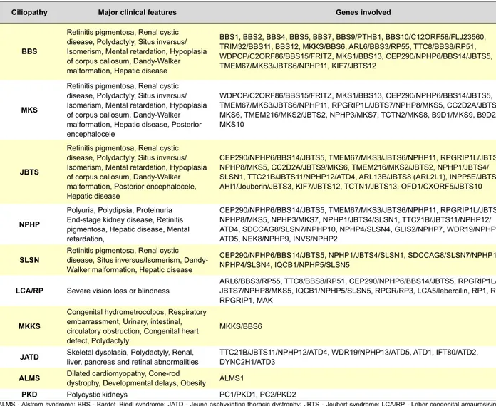
Table 2 - Ciliopathies, major clinical features and genes involved.
Ciliopathy Major clinical features Genes involved
BBS
Retinitis pigmentosa, Renal cystic disease, Polydactyly, Situs inversus/ Isomerism, Mental retardation, Hypoplasia of corpus callosum, Dandy-Walker malformation, Hepatic disease
BBS1, BBS2, BBS4, BBS5, BBS7, BBS9/PTHB1, BBS10/C12ORF58/FLJ23560, TRIM32/BBS11, BBS12, MKKS/BBS6, ARL6/BBS3/RP55, TTC8/BBS8/RP51, WDPCP/C2ORF86/BBS15/FRITZ, MKS1/BBS13, CEP290/NPHP6/BBS14/JBTS5, TMEM67/MKS3/JBTS6/NPHP11, KIF7/JBTS12
MKS
Retinitis pigmentosa, Renal cystic disease, Polydactyly, Situs inversus/ Isomerism, Mental retardation, Hypoplasia of corpus callosum, Dandy-Walker malformation, Hepatic disease, Posterior encephalocele
WDPCP/C2ORF86/BBS15/FRITZ, MKS1/BBS13, CEP290/NPHP6/BBS14/JBTS5, TMEM67/MKS3/JBTS6/NPHP11, RPGRIP1L/JBTS7/NPHP8/MKS5, CC2D2A/JBTS9/ MKS6, TMEM216/MKS2/JBTS2, NPHP3/MKS7, TCTN2/MKS8, B9D1/MKS9, B9D2/ MKS10
JBTS
Retinitis pigmentosa, Renal cystic disease, Polydactyly, Situs inversus/ Isomerism, Mental retardation, Hypoplasia of corpus callosum, Dandy-Walker malformation, Posterior encephalocele, Hepatic disease
CEP290/NPHP6/BBS14/JBTS5, TMEM67/MKS3/JBTS6/NPHP11, RPGRIP1L/JBTS7/ NPHP8/MKS5, CC2D2A/JBTS9/MKS6, TMEM216/MKS2/JBTS2, NPHP1/JBTS4/ SLSN1, TTC21B/JBTS11/NPHP12/ATD4, ARL13B/JBTS8 (ARL2L1), INPP5E/JBTS1, AHI1/Jouberin/JBTS3, KIF7/JBTS12, TCTN1/JBTS13, OFD1/CXORF5/JBTS10
NPHP
Polyuria, Polydipsia, Proteinuria End-stage kidney disease, Retinitis pigmentosa, Hepatic disease, Mental retardation,
CEP290/NPHP6/BBS14/JBTS5, TMEM67/MKS3/JBTS6/NPHP11, RPGRIP1L/JBTS7/ NPHP8/MKS5, NPHP3/MKS7, NPHP1/JBTS4/SLSN1, TTC21B/JBTS11/NPHP12/ ATD4, SDCCAG8/SLSN7/NPHP10, NPHP4/SLSN4, GLIS2/NPHP7, WDR19/NPHP13/ ATD5, NEK8/NPHP9, INVS/NPHP2
SLSN Retinitis pigmentosa, Renal cystic disease,Situs inversus/Isomerism, Dandy-Walker malformation, Hepatic disease
CEP290/NPHP6/BBS14/JBTS5, NPHP1/JBTS4/SLSN1, SDCCAG8/SLSN7/NPHP10, NPHP4/SLSN4, IQCB1/NPHP5/SLSN5
LCA/RP Severe vision loss or blindness ARL6/BBS3/RP55, TTC8/BBS8/RP51, CEP290/NPHP6/BBS14/JBTS5, RPGRIP1L/JBTS7/NPHP8/MKS5, IQCB1/NPHP5/SLSN5, RPGR/RP3, LCA5/lebercilin, RP1, RP2, RPGRIP1, MAK
MKKS
Congenital hydrometrocolpos, Respiratory embarrassment, Urinary, intestinal, circulatory obstruction, Congenital heart defect, Polydactyly
MKKS/BBS6
JATD Skeletal dysplasia, Polydactyly, Renal,
liver, pancreas and retinal abnormalities TTC21B/JBTS11/NPHP12/ATD4, WDR19/NPHP13/ATD5, ATD1, IFT80/ATD2, DYNC2H1/ATD3 ALMS Dilated cardiomyopathy, Cone-rod dystrophy, Developmental delays, Obesity ALMS1
PKD Polycystic kidneys PC1/PKD1, PC2/PKD2
Revista Científica da Ordem dos Médicos www.actamedicaportuguesa.com 589
ARTIGO DE REVISÃO
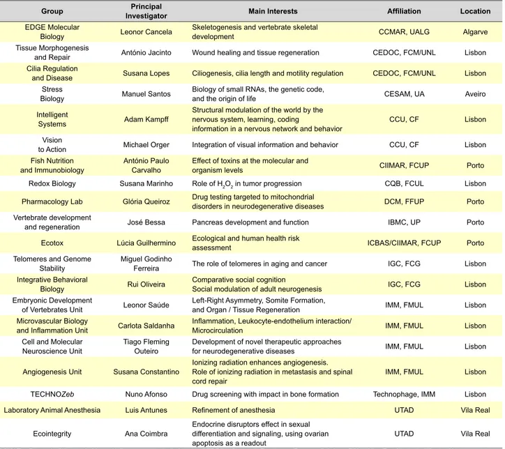
Table 3 - Main Portuguese fundamental and biomedical research groups working with zebrafish model.
Group InvestigatorPrincipal Main Interests Affiliation Location EDGE Molecular
Biology Leonor Cancela Skeletogenesis and vertebrate skeletal development CCMAR, UALG Algarve Tissue Morphogenesis
and Repair António Jacinto Wound healing and tissue regeneration CEDOC, FCM/UNL Lisbon Cilia Regulation
and Disease Susana Lopes Ciliogenesis, cilia length and motility regulation CEDOC,FCM/UNL Lisbon Stress
Biology Manuel Santos Biology of small RNAs, the genetic code, and the origin of life CESAM, UA Aveiro Intelligent
Systems Adam Kampff
Structural modulation of the world by the nervous system, learning, coding
information in a nervous network and behavior CCU, CF Lisbon Vision
to Action Michael Orger Integration of visual information and behavior CCU, CF Lisbon Fish Nutrition
and Immunobiology António Paulo Carvalho Effect of toxins at the molecular and organism levels CIIMAR, FCUP Porto Redox Biology Susana Marinho Role of H2O2 in tumor progression CQB, FCUL Lisbon
Pharmacology Lab Glória Queiroz Drug testing targeted to mitochondrial disorders in neurodegenerative diseases DCM, FFUP Porto Vertebrate development
and regeneration José Bessa Pancreas development and function IBMC, UP Porto Ecotox Lúcia Guilhermino Ecological and human health risk assessment ICBAS/CIIMAR, FCUP Porto Telomeres and Genome
Stability Miguel Godinho Ferreira The role of telomeres in aging and cancer IGC, FCG Lisbon Integrative Behavioral
Biology Rui Oliveira Comparative social cognitionSocial modulation of adult neurogenesis IGC, FCG Lisbon Embryonic Development
of Vertebrates Unit Leonor Saúde Left-Right Asymmetry, Somite Formation, and Organ / Tissue Regeneration IMM, FMUL Lisbon Microvascular Biology
and Inflammation Unit Carlota Saldanha Inflammation, Leukocyte-endothelium interaction/Microcirculation IMM, FMUL Lisbon Cell and Molecular
Neuroscience Unit Tiago Fleming Outeiro Development of novel therapeutic approaches for neurodegenerative diseases IMM, FMUL Lisbon
Angiogenesis Unit Susana Constantino Ionizing radiation enhances angiogenesis. Role of ionizing radiation in metastasis and spinal
cord repair IMM, FMUL Lisbon
TECHNOZeb Nuno Afonso Drug screening with impact in bone formation Technophage, IMM Lisbon
Laboratory Animal Anesthesia Luis Antunes Refinement of anesthesia UTAD Vila Real
Ecointegrity Ana Coimbra Endocrine disruptors effect in sexual differentiation and signaling, using ovarian
apoptosis as a readout UTAD Vila Real
CCMAR – Centro de Ciências do Mar; UALG – Universidade do Algarve; CEDOC – Centro de Estudos de Doenças Crónicas; FCM/UNL – Faculdade de Ciências Médicas da Univer-sidade de Lisboa; CESAM – Centro de Estudos do Ambiente e do Mar; UA – UniverUniver-sidade de Aveiro; CCU – Champalimaud Center for the Unknown; CF – Champalimaud Foundation; CIIMAR – Centro Interdisciplinar de Investigação Marinha e do Ambiente; FCUP – Faculdade de Ciências da Universidade do Porto; CQB – Centro de Química e de Bioquímica; FCUL – Faculdade de Ciências da Universidade de Lisboa; IBMC – Instituto de Biologia Molecular e Celular; UP – Universidade de Porto; ICBAS – Instituto de Ciências Biomédicas de Abel Salazar; DMC – Departamento de Ciências do Medicamento; FFUP – Faculdade de Farmácia da Universidade do Porto; IGC – Instituto Gulbenkian de Ciência; FCG – Fundação Calouste Gulbenkian; IMM – Instituto de Medicina Molecular; FMUL – Faculdade de Medicina da Universidade de Lisboa; UTAD – Universidade de Trás-os-Montes e Alto Douro.
Zebrafish and Drug Screening
Over all zebrafish’s characteristics make them ideal for fast, reliable and low-cost drug screenings during pre-reg-ulatory phases of drug development or repositioning and they can now be used in high-throughput screening (HTS) of drug libraries.65–67 This screening procedure involves
ob-taining zebrafish embryos or larvae, at the same develop-ment stage, loading them into multiwell plates, dosing the plates with chemical compounds, and then checking for changes elicited by the drugs at different concentrations, with the aim of improving the zebrafish phenotype that mim-ics a specific human disease.65,67,68 True HTS use robotics
and automated fluid handling systems. These technologies have been adapted in the vertebrate automated screening technology (VAST), which loads live larvae from a reservoir
and positions it into a capillary-based imaging chamber that can be rotated for an optimal field-of-view. After imaging, further manipulations can be performed, and then the lar-vae are automatically returned to their original container.67,68
This basic model of HTS, with or without the automatiza-tion provided by robotics, has allowed for many types of studies, such as new or repositioning drug screens for a variety of diseases like cardiovascular, polycystic kidneys, cancer69,70 and obesity; screening for regeneration,
psycho-tropic, antimicrobial and immunosuppressant drugs; sen-sory organ and behavioral screens; screening of bioactive natural products; and toxicology studies.67,68,71–74 Recently,
ARTIGO DE REVISÃO prostaglandin E2 (PGE2) usually used to treat stomach ul-cers. This drug was found to boost the production of blood stem cells and has just successfully concluded Phase 1 of Clinical trials.75
Biomedical Research with Zebrafish model in Portugal
Several Portuguese groups currently use zebrafish as a model for fundamental and biomedical research. Some of these groups and their research focus are summarized in Table 3. The variety in the fields of research is a good indication of the amenity and eclectic usefulness of the zebrafish as a biologic model. For example, zebrafish ex-ceptional ability to regenerate has allowed researchers like Cancela, Jacinto, Saúde and Constantino (Table 3) to study skeletal biomineralization, wound healing, tissue and organ regeneration, and the role of ionizing radiation in spinal cord repair, respectively. The zebrafish embryo/larval transpar-ency has been useful in studies of development of neuro-logical networks, necessary for understanding learning and memory (Kampff and Orger, Table 3). In addition, zebrafish is by far the most versatile vertebrate model organism for studying cilia biology in vivo. Lopes’ group is interested in cilia length and motility regulation to understand ciliopa-thies, whereas Saúde’s group is exploiting the role of cilia in tissue regeneration (Table 3).
Cancer is currently being investigated with zebrafish in several Portuguese labs (Marinho, Godinho Ferreira and Constantino, Table 3), namely the role of H2O2 as a signaling molecule in tumor microenvironment and progression, the role of telomeres in cancer, or the mechanisms of tumoral angiogenesis and metastization under ionizing radiation. Behavior and neurogenesis are being studied by Oliveira’s group using as a readout zebrafish’s natural behavioral pat-terns (Table 3). Important work is also being done in the role of small RNAs related with environmental stress, genome translation fidelity, proteotoxic stress and human diseases (Santos, Table 3). Saldanha’s group is interested in under-standing the mechanisms that govern leukocyte recruitment
and cell-cell interaction in inflammationand the role of spe-cific inflammatory mediators, such as H2O2 and Cxcl-8 in this process (Table 3).
Research on neurodegenerative disorders is also being undertaken. Outeiro’s group focus in developing novel ze-brafish models for Parkinson’s or Alzheimer’s, and intends to test novel drugs for therapeutic value. In the meanwhile Queiroz’s group uses zebrafish to test drugs to target mito-chondria-associated disorders (Table 3).
Bessa’s group uses zebrafish to research the impact that non-coding mutations have in pancreas development and in recapitulating human disorders. Antunes’ group use zebrafish in order to refine anesthesia and Coimbra’s team studies the role of endocrine disruptors in sexual differentia-tion (Table 3).
Furthermore, environmental pollution and the effects of chemicals and radiation at the molecular and organism levels are being studied by Carvalho’s group, while the im-pact of these stressors in human health is being addressed through toxicology assays by Guilhermino’s group (Table 3). Lastly, the biotech company Technophage has also taken advantage of zebrafish model with the unit TECHNOZeb, which uses larvae and zebrafish adults to screen drug li-braries and identify molecules with application in bone dis-orders like osteoporosis and cancer (Afonso’s group, Table 3).
Taken together, these studies prove zebrafish as a suc-cessful model system for studying a comprehensive spec-trum of diseases that affect millions of humans, and point to a very auspicious future of inexpensive and rapid drug discovery based on the zebrafish model.
CONFLICT OF INTERESTS
None stated.
FUNDING SOURCE
PTDC/SAU-OBD/103981/2008 and SFRH/BPD/77258 /2011
REFERENCES
1. Spence R, Gerlach G, Lawrence C, Smith C. The behaviour and ecol-ogy of the zebrafish, Danio rerio. Biol Rev Philos Soc. 2008;83:13–34. 2. Major RJ, Poss KD. Zebrafish heart regeneration as a model for cardiac
tissue repair. Drug Discov Today Dis Models. 2007;4:219–25. 3. Patton EE, Zon LI. The art and design of genetic screens: zebrafish. Nat
Rev Genet. 2001;2:956–66.
4. Woods IG, Schier AF. Targeted mutagenesis in zebrafish. Nat Biotech-nol. 2008;26:650–1.
5. Huang P, Xiao A, Zhou M, Zhu Z, Lin S, Zhang B. Heritable gene targeting in zebrafish using customized TALENs. Nat Biotechnol. 2011;29:699–700.
6. Hwang WY, Fu Y, Reyon D, Maeder ML, Tsai SQ, Sander JD, et al. Ef-ficient genome editing in zebrafish using a CRISPR-Cas system. Nat Biotechnol. 2013;31:227–9.
7. Jaffe KM, Thiberge SY, Bisher ME, Burdine RD. Imaging cilia in zebraf-ish. Methods in cell biology. Elsevier Inc. 2010;97:415–35.
8. Bradford Y, Conlin T, Dunn N, Fashena D, Frazer K, Howe DG, et al. ZFIN: enhancements and updates to the zebrafish model organism da-tabase. Nucleic Acids Res. 2011;39:D822-9.
9. White RM, Sessa A, Burke C, Bowman T, LeBlanc J, Ceol C, et al. Transparent adult zebrafish as a tool for in vivo transplantation analysis. Cell Stem Cell. 2008;2:183–9.
10. Jones R. Let sleeping zebrafish lie: a new model for sleep studies. PLoS Biol. 2007;5:e281.
11. Stewart S, Tsun Z-Y, Izpisua Belmonte JC. A histone demethylase is necessary for regeneration in zebrafish. Proc Natl Acad Sci U.S.A. 2009;106:19889–94.
12. Bernardos RL, Barthel LK, Meyers JR, Raymond PA. Late-stage neuro-nal progenitors in the retina are radial Müller glia that function as retineuro-nal stem cells. J Neurosci. 2007;27:7028–40.
13. Goldshmit Y, Sztal TE, Jusuf PR, Hall TE, Nguyen-Chi M, Currie PD. Fgf-dependent glial cell bridges facilitate spinal cord regeneration in ze-brafish. J Neurosci. 2012;32:7477–92.
14. Hughes V. Mapping brain networks: Fish-bowl neuroscience. Nature. 2013;493:466–8.
15. Stanton MF. Diethylnitrosamine-induced hepatic degeneration and neoplasia in the aquarium fish, brachydanio rerio. J Natl Cancer Inst. 1965;34:117–30.
16. Spitsbergen JM, Tsai H-W, Reddy A, Miller T, Arbogast D, Hen-dricks JD, et al. Neoplasia in Zebrafish (Danio rerio) Treated with 7,12-Diniethylbenz[a]anthracene by two exposure routes at different developmental stages. Toxicol Pathol. 2000;28:705–15.
N-methyl-N’nitro-Revista Científica da Ordem dos Médicos www.actamedicaportuguesa.com 591
ARTIGO DE REVISÃO
N-nitrosoguanidine by three exposure routes at different developmental stages. Toxicol Pathol. 2000;28:716–25.
18. Bourque C, Houvras Y. Hooked on zebrafish: insights into development and cancer of endocrine tissues. Endocr Relat Cancer. 2011;18:R149– 64.
19. Taylor AM, Zon LI. Zebrafish tumor assays: the state of transplantation. Zebrafish. 2009;6:339–46.
20. Jing L, Zon LI. Zebrafish as a model for normal and malignant hemato-poiesis. Dis Model Mech. 2011;4:433–8.
21. Teittinen KJ, Grönroos T, Parikka M, Rämet M, Lohi O. The zebrafish as a tool in leukemia research. Leuk Res. 2012;36:1082–8.
22. Chu J, Sadler KC. New school in liver development: lessons from ze-brafish. Hepatology. 2009;50:1656–63.
23. Lu JW, Hsia Y, Tu H-C, Hsiao Y-C, Yang W-Y, Wang H-D, et al. Liver development and cancer formation in zebrafish. Birth Defects Res C Embryo Today. 2011;93:157-72.
24. Tobia C, De Sena G, Presta M. Zebrafish embryo, a tool to study tumor angiogenesis. Int J Dev Biol. 2011;55:505–9.
25. Moshal KS, Ferri-Lagneau KF, Leung T. Zebrafish model: worth con-sidering in defining tumor angiogenesis. Trends Cardiovasc Med. 2010;20:114–9.
26. Jensen LD, Rouhi P, Cao Z, Länne T, Wahlberg E, Cao Y. Zebrafish models to study hypoxia-induced pathological angiogenesis in malig-nant and nonmaligmalig-nant diseases. Birth Defects Res C Embryo Today. 2011;93:182–93.
27. Liu S, Leach SD. Zebrafish models for cancer. Annu Rev Pathol. 2011;6:71–93.
28. Zilfou JT, Lowe SW. Tumor suppressive functions of p53. Cold Spring Harb Perspect Biol. 2009;1:a001883.
29. Berghmans S, Murphey RD, Wienholds E, Neuberg D, Kutok JL, Fletch-er CD, et al. tp53 mutant zebrafish develop malignant pFletch-eriphFletch-eral nFletch-erve sheath tumors. Proc Natl Acad Sci U.S.A. 2005;102:407–12.
30. Lee K-C, Goh WL, Xu M, Kua N, Lunny D, Wong JS, et al. Detection of the p53 response in zebrafish embryos using new monoclonal antibod-ies. Oncogene. 2008;27:629–40.
31. Langheinrich U, Hennen E, Stott G, Vacun G. Zebrafish as a model or-ganism for the identification and characterization of drugs and genes affecting p53 signaling. Curr Biol. 2002;12:2023–8.
32. Ghosh P, Chin L. Genetics and genomics of melanoma. Expert Rev Der-matol. 2009;4:131.
33. Patton EE, Widlund HR, Kutok JL, Kopani KR, Amatruda JF, Murphey RD, et al. BRAF mutations are sufficient to promote nevi formation and cooperate with p53 in the genesis of melanoma. Curr Biol. 2005;15:249– 54.
34. Dovey M, White RM, Zon LI. Oncogenic NRAS cooperates with p53 loss to generate melanoma in zebrafish. Zebrafish. 2009;6:397–404. 35. Anelli V, Santoriello C, Distel M, Köster RW, Ciccarelli FD, Mione M.
Global repression of cancer gene expression in a zebrafish model of melanoma is linked to epigenetic regulation. Zebrafish. 2009;6:417–24. 36. Sabaawy HE, Azuma M, Embree LJ, Tsai H-J, Starost MF, Hickstein DD. TEL-AML1 transgenic zebrafish model of precursor B cell acute lympho-blastic leukemia. Proc Natl Acad Sci U.S.A. 2006;103:15166–71. 37. Langenau DM, Traver D, Ferrando AA, Kutok JL, Aster JC, Kanki JP,
et al. Myc-induced T cell leukemia in transgenic zebrafish. Science. 2003;299:887–90.
38. Chen J, Jette C, Kanki JP, Aster JC, Look AT, Griffin JD. NOTCH1-in-duced T-cell leukemia in transgenic zebrafish. Leukemia. 2007;21:462– 71.
39. Tiso N, Moro E, Argenton F. Zebrafish pancreas development. Mol Cell Endocrinol. 2009;312:24–30.
40. Kinkel MD, Prince VE. On the diabetic menu: zebrafish as a model for pancreas development and function. BioEssays. 2009;31:139–52. 41. Gleeson M, Connaughton V, Arneson LS. Induction of hyperglycaemia
in zebrafish (Danio rerio) leads to morphological changes in the retina. Acta Diabetol. 2007;44:157–63.
42. Elo B, Villano CM, Govorko D, White LA. Larval zebrafish as a model for glucose metabolism: expression of phosphoenolpyruvate carboxyki-nase as a marker for exposure to anti-diabetic compounds. J Mol Endo-crinol. 2007;38:433–40.
43. Hölttä-Vuori M, Salo VT V, Nyberg L, Brackmann C, Enejder A, Pan-ula P, et al. Zebrafish: gaining popPan-ularity in lipid research. Biochem J. 2010;429:235–42.
44. Anderson JL, Carten JD, Farber SA. Zebrafish lipid metabolism: from mediating early patterning to the metabolism of dietary fat and choles-terol. Methods Cell Biol. 2011;101:111–41.
45. Stoletov K, Fang L, Choi S-H, Hartvigsen K, Hansen LF, Hall C, et al.
Vascular lipid accumulation, lipoprotein oxidation, and macrophage lipid uptake in hypercholesterolemic zebrafish. Circ Res. 2009;104:952–60. 46. Song Y, Cone RD. Creation of a genetic model of obesity in a teleost.
FASEB J. 2007;21:2042–9.
47. Jones KS, Alimov AP, Rilo HL, Jandacek RJ, Woollett LA, Penberthy WT. A high throughput live transparent animal bioassay to identify non-toxic small molecules or genes that regulate vertebrate fat metabolism for obesity drug development. Nutr Metab. 2008;5:23.
48. Archer A, Lauter G, Hauptmann G, Mode A, Gustafsson J-A. Transcrip-tional activity and developmental expression of liver X receptor (lxr) in zebrafish. Dev Dyn. 2008;237:1090–8.
49. Schlombs K, Wagner T, Scheel J. Site-1 protease is required for cartilage development in zebrafish. Proc Nat Acad Sci USA. 2003;100:14024–9. 50. Holden BJ, Bratt DG, Chico TJ. Molecular control of vascular develop-ment in the zebrafish. Birth Defects Res C Embryo Today. 2011;93:134-40.
51. Sabeh MK, Kekhia H, Macrae CA. Optical mapping in the developing zebrafish heart. Pediatric Cardiol. 2012;33:916–22.
52. Dahme T, Katus HA, Rottbauer W. Fishing for the genetic basis of car-diovascular disease. Dis Model Mech. 2009;2:18–22.
53. Liu J, Stainier DY. Zebrafish in the study of early cardiac development. Circ Res. 2012;110:870–4.
54. Kikuchi K, Poss KD. Cardiac regenerative capacity and mechanisms. Annu Rev Cell Dev Biol. 2012;28:719–41.
55. Xi Y, Noble S, Ekker M. Modeling neurodegeneration in zebrafish. Cur Neurol Neurosc Reports. 2011;11:274–82.
56. Vincensini L, Blisnick T, Bastin P. 1001 model organisms to study cilia and flagella. Biol Cell. 2011;103:109–30.
57. Satir P, Christensen ST. Structure and function of mammalian cilia. His-tochemist Cell Biol. 2008;129:687–93.
58. Eliasson R, Mossberg B, Camner P, Afzelius BA. The immotile-cilia syn-drome. A congenital ciliary abnormality as an etiologic factor in chronic airway infections and male sterility. N Engl J Med. 1977;297:1–6. 59. Ibañez-Tallon I, Pagenstecher A, Fliegauf M, Olbrich H, Kispert A,
Ke-telsen U-P, et al. Dysfunction of axonemal dynein heavy chain Mdnah5 inhibits ependymal flow and reveals a novel mechanism for hydrocepha-lus formation. Hum Mol Genet. 2004;13:2133–41.
60. Nonaka S, Tanaka Y, Okada Y, Takeda S, Harada A, Kanai Y, et al. Ran-domization of left-right asymmetry due to loss of nodal cilia generating leftward flow of extraembryonic fluid in mice lacking KIF3B motor pro-tein. Cell. 1998;95:829–37.
61. Pazour GJ, Dickert BL, Vucica Y, Seeley ES, Rosenbaum JL, Witman GB, et al. Chlamydomonas IFT88 and its mouse homologue, polycystic kidney disease gene tg737, are required for assembly of cilia and fla-gella. J Cell Biol. 2000;151:709–18.
62. Salomon R, Saunier S, Niaudet P. Nephronophthisis. Pediatr Nephrol. 2009;24:2333–44.
63. Zaghloul NA, Katsanis N. Functional modules, mutational load and hu-man genetic disease. Trends Genet: TIG. 2010;26:168–76.
64. Swanhart LM, Cosentino CC, Diep CQ, Davidson AJ, De Caestecker M, Hukriede NA. Zebrafish kidney development: basic science to trans-lational research. Birth Defects Res C Embryo Today. 2011;93:141-56. 65. Ali S, Champagne DL, Spaink HP, Richardson MK. Zebrafish embryos
and larvae: a new generation of disease models and drug screens. Birth Defects Res C Embryo Today. 2011;93:115–33.
66. Lee H, Inselman AL, Kanungo J, Hansen DK. Alternative models in de-velopmental toxicology. Syst Biol Reproductive Med. 2012;58:10–22. 67. Lessman CA. The developing zebrafish (Danio rerio): a vertebrate
mod-el for high-throughput screening of chemical libraries. Birth Defects Res C Embryo Today. 2011;93:268–80.
68. Peterson RT, Macrae C. Systematic approaches to toxicology in the ze-brafish. Ann Rev Pharmacol Toxicol. 2012;52:433–53.
69. Konantz M, Balci TB, Hartwig UF, Dellaire G, André MC, Berman JN, et al. Zebrafish xenografts as a tool for in vivo studies on human cancer. Ann NY Acad Sci. 2012;1266:124–37.
70. Rodrigues FS, Yang X, Nikaido M, Liu Q, Kelsh RN. A simple, highly visual in vivo screen for anaplastic lymphoma kinase inhibitors. ACS Biol Chem. 2012;7:1968–74.
71. Mandrekar N, Thakur NL. Significance of the zebrafish model in the discovery of bioactive molecules from nature. Biotechnol Lett. 2009;31:171–9.
72. Sipes NS, Padilla S, Knudsen TB. Zebrafish-As an integrative model for twenty-first century toxicity testing. Birth Defects Res C Embryo Today. 2011;93:256–67.
ARTIGO DE REVISÃO 74. De Esch C, Slieker R, Wolterbeek A, Woutersen R, De Groot D. Zebraf-Birth defects research. Part C, Embryo Today: Rev. 2011;93:173–81. ish as potential model for developmental neurotoxicity testing: a mini review. Neurotoxicol Teratol. 2012;34:545–53.