Prevalence of
Helicobacter pylori
in children in eastern Turkey
and molecular typing of isolates
Gokben Ozbey
1, Yasar Dogan
2, Kaan Demiroren
2, Ibrahim Hanifi Ozercan
3 1Vocational School of Health Services, Firat University, Elazig, Turkey. 2
Department of Pediatric Gastroenterology, Faculty of Medicine, Firat University, Elazig, Turkey. 3
Department of Pathology, Faculty of Medicine, Firat University, Elazig, Turkey.
Submitted: June 18, 2013; Approved: September 30, 2014.
Abstract
The objectives of the present study were to determineHelicobacter pylorivia culture, polymerase chain reaction and histopathological diagnosis in 101 children ranging in age from 4 to 18 years, to identify the association among restriction fragment length polymorphism types and clinical disease and to investigate the relationships among different isolates ofH. pyloriin different age groups. We observed a high prevalence ofH. pyloriinfections in children between the ages of 13 and 18 (75.8%), while children aged 4 to 6 years had the lowest prevalence of infection (40%).H. pyloriwas detected in 30.7% (31 of 101), 66.3% (67 of 101) and 63.2% (60 of 95) of children as determined by culture methods, PCR and histological examination, respectively.H. pyloriisolates with RFLP types I and III were the most common among children with antral nodularity, whereas RFLP types II and IV were the least detected types. Interestingly, all isolates from peptic ulcer patients were type III. Al-though our results show a high prevalence ofH. pyloriinfections in the pediatric population in east-ern Turkey, no association was identified between H. pyloriinfection with antral nodularity and recurring abdominal pain. In addition, we found low genetic variation amongH. pyloriisolates from children and no association between RFLP types and antral nodularity (p > 0.05). Additionally, we found thatH. pyloriisolates with specific RFLP types were predominant in different age groups.
Key words:children, culture,Helicobacter pylori, PCR-RFLP.
Introduction
MostHelicobacter pyloriinfections are thought to be acquired in childhood or adolescence, and infection with this bacterium at a young age increases the risk of associ-ated complications later in life (Vinetteet al., 2004).
Culture methods have been the “gold standard" for the detection of bacterial pathogens, yet for bacteria such as H. pylori, this technique is often difficult and time-con-suming (Singhet al., 2008). Serological tests also have lim-itations such as low specificity and failure to differentiate between active and past infections (Hestviket al., 2010). Polymerase chain reaction (PCR)-based techniques have successfully been used to detect pathogens that may be dif-ficult to culture, identify and/or isolate from clinical sam-ples (Singhet al., 2008). PCR-based restriction fragment
length polymorphism (RFLP) analysis, used in this study, has sufficient discriminatory power to differentiate among H. pyloristrains, in addition to being a relatively simple, fast and low cost sub-typing method (Andresonet al., 2007; Liet al., 1997).
Currently, little is reported about the prevalence of active H.pylori infections among children in the Elazig Province of eastern Turkey. The aims of this study were to: (i) identify the prevalence of active H.pylori infection among children and determine if prevalence differs with age, (ii) evaluate any correlation betweenH. pylori infec-tion and gastroduodenal disease and (iii) determine ifH. pylori RFLP sub-types are associated with antral nodu-larity, peptic ulcer, specific age groups and/or clinical out-comes.
DOI: http://dx.doi.org/10.1590/S1517-838246220140234
Send correspondence to G. Ozbey. Vocational School of Health Services, Firat University, Elazig, Turkey. E-mail: [email protected].
Materials and Methods
Patients
A total of 101 patients were enrolled in this study, in-cluding 53 girls and 48 boys ages 4 to 18 years (Ozbeyet al., 2013). Symptoms included recurrent abdominal pain, vomiting with or without blood, bloody stools and growth retardation. The children underwent endoscopy at the clinic of Pediatric Gastroenterology Department at the Firat Uni-versity Hospital in the period of March 2011 to September 2012 (Ozbeyet al., 2013) Ethical clearance for this study was provided by the Medical Ethics Committee of Firat University. Informed consent was obtained from each pa-tient and signed by the children’s parents prior to the endos-copy procedure.
Bacterial culture
Bacterial culturing of the antral biopsy was per-formed as described elsewhere (Chomvarinet al., 2006). Briefly, the antral biopsies were placed directly into sterile Eppendorf tubes containing 0.5 mL of Brain Heart Infusion broth (Oxoid, Basingstoke, UK) with 15% glycerol and processed for culture within 2 h. Each sample was smeared onto Columbia agar base (Oxoid, UK) added with 7% laked horse blood (SR0048C, Oxoid, UK) andH. pyloriDent’s supplement (Oxoid, UK). Plates were incubated at 37 °C in a microaerobic atmosphere using the Campygen gas gener-ating kit (Oxoid, UK) for up to 10 days (Frenck et al., 2006). Typical small, round colonies that were gram nega-tive and urease, catalase and oxidase posinega-tive were pre-sumed to beH. pylori(Goodwin and Wesley, 1993). All isolates were stored at -80 °C in Brain Heart Infusion broth added with 15% glycerol until further analysis. Reference H. pyloristrains, including some clinical strains, were pro-vided by the Department of Medical Biology, Faculty of Medicine, Pamukkale University, Denizli, Turkey. The his-tological evaluation of each antral biopsy sample was con-ducted by a pathologist according to the Sydney classification system (Dixonet al., 1996).
Primers and PCR conditions
DNA from antral biopsy samples and suspensions of H. pyloricolonies were purified using the QIAamp DNA mini kit (Qiagen, Germany). The forward [glmM-F (5’-AAGCTTTTAGGGGTGTTAGGGGTTT-3’)] and re-verse [glmM-R (5’-AAGCTTACTTTCTAACACTAAC GC-3’)] primers (Luet al., 1999) amplify a region of the glmM gene (formerlyureC) ofH. pylorito yield a 294 bp PCR product. The thermal cycling was as follows: 35 cy-cles of denaturation at 93 °C for 1 min, 1 min at an anneal-ing temperature of 55 °C, and a 1 min extension step at 72 °C (Luet al., 1999).
PCR-based amplification and RFLP analysis of PCR amplicons
For PCR-RFLP analysis, theureCgene was ampli-fied using the forwardureC-U (5’- AAG AAG TCA AAA ACG CCC CAA AAC -3’) and reverseureC-L (5’- CTT ATC CCC ATG CAC GAT ATT CCC -3’) primers to yield a PCR product size of 1169 bp (Liet al., 1997). The PCR cycling consisted of the following steps: a denaturation step at 94 °C for 5 min, 45 cycles at 94 °C for 45 s, 59 °C for 30 s, 72 °C for 1 min 30 s, and a final extension step at 72 °C for 10 min (Andresonet al., 2007). Ten microliters of PCR product was restricted with the restriction enzyme HhaI (Fermentas, Lithuania) according to the manufacturer’s in-structions.
Statistical analysis
Statistical analysis was performed using the statistical software program SPSS 12.00 (SPSS, Chicago, IL, USA). Relationships among the prevalence ofH. pyloriin children and the RFLP types with clinical outcomes were analyzed using Fisher’s exact and Pearson’sc 2tests. A p value < 0.05 is statistically significant.
Results
Of the 101 patients analyzed, 58 (57.4%) were experi-encing abdominal pain, 22 (21.8%) had growth retardation, 12 (11.9%) had bloody vomit and/or blood in stools, 8 (7.9%) were vomiting, and 1 (1%) was diagnosed with ane-mia. Endoscopic findings revealed antral nodularity, antral hyperemia, hyperemia in duodenal mucosa, duodenal ul-cers and gastric ulul-cers in 54.5%, 12.9%, 23.8%, 3% and 5.9% of cases, respectively.
Culture and PCR results
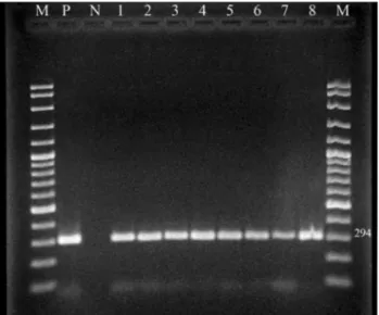
Helicobacter pyloriwas detected in antral gastric bi-opsies by culture, PCR and histology in 30.7%, 66.3% and 63.2% of patients, respectively (Table 1). AllH. pylori iso-lates examined generated the expected 294 bp fragment of theglmM gene (Figure 1). Patients 13 to 18 years of age showed the highest prevalence of infection (75.8%), while children 4 to 6 years of age had the lowest prevalence (40%) (Table 2). There was no statistically significant dif-ference in the prevalence of H. pylori between males (66.7%, 32/48) and females (67.9%, 36/53).H. pyloriwas detected by PCR in 76.4% (42/55) of cases presenting with
Table 1- Prevalence ofH. pylorias determined by culture, PCR and histopathological findings.
Tests H. pylori(+) n (%) H. pylori(-) n (%) Total (n)
culture 31 (30.7) 70 (69.3) 101
PCR 67 (66.3) 34 (33.7) 101
antral nodularity, 46.2% (6/13) presenting with antral hy-peremia, 50% (12/24) presenting with hyperemia in duode-nal mucosa, 66.7% (2/3) presenting with duodeduode-nal ulcers and 83.3% (5/6) presenting with gastric ulcers (Table 3). Statistical analysis was not performed because the number of cases for a particular symptom or diagnosis was rela-tively small. However, the number of cases of recurring ab-dominal pain and antral nodularity were relatively high.H. pyloriwas detected in 68.9% (40/58) of cases reporting re-curring abdominal pain, but no correlation was found be-tween the prevalence ofH. pyloriinfection with recurring abdominal pain and that with antral nodularity.
Histopathology results
Six cases were excluded because of insufficient tissue for histopathological examination. Of the remaining 95 antrum biopsy samples, histological examination showed that 60 (63.2%) were positive forH. pylori.Pathological analysis of the biopsy material showed that intestinal meta-plasia was present in only a single child (a 13-year old male) who also had chronic gastritis and wasH. pylori posi-tive.None of the cases presented with gastric atrophy.
RFLP analysis results
PCR amplification ofH. pyloriDNA using theureC primer set produced an amplicon of the expected size (1169 bp) for allH. pyloriisolates examined. The RFLP analysis of our isolates revealed a low heterogeneity among isolates. Table 4 shows the distribution ofH. pyloriRFLP types. Four profiles (types I, II, III and IV) were identified (Figure 2). From the cases with antral nodularity, types I (44.8%) and III (27.6%) were the predominant strains while types II and IV were detected at in lower numbers (10.4% and 17.2%, respectively). From cases presenting with peptic ulcers, only type IIIH. pyloriwas detected.
Only two isolates from culture were obtained from the 4 to 6 year old age group; one was RFLP type I and the other type II. In the 7 to 12 year old age group (17 isolates positive by culture), 47% of isolates were type I (8/17), 5.9% type II (1/17), 35.3% type III (6/17) and 11.8% type IV (2/17). In addition, both types I and III were detected in 33.3% (4/12) of 13 to 18 year olds. Likewise, types II and IV were detected in the same percentage of children (16.7%, 2/12) within this age group (Table 5). However, due to the relatively small number of children with peptic ulcers, the statistical analysis required to associate RFLP types with this medical condition could not be performed. No relationship was identified between RFLP type and antral nodularity in the present work (p > 0.05).
Discussion
Acquiring knowledge onH. pyloriinfections is im-portant for human pathogen studies because the lack of cur-rent knowledge is hindering actions to protect human health. Although RFLP analysis of H. pylori isolates in Figure 1- An agarose gel of PCR products ofH. pyloriisolates from antral
biopsy specimens (M: DNA Ladder (100 bp), P:H. pyloripositive control, N: negative control, 1-8:H. pyloripositive samples).
Table 2-H. pyloriprevalence according to different age groups.
Years H. pylori(+) n (%) H. pylori(-) n (%) Total (n)
4-6 2 (40) 3 (60) 5
7-12 40 (63.5) 23 (36.5) 63
13-18 25 (75.8) 8 (24.2) 33
Table 3- Prevalence ofH. pyloriisolates in children according to endo-scopic findings as detected by PCR.
Endoscopic findings H. pylori(+)n (%) H. pylori(-)n (%) Antral nodularity (n = 55) 42 (76.4) 13 (23.6)
Antral hyperemia (n = 13) 6 (46.2) 7 (53.8)
Hyperemia in duodenal mucosa (n = 24)
12 (50) 12 (50)
Duodenal ulcer (n=3) 2 (66.7) 1 (33.3) Gastric ulcer (n=6) 5 (83.3) 1 (16.7)
Total (101) 67 (66.3) 34 (33.7)
n: number ofH. pyloriisolates.
Table 4- RFLP types of 31 clinicalH. pyloriisolates obtained after diges-tion withHhaIenzyme.
RFLP types Antral nodularity n (%) Peptic ulcer n (%) Total n (%)
I 13 (44.8) - 13 (41.9)
II 3 (10.4) - 3 (9.7)
III 8 (27.6) 2 (100) 10 (32.3)
IV 5 (17.2) - 5 (16.1)
Total 29 2 31
adults has been performed in eastern Turkey (Ozbeyet al., 2012), there are currently no studies that report the isolation and genotyping of H. pylori in children residing in this same region. The current study was undertaken to examine the prevalence and genetic diversity ofH. pyloriisolates from children in the Elazig Province of Turkey. These re-sults suggest that appropriate and effective treatment strate-gies should be implemented againstH. pyloriinfections in the future.
The majority of studies investigating the prevalence of H. pylori among children in Turkey have employed serological methods (Selimogluet al., 2002; Yucelet al., 2009). Other studies show varied prevalence ofH. pyloriin
asymptomatic children including 7.1% in the Czech Re-public (0-15 year olds), 24.7% in Israel (0-5 year olds), 30.9% in Turkey (2-12 year olds), 31.6% in Portugal (0-15 year olds) and 82% in Iran (0-15 year olds) (Alborziet al., 2006; Koriet al., 2009; Oleastroet al., 2011; Sykoraet al., 2009; Yucelet al., 2009). We found that the highest preva-lence of infection was found in children from 13 to 18 years of age (75.8%). Our study also showed that a total of 66.3% of symptomatic children were infected withH. pylorias de-tected by PCR. A lower prevalence ofH. pyloriin children with dyspepsia (38%) was reported in the United States (Soodet al., 2005). However, our findings match previous data reported by Selimogluet al.(2002) in randomly se-lected healthy children between the ages of 6 and 17 in Tur-key (64.4% infected). This higherH. pyloriprevalence may be due to sociodemographic factors such as low socioeco-nomic status, poor sanitary conditions, higher percentage of low-income families, higher percentage of parents with a low educational background, poor living conditions, high density living quarters and higher rates of immigrant chil-dren from the surrounding cities (Azevedoet al., 2009).
Culture-positive biopsies were relatively low at 30.7% (31/101) as compared to histological and PCR re-sults but were similar to the number of positive biopsies de-tected in Turkey’s neighbor, Iran, which reported a H. pyloriprevalence of 39.8% using the culture method (Ira-nikhahet al., 2013). Negative culture results may be due to low colonization because of patchy localization of bacteria in the stomach, decreased bacterial viability, overgrowth of potential contaminants, technical problems during tissue transport, increased transportation times and varied cultur-ing methods (Ozcayet al., 2004; Torreset al., 2001).
In this study, most of the children reported recurrent abdominal pain, but the role ofH. pyloriin causing chronic abdominal pain is still disputable (Zeyrek et al., 2008; Hestviket al., 2010). Our data showed no significant corre-lation between abdominal symptoms andH. pylori infec-tion.
Bahuet al.(2003) noted a significant relationship be-tween H. pylori and endoscopic nodular gastritis. Here, antral nodularity was observed in 55 children (54.5% of those tested) of which 76.4% were positive forH. pylori. This observation is similar to previous data (73.8%) re-ported by Doganet al.(2007) but higher than that reported in Turkey (64.7%) by Ozcayet al.(2004) and lower than re-ported in Japan where 98.5% ofH. pyloricases were asso-ciated with antral nodularity (Katoet al., 2004).
Reports show that the peptic ulcer prevalence in chil-dren in different countries varies between 1.8% to 19.5% (Elitsur and Lawrence, 2001; Katoet al., 2004). In the pres-ent study, 8.9% of symptomatic children had peptic ulcers, similar to results reported by Megraud (2005) in European children (8.6%) but lower than those reported by Ugras and Pehlivanoglu (2011) in Turkish children (13.2%). How-ever, in the current study, the prevalence ofH. pyloriin pa-Figure 2- PCR-RFLP analysis ofH. pyloriisolates using restriction
en-zymes in agarose gel (2.5% w/v) (M: 100 bp DNA ladder, lanes I, II, III and IV: RFLP band profiles).
Table 5- RFLP types of 31H. pyloriisolates determined after culturing and grouped according to patient age.
RFLP types 4-6 n (%) 7-12 n (%) 13-18 n (%)
I 1 (50) 8 (47) 4 (33.3)
II - 1 (5.9) 2 (16.7)
III - 6 (35.3) 4 (33.3)
IV 1 (50) 2 (11.8) 2 (16.7)
Total 2 17 12
tients also presenting with ulcers was 66.7% for those with duodenal ulcers and 83.3% for those with gastric ulcers. The percentage for those with duodenal ulcers is lower than the earlier reports from Japan and Turkey where 83% and 76.9%, respectively, of patients with duodenal ulcers were positive forH. pylori(Katoet al., 2004; Ugras and Pehli-vanoglu, 2011). In this study, a similar number of children with gastric ulcers tested positive forH. pylori(83.3%), as was reported (85.2%) previously in Turkey (Ugras and Pehlivanoglu, 2011).
In areas with high incidence rates of gastric cancer, gastric atrophy is common amongH.pylori-infected chil-dren (Ricuarteet al., 2005). The prevalence of gastric atro-phy varies between countries, and it has been shown that intestinal metaplasia alters with respect to geographic/ge-netic origins and environmental factors (Katoet al., 2006: Pacifico et al., 2010; Ricuarte et al., 2005; Tutar et al., 2009; Ustaet al., 2004). Only one of the 95 symptomatic children (1.1%) had intestinal metaplasia and was positive forH. pylori. These data are in concordance with a previous report(Ustaet al., 2004) that found intestinal metaplasia in only one of 175 Turkish children infected withH. pylori. However, only 4.6% of children in Japan with intestinal metaplasia were positive forH. pylori(Katoet al.2006). In contrast, the prevalence of gastric atrophy is high in Colum-bian children (16%) and much higher in Japanese children (51.9%) (Katoet al., 2006; Ricuarteet al., 2005).
PCR-RFLP has been frequently used for genotyping and discrimination of H. pylori strains because it is low cost, rapid, easy to perform and can be used to analyzeH. pylorigenotype diversity (Röesleret al., 2009). The two re-striction endonucleasesHhaI and MboI were chosen for RFLP analysis of theureC gene (Liet al., 1997). RFLP analysis of the PCR products of our H. pylori isolates yielded four different RFLP types (I, II, III and IV). RFLP types I and III were the most frequently detected among children with antral nodularity whereas RFLP types II and IV were detected less frequently. Furthermore, type III was the most predominant type in isolates from patients with peptic ulcers. Our findings confirmed the results of Mishra et al.(2002), Saribasaket al.(2004) and Kulsantiwonget al.(2012) who reported no correlation between the RFLP patterns of all strains and the patients’ clinical outcome. In addition, certain RFLP profiles predominated according to different age groups; types I and IV (50%) in 4-6 year olds, type I (47%) in 7-12 year olds and types I and III (33.3%) in 13-18 year olds. These percentages show that children within different age groups were infected with variousH. pyloristrains, which might be related to differences in im-mune responses towardsH. pyloriduring childhood devel-opment. Further study is needed to better establish this relationship.
High genetic variation in H. pylori strains isolated from various patients has been demonstrated worldwide (Raymondet al., 2005; Kulsantiwonget al., 2012; Menoni
et al., 2013). This result is in contrast to the current study whereH. pyloriisolates from children showed a consider-ably low genetic diversity. However, limited PCR-RFLP profiles might be due to the small numbers ofH. pylori iso-lates in the present study (31 total isoiso-lates).
In conclusion, the results of the current study show a high prevalence ofH. pyloriinfection in children in eastern Turkey. Although the number ofH. pyloriisolates is too small to perform any epidemiological analysis, the bacte-rial isolates from children in the present study yielded a rel-atively small number of genetically distinct RFLP types. A different distribution of RFLP types was evident among children of different age groups. Therefore, it is necessary to conduct future work on larger populations of children in order to fully confirm the results of this study and to iden-tify the genetic heterogeneity ofH. pyloriisolates in chil-dren from the Elazig Province.
Acknowledgments
The abstract form of this study was presented as poster in 1st International Organic Electronic Material Tec-hnologies Conference (OEMT 2015), March 25-28, 2015, Elazig, Turkey. The authors thank Dr. Vildan Caner, De-partment of Medical Biology, Faculty of Medicine, Pamuk-kale University, Denizli, Turkey, for kindly providing some clinical strains and the management of the Elazig Veterinary Control and Research Institute for securing lab-oratory facilities for this study. We are also grateful to Dr. Francis Megraud, INSERM U853, Laboratoire de Bacte-riologie - C.H.U. Pellegrin, France; Dr. Alfizah Hanafiah, Department of Medical Microbiology and Immunology, Faculty of Medicine, UKM Medical Centre, Kuala Lum-pur, Malaysia and Dr. Emma Sproston, Bureau of Micro-bial Hazards, Health Canada, Ottawa, Canada for critical review of the manuscript.
References
Alborzi A, Soltani J, Pourabbas Bet al.(2006) Prevalence of
Helicobacter pylori infection in children (south of Iran). Diagn Microbiol Infect Dis 54:259-261.
Andreson H, Sillakivi T, Peetsalu Met al.(2007) Persistence of
Helicobacter pyloriinfection in patients with peptic ulcer perforation. Scand J Gastroenterol 42:324-329.
Azevedo NF, Huntington J, Goodman KJ (2009) The epidemiol-ogy ofHelicobacter pyloriand public health implications. Helicobacter 14:1-7.
Bahú MG, Silveira TR, Maguilnick Iet al.(2003) Endoscopic nodular gastritis: an endoscopic indicator of high-grade bac-terial colonization and severe gastritis in children with
Helicobacter pylori. J Pediatr Gastroenterol Nutr 36:217-222.
Chomvarin C, Kulsantiwong P, Chantarasuk Yet al.(2006) Com-parison of media and antibiotic supplements for isolation of
Dixon MF, Genta RM, Yardely JHet al.(1996) Classification and grading of gastritis. The Updated Sydney System. Am J Surg Pathol 20:1161-1181.
Dogan Y, Baris S, Erkan Tet al.(2007)Helicobacter pylori infec-tion in children: Evaluainfec-tion of complaints, endoscopic find-ings, diagnostic methods and post-treatment eradication rates. Turk Arch Ped 42:98-102.
Elitsur Y, Lawrence Z (2001) Non-Helicobacter pylorirelated du-odenal ulcer disease in children. Helicobacter 6:239-243. Frenck RW Jr, Fathy HM, Sherif Met al.(2006) Sensitivity and
specificity of various tests for the diagnosis ofHelicobacter pyloriin Egyptian children. Pediatrics 118:e1195-1202. Goodwin CS, Wesley BW (1993) Microbiology ofHelicobacter
pylori. Gastroenterol Clin North Am 22:5-19.
Hestvik E, Tylleskar T, Kaddu-Mulindwa DH et al. (2010)
Helicobacter pyloriin apparently healthy children aged 0-12 years in urban Kampala, Uganda: a community-based cross sectional survey. BMC Gastroenterol 10:62.
Iranikhah A, Ghadir M-R, Sarkeshikian Set al.(2013) Stool anti-gen tests for the detection ofHelicobacter pyloriin children. Iran J Pediatr 23:138-142.
Kato S, Nishino Y, Ozawa Ket al.(2004) The prevalence of
Helicobacter pylori in Japanese children with gastritis or peptic ulcer disease. J Gastroenterol 39:734-738.
Kato S, Nakajima S, Nishino Yet al.(2006). Association between gastric atrophy andHelicobacter pyloriinfection in Japa-nese children: a retrospective multicenter study. Dig Dis Sci 51:99-104.
Kori M, Goldstein E, Granot E (2009)Helicobacter pylori infec-tion in young children detected by a monoclonal stool anti-gen immunoassay. Pediatr Infect Dis J 28:157-159. Kulsantiwong P, Chomvarin C, Mairiang Pet al.(2012)
PCR-RFLP and Antimicrobial susceptibility profilrs of
Helicobacter pyloriisolated from antrum and corpus in dys-peptic patients in Thailand. Southeast Asian J Trop Med Public Health 43:933-942.
Li C, Ha T, Chi DSet al.(1997) Differentiation ofHelicobacter pylori strains directly from gastric biopsy specimens by PCR-based restriction fragment length polymorphism anal-ysis without culture. J Clin Microbiol 35:3021-3025. Lu JJ, Perng CL, Shyu RYet al.(1999) Comparison of five PCR
methods for detection ofHelicobacter pyloriDNA in gastric tissues.J Clin Microbiol 37:772-774.
Megraud F (2005) On behalf of the European pediatric task force on Helicobacter pylori comparison of non-invasive tests to detect Helicobacter pylori infection in children and adoles-cents results of a multicenter European study. J Pediatr 146:198-203.
Menoni SMF, Bonon SHA, Zeitune JMRet al.(2013) PCR-based detection and genotyping of Helicobacter pyloriin endo-scopic biopsy samples from Brazilian patients. Gastro-enterol Res Pract 2013:1-8.
Mishra KK, Srivastava S, Prabhat Pet al. (2002) PCR-based RFLP analysis of ureC gene for typing of Indian
Helicobacter pylori strains from gastric biopsy specimens and culture. J Microbiol 40:282-288.
Oleastro M, Cabral J, Ramalho PMet al.(2011) Primary antibi-otic resistance ofHelicobacter pyloristrains isolated from Portuguese children: a prospective multicentre study over a 10 year period. J Antimicrob Chemother 66:2308-2311.
Ozbey G, Bahcecioglu IH, Ceribasi Set al.(2012) Polymerase chain reaction-restriction fragment length polymorphism analysis ofHelicobacter pyloriisolates from patients with gastrointestinal complaints in Eastern Turkey. Afr J Microbiol Res 6:4068-4072.
Ozbey G, Dogan Y, Demiroren K (2013) Prevalence of
Helicobacter pylorivirulence genotypes among children in Eastern Turkey. World J Gastroenterol 19:6585-6589. Ozcay F, Kocak N, Saltik INet al.(2004)Helicobacter pylori
in-fection in Turkish children: comparison of diagnostic tests, evaluation of eradication rate, and changes in symptoms af-ter eradication. Helicobacaf-ter 9:242-248.
Pacifico L, Anania C, Osborn JFet al.(2010) Consequences of
Helicobacter pylori infection in children. World J Gastroenterol 16:5181-5194.
Raymond J, Nguyen B, Bergeret Met al.(2005). Heterogeneous susceptibility to metronidazole and clarithromycin of
Helicobacter pyloriisolates from a single biopsy in adults is confirmed in children. Int J Antimicrob Agents 26:272-278. Ricuarte O, Gutierrez O, Cardona Het al.(2005) Atrophic
gastri-tis in young children and adolescents. J Clin Pathol 58:1189-1193.
Röesler BM, de Oliveira TB, Bonon SHet al.(2009). Restriction fragment length polymorphism of urease C and urease B genes ofHelicobacter pyloristrains isolated from Brazilian patients with peptic ulcer and chronic gastritis. Dig Dis Sci 54:1487-93.
Saribasak H, Salih BA, Yamaoka Yet al. (2004) Analysis of
Helicobacter pylorigenotypes and correlation with clinical outcome in Turkey. J Clin Microbiol 42:1648-1651. Selimoglu MA, Ertekin V, Inandi T (2002) Seroepidemiology of
Helicobacter pyloriinfection in children living in eastern Turkey. Pediatr Int 44:666-669.
Singh V, Mishra S, Rao GRKet al.(2008) Evaluation of Nested PCR in detection ofHelicobacter pyloritargeting a highly conserved gene: HSP60. Helicobacter 13:30-34.
Sood MR, Joshi S, Akobeng AKet al.(2005) Growth in children withHelicobacter pyloriinfection and dyspepsia. Arch Dis Child 90:1025-1028.
Sykora J, Siala K, Varvarovska Jet al.(2009) Epidemiology of
Helicobacter pyloriinfection in asymptomatic children: a prospective population-based study from the Czech Repub-lic. Application of a monoclonal-based antigen in-stool en-zyme immunoassay. Helicobacter 14:286-297.
Torres J, Camorlinga-Povce M, Perez-Perez Get al.(2001) In-creasing multidrug resistance inHelicobacter pyloristrains isolated from children and adults in Mexico. J Clin Micro-biol 39:2677-2680.
Tutar E, Ertem D, Kotiloglu Karaa Eet al.(2009) Endoscopic and histopathologic findings associated withH. pyloriinfection in very young children. Dig Dis Sci 54:111-117.
Ugras M, Pehlivanoglu E (2011)Helicobacter pyloriinfection and peptic ulcer in eastern Turkish children: is it more com-mon than known? Turk J Pediatr 53:632-637.
Usta Y, Saltk-Temizel IN, Ozen H (2004) Gastric atrophy and in-testinal metaplasia in Helicobacter pylori infection. J Pediatr Gastroenterol Nutr 38:548.
Vinette KM, Gibney KM, Proujansky Ret al.(2004) Comparison of PCR and clinical laboratory tests for diagnosingH. pylori
Yucel O, Sayan A, Yildiz M (2009) The factors associated with asymptomatic carriage of Helicobacter pylori in children and their mothers living in three socio-economic settings. Jpn J Infect Dis 62:120-124.
Zeyrek D, Zeyrek F, Cakmak Aet al. (2008) Association of
Helicobacter pyloriand giardiasis in children with recurrent abdominal pain. Turk Parazitol Derg 32:4-7.
Associate Editor: Roxane Maria Fontes Piazza