Brazilian Journal of Microbiology (2012): 1332-1339 ISSN 1517-8382
PREVALENCE OF GENOTYPESIN HELICOBACTER PYLORI ISOLATES FROM PATIENTS IN EASTERN
TURKEY AND THE ASSOCIATION OF THESE GENOTYPES WITH CLINICAL OUTCOME
Gokben Ozbey1*, Cem Aygun2
1
Vocational School of Health Services, Firat University, 23119, Elazig, Turkey; 2Department of Gastroenterology, Faculty of Medicine, Firat University, 23119, Elazig, Turkey.
Submitted: July 25, 2011; Returned to authors for corrections: December 22, 2011; Approved: June 07, 2012.
ABSTRACT
There is not much information available regarding the prevalence of the genotypes of Helicobacter pylori
isolates in Turkey, particularly in eastern Turkey. The aims of this study were to detect the prevalence of
different genotypes of H. pylori in Turkish patients with gastrointestinal complaints and to determine the
relationship of these genotypes with clinical outcome and sex. One hundred forty H. pylori isolates were
examined for the presence of its genotypes by the PCR. We found that the prevalence of vacAs1, vacAs2,
cagA, cagE, iceA1, iceA2 and babA2 genes were 88.6%, 11.4%, 71.4%, 35.7%, 41.4%, 58.6%, and 62.1%,
respectively. The most predominant vacA subtype was s1a (81.4%). The most vacA allelic combination
detected were vacAs1m1 (65.2%) and s1m2 (53.9%) in patients with peptic ulcer and gastritis, respectively.
The only vacAs1 isolate was significantly associated with gastritis and peptic ulcer (p<0.05). The vacAs1a,
ml, slml and babA2 genes were significantly associated with peptic ulcer (p<0.05), whereas m2 gene was
significantly associated with only gastritis (p<0.05). The difference between sex and genotypes was
statistically significant among the cagA, vacAs1, iceA2 and babA2 genes. This study reported for the first
time the prevalence of H. pylori genotypes in patients with gastrointestinal complaints in eastern Turkey.
Further studies are needed to understand epidemiological importance of the genotypes of H. pylori isolates
in this region and the association between the virulence genes and clinical outcome in different regions.
Key words: Helicobacter pylori, prevalence, patient, genotypes, PCR.
INTRODUCTION
Helicobacter pylori (H. pylori) which infects more than
half of the world’s population, a major etiological agent in
development of gastritis (G), peptic ulcer (PU) and gastric
carcinoma (3). Scientists have been shown that several genes,
such as the vacuolating cytotoxin (vacA), cytotoxin associated
gene A (cagA), cytotoxin associated gene E (cagE), induced by
contact with epithelium (iceA) and blood adhesion binding
antigen (babA2) have been determined and these genes may
play important roles in the pathogenesis of H. pylori infection
(22, 24).
Ozbey, G. and Aygun, C. H. pylori genotypes
The cagA gene being a marker for the presence of the cag
pathogenicity island (cagPAI) of approximaely 40 kb was the
first gene found to be present in H. pylori strains (7) and its
presence is associated with a more severe clinical outcome,
such as PU, atrophic G, and gastric cancer (GC) (4, 5). The
cagA induces interleukin-8 (IL-8) production and mucosal
inflamation (4, 5). The cagPAI contains a gene known as cagE,
is one of the marker genes in cagI of the cagPAI and it is
required for translocation and phosphorylation (9, 28). The
cagE gene was found to be associated with a more severe
clinical outcome (11).
The vacA gene is present in all H. pylori strains and
contains at least two variable parts (a hypervariable signal
sequence and a middle region allele) (2). Among the vacA
subtypes, subtypes s1a, s1b, s1c and s2, and m1, m2a and m2b
have been identified (30). Although all strains of H. pylori
include the vacA gene, they vary in their ability to produce
cytotoxin (8). Type m1 strains show more toxic activity than
m2, type s1a is more active than s1b, and type s2 is less active
than s1 (2).
The recently discovered iceA gene exists in two main
allelic variants of the gene, iceA1 and iceA2 (21). iceA1 is
upregulated upon contact of H. pylori with the gastric
epithelium (21) and is a marker for PU disease (31).
Blood adhesion binding antigen A, encoded by the babA2
gene has been exhibited to mediate binding activity between
bacterial adhesin and human Lewis-b blood group antigens on
gastric epithelial cells (14). Although three babA alleles have
been identified (babA1, babA2 and babB), only the babA2 gene
product is necessary for Lewis b binding activity (14).
There is not much information available on determination
of the genotypes of H. pylori in Turkey, particularly in eastern
Turkey where the overall incidence of G and PU are high. This
study aimed 1) to detect the prevalence of the vacA, cagA,
cagE, iceA1, iceA2 and babA2 genotypes in patients with G
and PU, 2) to determine a possible association between clinical
outcome and genotypes and 3) to identify any association
between the genotypes and sex.
MATERIALS AND METHODS
Patients
A total of 140 H. pylori isolates (115 with G, 23 with PU,
2 with GC) identified by PCR from antral biopsies of 184
Turkish patients [17-92 years of age (average 49)] who
underwent endoscopy at Firat University Hospital,
Gastroenterology Department during 2009 and 2010. Approval
of this study was obtained from the Medical Ethics Committee
of Firat University. We received informed consent from all
patients.
DNA Extraction
DNA samples was extracted by QIAamp DNA mini kit
(Qiagen, Lot No: 11872534, Cat No: 51306) according to the
manufacturer’s instructions. The extracted DNA was stored at
-20 ºC until used as template in PCR.
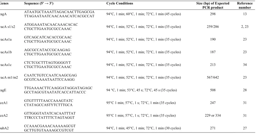
PCR analysis of genotypes in H. pylori isolates
The primers used in this study and PCR conditions are
shown in Table 1. For the cagA gene subtypıng, primers
CAGA-F and CAGA-R yielded a fragment of 298 bp of the
cagA gene were used (13). For analysis of the vacA s region,
primers VA1-F, VA1-R, SS2-F, and SS3-F were used (2, 23,
34). Primers VA1-F and VA1-R yielded a fragment of 259 bp
or 286 bp in size for type s1 or s2 strains, respectively. For
detection of the vacA m region, primers VAG-F and VAG-R
generated a fragment of 567 bp for m1 variants and a fragment
of 642 bp for m2 variants (23). For analysis of the cagE gene,
primers CagE-F and CagE-R yielded a fragment of 508 bp of
the cagE gene described by Tomasini et al. (28) were used. For
detection of the iceA gene, primers iceA1-F, iceA1-R, iceA2-F,
and iceA2-R were used (31). Primers iceA1-F and iceA1-R
generated a fragment of 247 bp for the iceA1 gene, and primers
iceA2-F and iceA2-R generated a fragment of 229 or 334 bp for
the iceA2 gene (31). For analysis of the babA2 gene, primers
BABA2-F and BABA2-R described by Sheu et al. (27) were
Ozbey, G. and Aygun, C. H. pylori genotypes
Amplification was performed in a reaction mixture (50 µl
final volume) containing 25 µl 2XPCR Master Mix (Fermentas,
K01071), 15 µl distilled water, 2.5 µl of each primer and 400 ng
genomic DNA. The thermal cycling conditions performed with a
touchdown thermal cycler (Hybaid, Middlesex, England). PCR
product was analyzed on 1.5% agarose gel containing 0.5 µg/ml of
ethidium bromide.
The DNAs of the HP 26695, HP J99 and some clinical
isolates, provided by Dr. Yoshio Yamaoka from Michael E.
DeBakey Veterans Affairs Medical Center, Houston, TX 77030,
USA was used to confirm the PCR test as positive controls.
Distilled water used as a negative control.
Table 1. Primers and PCR conditions used in our study.
Genes Sequence (5′→ 3′) Cycle Conditions Size (bp) of Expected PCR product
Reference number
cagA
vacA s1/s2
vacAs1a
vacAs1b
vacAs1c
vacA m1/m2
cagE
iceA1
iceA2
babA2
ATAATGCTAAATTAGACAACTTGAGCGA TTAGAATAATCAACAAACATCACGCCAT ATGGAAATACAACAAACACAC
CTGCTTGAATGCGCCAAAC GTCAGCATCACACCGCAAC CTGCTTGAATGCGCCAAAC AGCGCCATACCGCAAGAG CTGCTTGAATGCGCCAAAC CTCTCGCTTTAGTGGGGYT CTGCTTGAATGCGCCAAAC CAATCTGTCCAATCAAGCGAG GCGTCAAAATAATTCCAAGG
TTGAAAACTTCAAGGATAGGATAGAGC GCCTAGCGTAATATCACCATTACCC GTGTTTTTAACCAAAGTATC CTATAGCCASTYTCTTTGCA GTTGGGTATATCACAATTTAT TTRCCCTATTTTCTAGTAGGT CCAAACGAAACAAAAAGCGT GCTTGTGTAAAAGCCGTCGT
94°C, 1 min; 60°C, 1 min; 72°C, 1 min (45 cycles)
94°C, 1 min; 52°C, 1 min; 72°C, 1 min (35 cycles)
94°C, 1 min; 52°C, 1 min; 72°C, 1 min (35 cycles)
94°C, 1 min; 52°C, 1 min; 72°C, 1 min (35 cycles)
94°C, 1 min; 52°C, 1 min; 72°C, 1 min (35 cycles)
94°C, 1 min; 52°C, 1 min; 72°C, 1 min (35 cycles)
94 °C, 1 min; 53°C, 45 s; 72°C, 45 s (35 cycles)
95°C 1 min; 57°C, 1 s; 72°C, 1 min (35 cycles)
95°C 1 min; 57°C, 1 s; 72°C, 1 min (35 cycles)
94°C, 1 min; 45°C, 1 min; 72°C, 1 min (30 cycles)
298
259/286
190
187
213
567/642
508
247
229 or 334
271
13
2, 23
23
23
34
23
28
31
31
27
Statistical analysis
The Fischer’s exact and χ2 tests were used to compare the differences between H. pylori genotypes and clinical outcome
and between the sex and genotypes. A p value of <0.05 was
taken statistically significant.
RESULTS
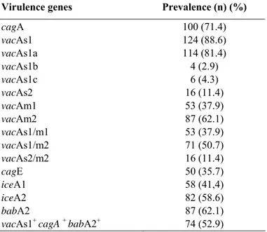
The prevalence of cagA vacA, cagE, iceA and babA2
genes are shown in Table 2. Because the number of patients
with GC is very low, the relationship between H. pylori
genotypes in patients with GC was not determined.
The cagA gene was determined in 100 (71.4%) of 140
isolates examined (Table 2). The vacA genes was found in all
isolates we studied. All vacAm1 genotypes from patients were
also vacAs1. The vacAs1a (81.4%) gene was found most
frequently than vacAs1b (2.9%) and vacAs1c (4.3%). The most
vacA allelic combination was s1/m2 (50.7%), followed by
s1/m1 (37.9%) and s2/m2 (11.4%). In addition, the most
common vacA allelic combination were vacAs1m1 (65.2%) and
s1m2 (53.9%) in patients with PU and G, respectively. No
vacAs2m1 genotype was observed in our study. Seventy-four
(52.9%) isolates were found to be “triple positive”
(vacAs1+cagA+babA2+) (Table 2).
The prevalence of the cagE genotype was 50 (35.7%), and
it was found more commonly in patients with PU. The iceA
Ozbey, G. and Aygun, C. H. pylori genotypes
iceA2 genes were detected in 58 (41.4%) and 82 (58.6%)
isolates, respectively. The iceA1 gene was most frequently
observed in patients (60.9%) with PU, whereas iceA2 was most
commonly found in patients (63.5%) with G. The iceA2
isolates classified in two types according to PCR product size:
229 and 334 bp. The distribution of these two types were
similar in the isolates examined (data not shown). The babA2
gene was observed in 87 (62.1%) of all isolates studied (Table
2 and 3).
The presence of the only vacAs1 isolate was significantly
associated with G and PU (p<0.05). The vacAs1a, ml, slml and
babA2 genes were significantly associated with PU (p<0.05),
whereas m2 gene was significantly associated with only G
(p<0.05). The cagA gene was significantly associated with s1,
s1m1 and babA2 genotypes (p<0.05) (Table 3).
The distribution of H. pylori genotypes and sex is shown
in Table 4. The difference between sex and genotypes was
statistically significant among the cagA, vacAs1, iceA2 and
babA2 genes (p<0.05) (Table 4).
Table 2. The prevalence of virulence genes in H. pylori
isolates.
Table 3. The prevalence of the genotypes in 140 H. pylori
positive patients with G and PU.
Genotypes G (n = 115) (%) PU (n = 23)(%)
cagA 79 (68.7) 19 (82.6)
vacAs1 99 (86.1)* 23 (100)*
vacAs1a 90 (78.3) 22 (95.7)*
vacAs1b 4 (3.5) 0 (0)
vacAs1c 5 (4.3) 1 (4.3)
vacAs2 16 (13.9) 0 (0)
vacAm1 37 (32.2) 15 (65.2)*
vacAm2 78 (67.8)* 8 (34.8)
vacAs1/m1 37 (32.2) 15 (65.2)*
vacAs1/m2 62 (53.9) 8 (34.8)
vacAs2/m2 16 (13.9) 0 (0)
cagE 39 (33.9) 10 (43.5)
iceA1 42 (36.5) 14 (60.9)
iceA2 73 (63.5) 9 (39.1)
babA2 68 (59.1) 17 (73.9)*
Table 4. The association between the sex of 140 H.
pylori-positive patients and its genotypes.
DISCUSSION
The number of studies related to genotypes of H. pylori in
Turkey is seldom, and the data on the relationship of the
genotypes and gastrointestinal diseases have been still
disputable (8).
The cagA prevalence is different in every part of the
world. The prevalence of cagA gene in this study was 71.4%.
Virulence genes Prevalence (n) (%)
cagA vacAs1 vacAs1a vacAs1b vacAs1c vacAs2 vacAm1 vacAm2 vacAs1/m1 vacAs1/m2 vacAs2/m2 cagE iceA1 iceA2 babA2
vacAs1+ cagA + babA2+
100 (71.4) 124 (88.6) 114 (81.4) 4 (2.9) 6 (4.3) 16 (11.4) 53 (37.9) 87 (62.1) 53 (37.9) 71 (50.7) 16 (11.4) 50 (35.7) 58 (41,4) 82 (58.6) 87 (62.1) 74 (52.9)
vacAs1+ cagA + babA2+ 59 (51.3) 13 (56.5)
G, gastritis; PU, peptic ulcer,* significant p<0.05
Genotypes Male (70) Female (70)
n (%) n (%)
cagA 53 (75.7)* 47 (67.1)
vacA s1 66 (94.3)* 58 (82.9)
vacA s2 4 (5.7) 12 (17.1)
vacA s1a 58 (82.9) 56 (80)
vacA s1b 3 (4.3) 1 (1.4)
vacA s1c 5 (7.1) 1 (1.4)
vacA m1 31 (44.3) 22 (31.4)
vacA m2 39 (55.7) 48 (68.6)
vacA s1/m1 31 (44.3) 22 (31.4)
vacA s1/m2 35 (50) 36 (51.4)
vacA s2/m2 4 (5.7) 12 (17.1)
cagE 28 (40) 22 (31.4)
iceA1 iceA2 babA2
vacAs1+ cagA + babA2+
20 (28.6) 50 (71.4)* 38 (54.3) 50 (71.4)* 32 (45.7) 37 (52.9) 29 (41.4) 45 (64.3)
Ozbey, G. and Aygun, C. H. pylori genotypes
This finding is in agrement with reports from Western
countries (22, 24) but lower than reports from East Asian
countries where the cagA are present in more than 90% of
cases (33). In addition, the results of this study are consistent
with previous reports (6, 29) which no association was found
between cagA gene and PU. However, some studies have been
reported that cagA gene was statistically associated with PU
(10, 20, 26).
The different results have also been reported in studies
related to the vacA gene of H. pylori strains. In the present
study, the vacA gene was observed in all strains. Our finding
was similar to previous reports from Turkey (10, 25, 26),
Northern and Eastern European countries (32) where s1a gene
was predominant genotype, but in contrast to a report from
Korea (16). We detected a low prevalence of vacAs1b in this
study which was contrast to pevious reports in Portugal,
Central and South America (32). No vacAs1c genotype was
determined except for the only one study reported in Turkey
(18). In this study, the prevalence of the vacAs1c gene was
found to be low. This may be related to low prevalence of GC
in Turkey as stated in a study carried out by Erzin et al. (10).
We found that no s1b, s1c and s2 genotypes associated
with G and PU. Our data similar to previous reports in Turkey
where vacAs1a strains were showed to be significantly
associated with PU (10, 18), but different from a study in
Turkey reported that vacAs1a strains were not found to be
statistically associated with PU (26).
Our results supported the findings of the studies
performed in Turkey and other countries (3, 6, 10, 18) where
the s1m2 genotype was the most common gene combinations
of the vacA, but contrary to studies (8, 26) reported that s1m1
genotype was the most predominant gene. The prevalence of
s2m2 genotype was determined as 11.4% and all of s2m2
positive isolates were cagA negative. No s2m1 genotype was
found in this study. This finding correlated confirm few data
reported from other geographic regions of Turkey (6, 10, 18).
On the other hand, this study confirmed the findings of
previous reports (6, 8, 10, 29) where there was no significant
association between vacAs1m2 genotype, G and PU disease.
The prevalence (35.7%) of cagE in this study is lower
than other studies conducted in USA (64%) (22), Turkey
(59.3%) (10), UK (71.2%) (15) and Thailand (88.4%) (8) but
higher than a previous study in Turkey (28.6%) (25). In an
attempt to detect association between the cagE gene and PU,
Fallone et al. (11), Day et al. (9) and Erzin et al. (10) have
reported a significant association between the cagE gene and
PU which is contrast to our findings.
In contrast to the results reported in China, Japan, Korea
and Netherlands where the iceA1 gene was predominant (16,
20, 31), the iceA2 gene was detected to be predominant
genotype in this study. This result is in agreement with
previous reports that the iceA2 gene was found to be prevalent
in Brazilian, European and American patients (1, 20, 22).
Our results were similar to a previous study (1) reported
that the distribution of iceA2 229 and 334-bp amplicon was
found to almost the same in H. pylori strains. The results of the
present study are similar to previous studies that no significant
association between the iceA1 gene and PU have found in
Brazilian and Turkish patients (1, 6, 10), but in contrast to
those reported by Peek et al. (21) and van Doorn et al. (31),
who showed the association between the presence of iceA1
gene and PU. The iceA2 was more frequently observed in
males than in females. Similar result have been reported by
Ashour et al. (1).
The prevalence of the babA2 genotype in H. pylori strains
varied in different countries of world. The prevalence of the
babA2 was 34-72% in Western countries (12, 19, 29) while it
was 80-100% in Asian countries (16). We found a higher
prevalence (62.1%) of babA2 than the result (53.8%) reported
in a previous study (10) in Turkey. Erzin et al. (10) had used
the primers described by Gerhard et al. (12), we used the
primers of Sheu et al. (27) which exhibited a high prevalence
of babA (8). A low prevalence of the babA2 gene was detected
when the primers described by Gerhard et al. (12) were used
(12, 20). These primers amplifies 832 bp (12) in a highly
Ozbey, G. and Aygun, C. H. pylori genotypes
variation (19). This study showed a highly significant
association between the babA2 gene and PU disease,
concurring with the previous studies (12, 29). This state may be
explained by allelic variation in the babA gene which could
have a variable affect in the different geographic regions of a
country (19).
Regarding the relationship with genotypes in each isolate,
we found to be an association between the cagA stratus and the
vacAs1 genotype in present study which was similar with
previous findings (12, 26, 29, 32). In addition, our study
showed significantly association between the babA2 and the
vacAs1 in contrast to the results reported by Erzin et al. (10)
and Torres et al. (29).
In regard to the association with the distribution of H.
pylori genotypes and sex in this study, the association was
statistically significant among the cagA, vacAs1, iceA2 and
babA2 genes. In a study conducted by Mansour and colleagues
(17), there was no significant association between cagA gene
and sex, but the association was statistically significant among
the vacA, iceA and oipA genes (17).
This study reported the prevalence of the genotypes in H.
pylori isolates in Elazig province located in the East of Turkey
for the first time. However, the multicenter and large scale
studies are needed to help us better understand epidemiological
importance of this disease and the association between H.
pylori genotypes and clinical outcome in different regions and
populations.
ACKNOWLEDGEMENTS
We acknowledge Dr. Yoshio Yamaoka (Michael E.
DeBakey Veterans Affairs Medical Center, Houston, TX
77030, USA) for supplying the DNAs of the HP 26695, HP J99
and some clinical isolates, Dr. Hans-Jurg Monstein (Division
of Clinical Microbiology, Department of Clinical and
Experimental Medicine, Faculty of Health Sciences, Linköping
University, Linköping, Sweden), Dr. Gireesh Rajashekara
(Food Animal Health Research Program, Ohio Agricultural
Research and Development Center, Department of Veterinary
Preventive Medicine, The Ohio State University, Wooster, OH,
USA) and Dr. Gilmara Coelho Meine (Postgraduation Program
in Gastroenterology, Universidade Federal do Rio Grande do
Sul, Brazil) for their suggestions and the constructive criticism
of the manuscript, and Dr. Yakut Akyon Yilmaz, Department
of Medical Microbiology, Faculty of Medicine, Hacettepe
University, Ankara, Turkey for her technicial support and help.
In addition, the authors thank to the management of the Elazig
Veterinary Control and Research Institute for providing
laboratory facilities during our study. This work was part of the
project supported by the Scientific Research Council ofFirat
University (FUBAP 1609).
REFERENCES
1. Ashour, A.A.R.; Collares, G.B.; Mendes, E.N.; de Gusmão, V.R.; Queiroz, D.M.M.; Magalhães, P.P.;Carvalho, A.S.T.;Oliveira, C.A.; Nogueira, A.M.M.F.; Rocha, G.A.; Rocha, A.M.C. (2001). iceA genotypes of Helicobacter pylori strains isolated from Brazilian children and adults. J. Clin. Microbiol. 39, 1746-1750.
2. Atherton, J.C.; Cao, P.; Peek, R.M. Jr.; Tummuru, M.K.; Blaser, M.J.; Cover T.L. (1995). Mosaicism in vacuolating cytotoxin alleles of Helicobacter pylori. Association of specific vacA types with cytotoxin
production and peptic ulceration. J. Biol. Chem. 270, 17771-17777. 3. Atherton, J.C. (1997). The clinical relevance of strain types of
Helicobacter pylori. Gut. 40, 701-703.
4. Atherton, J.C. (2000). CagA: a role at last. Gut. 47, 33-41.
5. Blaser, M.J.; Perez-Perez, G.I.; Kleanthous, H.; Cover, T.L.; Peek, R.M.; Chyou, P.H.; Stemmermann, G.N.; Nomura, A. (1995). Infection with Helicobacter pylori strains possessing cagA is associated with an
increased risk of developing adenocarcinoma of the stomach. Cancer Res. 55, 2111-2115.
6. Caner, V.; Yilmaz, M.; Yonetci, N.; Zincir, S.; Karagenc, N.; Kaleli, I.; Bagci, H. (2007). H. pylori iceA alleles are disease-specific virulence factors. World J. Gastroenterol. 13, 2581-2585.
7. Censini, S.; Lange, C.; Xiang, Z.; Crabtree, J.E.; Ghiara, P.; Borodovsky, M.; Rappuoli, R.; Covacci, A. (1996). cag, a pathogenicity island of Helicobacter pylori, encodes type I-specific and disease-associated
virulence factors. Proc. Nat. Acad. Sci. USA. 93, 14648-14653. 8. Chomvarin, C.; Namwat, W.; Chaicumpari K.; Mairiang, P.; Sangchan,
A.; Sripa, B.; Tor-Udom, S.; Vilaichone, R.K. (2008). Prevalence of Helicobacter pylori vacA, cagA, cagE, iceA and babA2 genotypes in
Thai dyspeptic patients. Int. J. Infect. Dis. 12, 30-36.
Ozbey, G. and Aygun, C. H. pylori genotypes
with Helicobacter pylori-induced duodenal ulceration in children. J. Infect. Dis. 181, 1370-1375.
10. Erzin, Y.; Koksal, V.; Altun, S.; Dobrucali, A.; Aslan, M.; Erdamar, S.; Dirican, A.; Kocazeybek, B. (2006). Prevalence of Helicobacter pylori vacA, cagA, cagE, iceA, babA2 genotypes and correlation with clinical
outcome in Turkish patients with dyspepsia. Helicobacter 11, 574-580. 11. Fallone, C.A.; Barkun, A.N.; Gottke, M.U.; Beech, R.N. (1998). A
review of the possible bacterial determinants of clinical outcome in Helicobacter pylori infection. Can. J. Microbiol. 44, 201-210.
12. Gerhard, M.; Lehn, N.; Neumayer, N.; Borén, T.; Rad, R.; Schepp, W.; Miehlke, S.; Classen, M.; Prinz, C. (1999). Clinical relevance of the Helicobacter pylori gene for blood-group antigen-binding adhesin. Proc.
Natl. Acad. Sci. USA. 96, 12778-12783.
13. Hamlet, A.; Thoreson, A.C.; Nilsson, O.; Svennerholm, A.M.; Olbe, L (1999). Duodenal Helicobacter pylori infection differs in cagA genotype between asymptomatic subjects and patients with duodenal ulcers. Gastroenterology 116, 259-268.
14. Ilver, D.; Arnqvist, A.; Ogren, J.; Frick, I.M.; Kersulyte, D.; Incecik, E.T.; Berg, D.E.; Covacci, A.; Engstrand, L.; Boren, T. (1998). Helicobacter pylori adhesin binding fucosylated histo-blood group
antigens revealed by retagging. Science. 279, 373-377.
15. Kauser, F.; Hussain, M.A.;Ahmed, I.;Srinivas, S.; Devi, S.M.; Majeed, A.A.; Rao, K.R.; Khan, A.A.; Sechi, L.A.; Ahmed, N. (2005). Comparative genomics of Helicobacter pylori isolates recovered from ulcer disease patients in England. BMC Microbiol. 5, 32.
16. Kim, S.Y.; Woo, C.W.; Lee, Y.M.; Son, B.R.; Kim, J.W.; Chae, H.B.; Youn, S.J.; Park, S.M. (2001). Genotyping cagA, vacA subtype, iceA1, and babA of Helicobacter pylori isolates from Korean patients, and their association with gastroduodenal diseases. J, Korean Med. Sci. 16, 579-584.
17. Mansour, K.B.; Fendri, C.; Zribi, M.; Masmoudi, A.;Labbene, M.; Fillali, A.; Mami, N.B.;Najjar, T.; Meherzi, A.; Sfar, T.;Burucoa, C. (2010). Prevalence of Helicobacter pylori vacA, cagA, iceA and oipA genotypes in Tunisian patients. Ann. Clin. Microbiol. Antimicrob. 9, 10. 18. Nagiyev, T.;Yula, E.; Abayli, B.; Koksal, F. (2009). Prevalence and
genotypes of Helicobacter pylori in gastric biopsy specimens from patients with gastroduodenal pathologies in the Cukurova region of Turkey. J. Clin. Microbiol. 47, 4150-4153.
19. Olfat, F.O.; Zheng, Q.; Oleastro, M.; Voland, P.; Boren, T.; Karttunen, R.; Engstrand, L.; Rad, R.; Prinz, C.; Gerhard, M. (2005). Correlation of the Helicobacter pylori adherence factor BabA with duodenal ulcer disease in four European countries. FEMS Immunol Med Microbiol 44, 151-156.
20. Oliveira, A.G.; Santos, A.; Guerra, J.B.; Rocha, G.A.; Rocha, A.M.; Oliveira, C.A.; Cabral, M.M.; Nogueira, A.M.; Queiroz, D.M. (2003). babA2- and cagA-positive Helicobacter pylori strains are associated with
duodenal ulcer and gastric carcinoma in Brazil. J. Clin. Microbiol. 41,
3964-3966.
21. Peek, R.M.J.; Thompson, S.A.; Donahue, J.P.; Tham, K.T.; Atherton, J.C.; Blaser, M.J.; Miller, G.G. (1998). Adherence to gastric epithelial cells induces expression of a Helicobacter pylori gene, iceA, that is associated with clinical outcome. Proc. Am. Assoc. Phys. 110, 531-544. 22. Podzorski, R.P.; Podzorski, D.S.; Wuerth, A.; Tolia, V. (2003). Analysis
of the vacA, cagA, cagE, iceA, and babA2 genes in Helicobacter pylori from sixty-one pediatric patients from the Midwestern United States. Diagn. Microbiol. Infect. Dis. 46, 83-88.
23. Qiao, W.; Hu, J.L.; Xiao, B.; Wu, K.C.; Peng, D.R.; Atherton, J.C.; Xue, H. (2003). cagA and vacA genotype of Helicobacter pylori associated with gastric diseases in Xi’an area. World J Gastroenterol 9, 1762-1766. 24. Ribeiro, M.L.; Godoy, A.P.; Benvengo, Y.H.; Mendonca, S.; Pedrazzoli,
J. (2003). Clinical relevance of the cagA, vacA and iceA genotypes of Helicobacter pylori in Brazilian clinical isolates. FEMS Immunol. Med.
Microbiol. 36, 181-185.
25. Salih, B.A.; Abasiyanik, M.F.; Ahmed, N. (2007). A preliminary study on the genetic profile of cag pathogenicity-island and other virulent gene loci of Helicobacter pylori strains from Turkey. Infect. Genet. Evol. 7, 509-512.
26. Saribasak, H.; Salih, B.A.; Yamaoka, Y.; Sander, E. (2004). Analysis of Helicobacter pylori genotypes and correlation with clinical outcome in
Turkey. J. Clin. Microbiol. 42, 1648-1651.
27. Sheu, B.S.; Sheu, S.M.; Yang, H.B.; Huang, A.H.; Wu, J.J. (2003). Host gastric Lewis expression determines the bacterial density of Helicobacter pylori in babA2 genopositive infection. Gut 52, 927-932.
28. Tomasini, M.L.; Zanussi, S.; Sozzi, M.; Tedeschi, R.; Basaglia, G.; De Paoli, P. (2003). Heterogeneity of cag genotypes in Helicobacter pylori isolates from human biopsy specimens. J. Clin. Microbiol. 41, 976-980. 29. Torres, L.E.; Melian, K.; Moreno, A.; Alonso, J.; Sabatier, C.A.;
Hernandez, M.; Bermúdez, L.; Rodríguez, B.L. (2009). Prevalence of vacA, cagA and babA2 genes in Cuban Helicobacter pylori isolates.
World J. Gastroenterol. 15, 204-210.
30. van Doorn, L.-J.; Figueiredo, C.; Sanna, R.; Pena, S.; Midolo, P.; Ng, E.K.W.; Atherton, J.C.; Blaser, M.J.; Quint, W.G.V. (1998a). Expanding allelic diversity of Helicobacter pylori vacA. J. Clin. Microbiol. 36, 2597-2603.
31. van Doorn, L.J.; Figueiredo, C, Sanna, R, Plaisier A, Schneeberger PW, de Boer W, Quint W (1998b) Clinical relevance of the cagA, vacA, and iceA status of Helicobacter pylori. Gastroenterology. 115:58-66.
32. van Doorn, L.J.; Figueiredo, C.; Megraud, F.; Pena, A.S.; Midolo, P.; Queroz, D.M.; Carnero, F.; Pegado, M.D.; Sanna, R. (1999). Geographic distribution of vacA allelic types of Helicobacter pylori. Gastroenterology. 116, 823-830.
Ozbey, G. and Aygun, C. H. pylori genotypes
countries. J. Clin. Microbiol. 37, 2274-2279.
34. Yamazaki, S.; Yamakawa, A.; Okuda, T.; Ohtani, M.; Suto, H.; Ito, Y.; Yamazaki, Y.; Keida, Y.; Higashi, H.; Hatakeyama, M.; Azuma, T.
(2005). Distinct diversity of vacA, cagA, and cagE genes of Helicobacter pylori associated with peptic ulcer in Japan. J. Clin. Microbiol. 43,
3906-3916.