Prevalence and phenotypic characterization of
Enterococcus
spp.
isolated from food in Brazil
Carlos Henrique Camargo
1,2, Ariane Bruder-Nascimento
1, Sarah Hwa In Lee
3,
Ary Fernandes Júnior
1, Ramon Kaneno
1, Vera Lúcia Mores Rall
1 1Departamento de Microbiologia e Imunologia, Instituto de Biociências de Botucatu, Universidade Estadual Paulista “Júlio de Mesquita Filho”, Botucatu, SP, Brazil.
2
Departamento de Clínica Médica, Faculdade de Medicina de Botucatu, Universidade Estadual Paulista “Júlio de Mesquita Filho”, Botucatu, SP, Brazil. 3
Departamento de Engenharia de Alimentos, Faculdade de Zootecnia e Engenharia de Alimentos, Universidade de São Paulo, Pirassununga, SP, Brazil.
Submitted: September 29, 2012; Approved: September 9, 2013.
Abstract
We evaluated the frequency of enterococci from food and found 95.2% of positivity, being E. faeciumandE. faecalisthe most frequent species. High-level streptomycin resistance was observed, as well as gelatinase and hemolysis activity, showing the potential role of environmental strains as reservoir of virulence and resistance traits.
Key words:Enterococcusspp., food, antimicrobial resistance.
Enterococci are lactic acid bacteria and although this genus comprises more than 25 species,E. faeciumandE. faecalisare the main species isolated from food (Giraffa, 2002; Foulquié Morenoet al., 2006) and clinical samples (Cetinkayaet al., 2000). These bacteria can also be used as starter in the food industry due to their capacity to produce lipase, protease and volatile compounds ensuring desirable organoleptic features in some specific kinds of food. De-spite their importance in food technology, certain features, such as the ability to growth over a wide range of tempera-ture, salinity and pH make these organisms able to multiply in several foods and even spoil them (Giraffa, 2002; Foul-quié Morenoet al., 2006).
Enterococcushave already been isolated from several kinds of food, such as vegetables, meat, milk and dairy foods (Giraffa, 2002; Hayeset al., 2003; Foulquié Moreno
et al., 2006; Gomeset al., 2008). The frequency of isolation ranged from 52.5 to 99% (Hayeset al., 2003; Johnston and Jaykus, 2004; Gomeset al., 2008), depending on the kind of food, as well as the seasonal and manufacture conditions during their processing. The later aspects influence the bac-terial survival, especially on cheese manufacturing and rip-ening (Foulquié Morenoet al., 2006).
Besides the high prevalence in food, several virulence factors and antimicrobial resistance have been identified in these enterococci (Franzet al., 2001; Barbosaet al., 2010).
The presence of virulence and resistance factors in enterococci is quite variable (Jettet al., 1994; Mundyet al., 2000) and the occurrence of these bacteria in food is a mat-ter of debate (Franzet al., 2003). Differentiation between safe and non-safe strains is not easy, due to their capacity to exchange genetic elements with each other (Eaton and Gasson, 2001; Messiet al., 2006). The real role of virulence and antimicrobial resistance factors of food enterococci is not well elucidate, but bacteria presenting such factors in the environment may be understood as a genetic reservoir of virulence (Hayeset al., 2003). In this study we aimed to isolate enterococci species from different kinds of food evaluating some virulence factors and antimicrobial resis-tance.
We analyzed samples of poultry (35), pork (20), cheese (35) and vegetables (15) commercially available in eight supermarkets located in Botucatu, SP, Brazil. Sam-ples were allocated in sterile plastic bags and transported in isothermal box under refrigeration during the way to the laboratory to be processed at the same day. Twenty-five
Send correspondence to C.H. Camargo. Departamento de Microbiologia e Imunologia, Instituto de Biociências de Botucatu, Universidade Estadual Paulista “Júlio de Mesquita Filho”, 18618-970 Botucatu, SP, Brazil. E-mail: [email protected].
grams of each sample was homogenized in 225 mL of bile esculin azide broth (BBL) in Stomacher Lab Blender 400 for 30 s. Next, 0.1 and 0.01 mL of the initial dilution were spread onto the surface of bile esculin azide agar plates (BBL) and incubated at 45 °C/24 and 48 h. The initial dilu-tions were also incubated at 45 °C/48 h, and a loop of the broth was streaked onto the surface of bile esculin azide agar plates, followed by incubation at 45 °C/24 and 48 h (Hayeset al., 2003). In order to guarantee the absence of contamination, typical enterococci colonies (black) on bile esculine azide agar (from either direct growth or after the enrichment step) were streaked onto blood agar (Oxoid) plates prepared with 5% of defibrinated sheep blood and in-cubated at 37 °C/24 and 48 h.
Identification of presumptive enterococci was con-firmed using the tests proposed by Facklamet al.(2007): Gram staining, hemolysins, catalase, salt tolerance, esculin hydrolysis, pyrrolidonyl arylamidase (PYR), arginine decarboxylation, mannitol, arabinose, sorbitol and rafinose fermentation, pigment production, motility and tetrazolium reduction test. For determination of virulence traits, hemo-lysins were detected in blood agar base plates with 5% of defibrinated sheep blood after incubation at 37 °C/24 h and 5 °C/48 h.a-Hemolysis was defined by the presence of a viridant halo around isolate colonies, whileb-hemolysis was defined by translucent halo. Gelatinase assay was car-ried out as described by Suet al.(1991). Briefly, a spot of freshly cultured enterococci was seeded onto the surface of gelatin agar and the plate was incubated at 37 °C/48 h; next the Petri dishes were kept at 4 °C/4 h, and a precipitation halo around the spot denoted a positive result. For anti-microbial susceptibility testing, vancomycin and high-level aminoglycoside (gentamicin and streptomycin) resistance was screened by disk diffusion assay (120-mg gentamicin disk and 300-mg streptomycin disk) assay and confirmed by minimal inhibitory concentration (MIC) determined by agar dilution (MIC above 500 and 2000mg/mL for genta-micin and streptomycin, respectively). Based on halos and MIC measures, the isolates were categorized as susceptible, intermediate and resistant according to the Clinical and Laboratory Standards Institute (2011). Categorical vari-ables were compared by chi-square or Fisher’s exact tests. Differences were considered statistically significant when p£0.05.
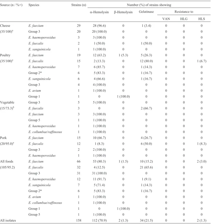
Hundred fifty-eight strains of enterococci were iso-lated from 100 samples out of 105 analyzed food samples (positivity ratio = 95.2%). Enterococci was less frequently isolated from vegetable (73.3%) than cheese (100%), poul-try (100%) and pork (95%) (p = 0.001). Samples of all the eight evaluated supermarkets were positive.E. faeciumwas the most frequent species isolated (41.8% of overall strains) followed byE. faecalis(20.3%). Other species were also less frequently identified (Table 1).
Antimicrobial susceptibility showed that all of strains were susceptible to vancomycin and high-level gentamicin. Only two strains presented high-level streptomycin resis-tant (confirmed MIC > 2000mg/mL), both of them were identified asE. faecalisand isolated from poultry and pork meats purchased at the same supermarket, but with a gap of almost three months between the evaluation of two sam-ples. Seventy-two percent of enterococci isolated from foods showed hemolysis; this characteristic was associated toE. faecium species (p = 0.016). Gelatinase production, however, was associated with E. faecalis species (p < 0.0001). More detailed data on source, frequencies and phenotypic characterization of enterococci strains are pre-sented at Table 1.
In this study, enterococci were isolated from 95.2% of the 105 food samples. High frequency of enterococci in foods has also been previously reported in North America (Hayeset al., 2003; Johnston and Jaykus, 2004), Europe (Koluman, 2009), and Latin America (Morales et al., 2004). In Brazil, there is no much available information about it: Fracalanzzaet al.(2007) analyzed 50 samples of meat and milk, and 86.6% of them were positive for entero-cocci. Later, Gomeset al.(2008) observed the presence of enterococci in 120 samples of raw and pasteurized cheese, meat, and vegetables and found out a lower frequency com-pared with our results (52.5% of positivity), probably due to the absence of an enrichment step.
We observed thatE. faecalisandE. faeciumtogether figured more than 60% of the overall isolated strains, but it is remarkable the diversity of species isolated from food samples (Table 1). Particularly E. haemoperoxidus (iso-lated from cheese, poultry, pork and lettuce samples) andE. sanguinicola(from cheese and poultry samples) species have rarely been recovered from food samples (Martínet al., 2009). Although more than 25 species have been de-scribed intoEnterococcusgenus,E. faeciumandE. faecalis
are the most widespread species isolated from food samples (Hayeset al., 2003, 2004; Abriouelet al., 2008; Gomeset al., 2008; Koluman, 2009). Our difficult to identify the spe-cies of a few strains was due to the similarity of phenotypic features; this limitation resulted in some isolates that have been only identified as “group” of strains according to Facklamet al.(2007) scheme. Hayeset al.(2004) have also had the same limitation to identify isolates from commer-cial poultry production environments.
of inhibition halos lower than 17 mm, the motility and pig-ment production tests were performed for all isolates. The strains presented neither motility nor pigment production, confirming the absence of E. casseliflavus and E. gallinarumin our samples.
Vancomycin resistance in enterococci isolated from food has a variable pattern both in Brazil and abroad (Franz
et al., 2001; Giraffa, 2002; Johnston and Jaykus, 2004; Fracalanzzaet al., 2007; Gomeset al., 2008). The source of vancomycin resistant strains appears to be different accord-ing to the geographic region where they emerge. In Europe it is attributed to the use of antimicrobial agents as growth promoter whereas in United States, it is attributed to the Table 1- Prevalence, species frequency and distribution of virulence and resistance traits inEnterococcusspp. isolated from Brazilian foods.
Source (n / %+) Species Strains (n) Number (%) of strains showing
a-Hemolysin b-Hemolysin Gelatinase Resistance to
VAN HLG HLS
Cheese E. faecium 29 28 (96.6) 0 1 (3.4) 0 0 0
(35/100)a Group 3 20 20 (100.0) 0 0 0 0 0
E. haemoperoxidus 3 3 (100.0) 0 0 0 0 0
E. faecalis 2 1 (50.0) 0 1 (50.0) 0 0 0
E. sanguinicola 1 1 (100.0) 0 0 0 0 0
Poultry E. faecium 19 12 (63.2) 1 (5.3) 5 (26.3) 0 0 0
(35/100)a E. faecalis 15 2 (13.3) 0 12 (80.0) 0 0 1 (6.7)
E. haemoperoxidus 7 6 (85.7) 0 1 (14.3) 0 0 0
Group 2* 6 5 (83.3) 0 1 (16.7) 0 0 0
E. sanguinicola 6 4 (66.6) 0 1 (16.7) 0 0 0
Group 3 4 4 (100.0) 0 0 0 0 0
E. avium 1 1 (100.0) 0 0 0 0 0
Group 1 1 0 1 (100.0) 0 0 0 0
Vegetable Group 3 5 5 (100.0) 0 0 0 0 0
(15/73.3)b E. faecalis 3 0 0 2 (66.7) 0 0 0
E. faecium 3 3 (100.0) 0 0 0 0 0
Group 5 1 1 (100.0) 0 0 0 0 0
E. haemoperoxidus 1 1 (100.0) 0 0 0 0 0
E. collumbae/raffinosus 1 1 (100.0) 0 0 0 0 0
Pork E. faecium 15 10 (66.7) 0 4 (26.7) 0 0 0
(20/95.0)a E. faecalis 12 1 (8.3) 0 6 (50.0) 0 0 1 (8.3)
Group 3 2 2 (100.0) 0 0 0 0 0
E. haemoperoxidus 1 1 (100.0) 0 0 0 0 0
All foods E. faecium 66 53 (80.3) 1 (1.5) 10 (15.2) 0 0 2 (3.0)
(105/95.2) E. faecalis 32 4 (12.5) 0 21 (65.6) 0 0 0
Group 3 31 31 (100.0) 0 0 0 0 0
E. haemoperoxidus 12 11 (91.7) 0 1 (9.1) 0 0 0
E. sanguinicola 7 5 (71.4) 0 1 (14.3) 0 0 0
Group 2* 6 5 (83.3) 0 1 (16.7) 0 0 0
E. avium 1 1 (100.0) 0 0 0 0 0
E. collumbae/raffinosus 1 1 (100.0) 0 0 0 0 0
Group 1 1 0 1 (100.0) 0 0 0 0
Group 5 1 1 (100.0) 0 0 0 0 0
All isolates 158 112 (70.9) 2 (1.3) 34 (21.5) 0 0 2 (1.3)
a, b: different letters represent significant statistical differences (p = 0.001).
VAN: vancomycin; HLG: High-level gentamicin [Minimal Inhibitory Concentration (MIC) > 500mg/mL]; HLS: High-level streptomycin (MIC > 2000 mg/mL).
wide hospital usage of vancomycin (Woodford, 1998; Wegeneret al., 1999).
The concern of high-level aminoglycoside resistance (HLAR) in enterococci is well documented. For instance, Donabedian et al. (2003) showed the transmission of HLAR strains among farm animals and humans. Although enterococci present intrinsic low level resistance to amino-glycosides, the association of high-levels of such drugs with a cell inhibitor antibiotic was showed to be efficient against enterococciin vivo(Murray, 1990). Resistance to high-level gentamicin (HLGR) and streptomycin (HLSR) was evaluated because HLGR predicts resistance to all aminoglycosides but streptomycin (Chow, 2000), which can be modified by other aminoglycoside acetyltransferase (Aac) family enzymes. By examining both aminoglyco-sides it is possible to predict high-level resistance to amino-glycosides (Chow, 2000; Clinical and Laboratory Standards Institute, 2011).
Resistance to high-level gentamicin was not ob-served, whereas only two (1.3%) strains isolated from poul-try and pork samples showed high-level streptomycin resis-tance. In previous reports, HLSR was also more frequent than HLGR (Franzet al., 2001; Johnston and Jaykus, 2004; Fracalanzza et al., 2007). In opposition, Teuber et al.
(2009) found out 80% of enterococci isolated from cheese with high-level gentamicin resistance, and, in Brazil, Gomes et al. (2008) reported 22% of E. faecalis with high-level gentamicin resistance. The low prevalence of HLAR we have observed may be explained by the fact that in Brazil, the use of antimicrobial agents was prohibited as growth promoters in farms (Brasil, 1998) until some years ago (Brasil, 2009).
Virulence markers are also cause of concern among enterococci from food (Eaton and Gasson, 2001; Foulquié Morenoet al., 2006). Several factors such as aggregation proteins to eukaryotic cells and adhesins, biofilm produc-tion, extracellular proteases (colagenases, gelatinase), cytolysins (bacteriocins and hemolysins), leukocytary eva-sion proteins and sex pheromones have been associated with enterococci pathogenicity (Jettet al., 1994; Eaton and Gasson, 2001). Hemolysin production seems to be associ-ated with virulence in experimental models, as well as the gelatinase (Mundy etal., 2000) and the expression of these characteristics was associated with E. faecalis and E. faecium, according to previous study (Mundy etal., 2000).
In this study we observed higher frequency of
a-hemolysin (79.8%) than b-hemolysin (12.6%) in the food enterococci, in agreement with previous reports (Go-meset al., 2008; Barbosaet al., 2010). The gelatinase pro-duction was present in about 20% of the strains, most of them belonging toE. faecalisspecies. Most of these strains were isolated from chicken meat, diverging from Franzet al.(2001) that found a high incidence of gelatinase produc-ingE. faecalisin cheese samples. It is remarkable that both foods are rich in protein contents and that gelatinase
pro-ducing strains can use these substrata as amino acid source (Franzet al., 2001). In our samples, 65.6% of theE. faecalis
strains were gelatinase producers, while 15.2% of the E. faecium strains showed this virulence factor. Gelatinase-producing enterococci were isolated from meat and it could cause the food degradation (Gomeset al., 2008).
Several studies have investigated the occurrence of virulence factors and antimicrobial resistance in entero-cocci from different sources as environment, water, food and infectious diseases (Semedoet al., 2003). For instance, Abriouelet al.(2008) have found virulence traits both in clinical and food, water and soil isolated strains. In the same way, Eaton and Gasson (2001) found a higher propor-tion of virulent enterococci strains in clinical isolates com-pared with those isolated from food or employed as starter culture in the food industry. The starter culture should pres-ent proteases and lipases in order to metabolize volatile compounds while they should be free of virulence determi-nants (Foulquié Moreno et al., 2006). The concomitant presence of virulence factors in clinical and environmental samples, however, hinders the classification as safe or non-safe strains (Eaton and Gasson, 2001; Franz et al., 2001).
In summary, we observed the presence of
Enterococcusspp. in almost all the samples evaluated as well as hemolysin and gelatinase production in those strains. Antimicrobial resistance was very rare. The re-markable finding of enterococci in foods is their ability to exchange virulence and drug resistance with potential pathogenic strains (Eaton and Gasson, 2001; Messiet al., 2006). It suggests us that environmental and food strains can represent a natural reservoir of these features, enhanc-ing the development of more dangerous clinical strains.
Acknowledgments
The authors thank FAPESP for the scientific initia-tion fellowship to Carlos Henrique Camargo.
References
Abriouel H, Omar N, Molinos A, López R, Grande M, Martí-nez-Viedma P, Galvez A (2008) Comparative analysis of genetic diversity and incidence of virulence factors and anti-biotic resistance among enterococcal populations from raw fruit and vegetable foods, water and soil, and clinical sam-ples. Int J Food Microbiol 123:38-49.
Barbosa J, Gibbs PA, Teixeira P (2010) Virulence factors among enterococci isolated from traditional fermented meat prod-ucts produced in the North of Portugal. Food Control 21:651-656.
Brasil (1998)Portaria Nº 193, de 12 de maio de 1998. Retrieved from
http://extranet.agricultura.gov.br/sislegis-consulta/consulta rLegislacao.do?operacao=visualizar&id = 1125.
Brasil (2009)Instrução Normativa Nº 26, de 09 de julho de 2009.
http://extranet.agricultura.gov.br/sislegis-consulta/consulta rLegislacao.do?operacao=visualizar&id = 20408.
Cetinkaya Y, Falk P, Mayhall C (2000) Vancomycin-resistant enterococci. Clin Microbiol Rev 13:686-707.
Chow JW (2000) Aminoglycoside resistance in enterococci. Clin Infect Dis 31:586-589.
Clinical and Laboratory Standards Institute (2011) Performance standards for antimicrobial susceptibility testing: twenty first informational supplement. M100-S21. CLSI, Wayne. Donabedian S, Thal L, Hershberger E, Perri M, Chow J, Bartlett P,
Zervos M (2003) Molecular characterization of gentamicin-resistant Enterococci in the United States: evi-dence of spread from animals to humans through food. J Clin Microbiol 41:1109-1113.
Eaton T, Gasson M (2001) Molecular screening ofEnterococcus
virulence determinants and potential for genetic exchange between food and medical isolates. Appl Environ Microbiol 67:1628-1635.
Facklam RR, Carvalho MGS, Teixeira LM (2007)Enterococcus. In: Murray, P.R., Baron, E.J., Pfaller, M.A., Jorgensen J.H., Landry, M.L. (eds.) Manual of Clinical Microbiology. Ame-rican Society for Microbiology, Washington, p. 430-442. Foulquié Moreno M, Sarantinopoulos P, Tsakalidou E, De Vuyst
L (2006) The role and application of enterococci in food and health. Int J Food Microbiol 106:1-24.
Fracalanzza S, Scheidegger E, Santos P, Leite P, Teixeira L (2007) Antimicrobial resistance profiles of enterococci iso-lated from poultry meat and pasteurized milk in Rio de Ja-neiro, Brazil. Mem Inst Oswaldo Cruz 102:853-859. Franz C, Muscholl-Silberhorn A, Yousif N, Vancanneyt M,
Swings J, Holzapfel W (2001) Incidence of virulence factors and antibiotic resistance among Enterococci isolated from food. Appl Environ Microbiol 67:4385-4389.
Franz C, Stiles M, Schleifer K, Holzapfel W (2003) Enterococci in foods—a conundrum for food safety. Int J Food Microbiol 88:105-122.
Giraffa G (2002) Enterococci from foods. FEMS Microbiol Rev 26:163-171.
Gomes B, Esteves C, Palazzo I, Darini A, Felis G, Sechi L, De Martinis E (2008) Prevalence and characterization of
Enterococcus spp. isolated from Brazilian foods. Food
Microbiol 25:668-675.
Hayes J, English L, Carr L, Wagner D, Joseph S (2004) Multi-ple-antibiotic resistance ofEnterococcusspp. isolated from commercial poultry production environments. Appl Environ Microbiol 70:6005-6011.
Hayes J, English L, Carter P, Proescholdt T, Lee K, Wagner D, White D (2003) Prevalence and antimicrobial resistance of
Enterococcusspecies isolated from retail meats. Appl
Envi-ron Microbiol 69:7153-7160.
Jett B, Huycke M, Gilmore M (1994) Virulence of enterococci. Clin Microbiol Rev 7:462-478.
Johnston L, Jaykus L (2004) Antimicrobial resistance of
Enterococcusspecies isolated from produce. Appl Environ
Microbiol 70:3133-3137.
Koluman A (2009) Occurrence and antimicrobial resistance of enterococci in retail foods. Food Control 20:281-283. Martín B, Corominas L, Garriga M, Aymerich T (2009)
Identifi-cation and tracing ofEnterococcusspp. by RAPD-PCR in traditional fermented sausages and meat environment. J Appl Microbiol 106:66-77.
Messi P, Guerrieri E, de Niederhäusern S, Sabia C, Bondi M (2006) Vancomycin-resistant enterococci (VRE) in meat and environmental samples. Int J Food Microbiol 107:218-222.
Morales G, Blanco L, Arias ML, Chaves C (2004) Evaluación de la calidad bacteriología de tilapia fresca (Orochoromis
niloticus)proveniente de la zona norte de Costa Rica. Arch
Latinoam Nutr 54:433-437.
Mundy L, Sahm D, Gilmore M (2000) Relationships between enterococcal virulence and antimicrobial resistance. Clin Microbiol Rev 13:513-522.
Murray B (1990) The life and times of theEnterococcus. Clin Microbiol Rev 3:46-65.
Semedo T, Santos M, Lopes M, Figueiredo Marques J, Barreto Crespo M, Tenreiro R (2003) Virulence factors in food, clin-ical and reference Enterococci: a common trait in the genus? Syst Appl Microbiol 26:13-22.
Su Y, Sulavik M, He P, Makinen K, Makinen P, Fiedler S, Clewell D (1991) Nucleotide sequence of the gelatinase gene (gelE)
from Enterococcus faecalis subsp. liquefaciens. Infect
Immun 59:415-420.
Teuber M, Meile L, Schwarz F (1999) Acquired antibiotic resis-tance in lactic acid bacteria from food. Antonie Van Leeuwenhoek 76:115-137.
Wegener H, Aarestrup F, Jensen L, Hammerum A, Bager F (1999) Use of antimicrobial growth promoters in food animals and
Enterococcus faecium resistance to therapeutic
anti-microbial drugs in Europe. Emerg Infect Dis 5:329-335. Woodford N (1998) Glycopeptide-resistant enterococci: a decade
of experience.J Med Microbiol 47:849-862.