577
DNA fragmentation and membrane damage of bocachico
Prochilodus magdalenae
(Ostariophysi: Prochilodontidae) sperm following
cryopreservation with dimethylsulfoxide and glucose
José Gregorio Martínez
1, Víctor Atencio García
2and Sandra Pardo Carrasco
1The endangered bocachico Prochilodus magdalenae is a native freshwater fish of Colombia, the most captured species locally and one of the most important species for ex-situ conservation (germplasm banks). The aim of this study was to examine the effect of three concentrations of Dimethylsulfoxide (DMSO) (5%, 10%, 15%) and three of glucose (305, 333, 361 mM) in the extender on spermatic DNA fragmentation (F-DNA) (by Halomax®, Chromatin dispersion) and membrane damage (D-Me) (by eosin-nigrosin staining). After assessment of sperm quality by computer analysis of motility, one part of semen from males was diluted separately with three parts of extender and filled into 0.5 ml straws. Freezing was carried out in liquid nitrogen vapor dry shipper for 30 minutes and thawed at 60°C for 8 seconds in a water bath and evaluated for the percentage of cells found with F-DNA and D-Me. The results demonstrated that cryopreservation causes greater F-DNA (13.62 ± 1.6% to 28.91 ± 3.25) and D-Me (24.27 ± 1.1% to 58.33 ± 2.81%) when compared with pre-freezing semen (PFS) (6.71 ± 1.54% and 2.34 ± 0.5%, respectively for F-DNA and D-Me). A significant interaction was found between DMSO and glucose concentration in this experiment. Use of extender: 10% DMSO + 305 mM glucose + 12% chicken egg yolk and, 10% DMSO + 333 mM glucose + 12% chicken egg yolk, allow for lower F-DNA and D-Me during cryopreservation of bocachico semen. A high correlation between F-DNA and D-Me was found (r = 0.771).
O curimba Prochilodus magdalenae, é uma espécie nativa de água doce da Colômbia ameaçada de extinção, sendo a mais capturada localmente e uma das mais importantes para a conservação ex-situ (bancos de germoplasma). O objetivo deste estudo foi avaliar o efeito de três concentrações de dimetilsulfóxido (DMSO) (5%, 10%, 15%) e três de glicose (305, 333, 361 mM) no diluente sobre a fragmentação do ADN espermático (F-DNA) (através de Halomax®, dispersão da cromatina) e danos em membrana (D-Me) (através da coloração eosina-nigrosina). Depois de avaliar a qualidade espermática por análise computacional da mobilidade, uma parte do sêmen dos machos foi diluída separadamente com três partes do diluente e colocado dentro de canudos de 0.5 ml. O congelamento foi feito em tanque de vapores secos de nitrogênio líquido por 30 minutos e descongelado a 60°C por 8 segundos em banho de água e avaliada a percentagem de células com F-DNA e D-Me. Os resultados demonstraram que a criopreservação causa grande F-DNA (13.62 ± 1.6% até 28.91 ± 3.25) e D-Me (24.27 ± 1.1% até 58.33 ± 2.81%) quando comparada com o sêmen pré-congelamento (PFS) (6.71 ± 1.54% e 2.34 ± 0.5%, respectivamente para F-DNA e D-Me). Uma interação significativa foi encontrada entre a concentração de DMSO e glicose neste experimento. O uso dos diluentes 10% DMSO + 305 mM glicose + 12% gema de ovo de galinha e 10% DMSO + 333 mM glicose + 12% gema de ovo de galinha permitem obter a menor F-DNA e D-Me durante a criopreservação do sêmen de curimba. Uma alta correlação entre F-DNA e D-Me foi encontrada (r = 0.771).
Key words: Characiformes, Cryobiology,Cytotoxicity, Genotoxicity, Germplasm.
1Universidad Nacional de Colombia, Facultad de Ciencias Agropecuarias, Departamento de Producción Animal, Grupo de Investigación Biodiversidad
y Genética Molecular (BIOGEM). Calle 59A No. 63-020, Código Postal 4-72 Medellín, Colombia. grema48@hotmail.com (JGM); coscpardoc@unal.edu.co (SPC)
2Universidad de Córdoba, Facultad de Medicina Veterinaria y Zootecnia, Departamento de Ciencias Acuícolas, Centro de Investigación
Piscícola, Carrera 6 No. 76-103, Montería, Colombia. vatencio@correo.unicordoba.edu (VAG)
Introduction
In South America overexploitation of fishing resources, deterioration of aquatic atmosphere and lack of authority on part of natural protective services have all contributed to the decrease of fishing resources and, in some cases, even continues to generate the risk of extinction for some species of
decade (2000-2009). This drop has precipitated placing the bocachico in extreme danger of extinction (Mojica et al., 2002). Its cultivation is important in rural zones, with capture volume reaching values of 2,545 tons in 2005 (CCI, 2009).
The cryopreservation has constituted a viable alternative to the solution of ecological problems such as extinction of the species and the loss of diversity (Wildt & Wemmer, 1999) and for aquaculture (Watson & Holt, 2001), by permitting the use of germplasm in the future. However, programs implementing the spermatic cryopreservation of commercially valuable fish in the neotropics, such as the bocachico, have only recently begun.
Among the most widely used variables in evaluating the success of the cryopreservation of the fish’s semen are the motility of the sperm and fertilization capacity (Bobe & Labbé, 2010) and, more recently, damage of DNA and cell membrane (Pérez-Cerezales et al., 2010), which have been found to greatly influence and even determine the first two variables (Li et al., 2008; Gwo et al., 2003; Flesch & Gadella, 2000), by which its importance as a biomarker of sperm quality has become a powerful tool for this type of study, despite the technical complexity and high cost for the producers and researchers in this area. Traditional methods such as the differential tinting of the eosin-nigrosin, which bases itself in the principle that sperm with membrane damage are permeable to the dye, has allowed for the development of a useful and simple tool which the estimates membrane damage in order to evaluate sperm quality (Chao et al., 1975). Contrary to the tinting method utilizing eosin-nigrosin, the evaluation of the DNA damage has developed through complex methods such as Comet Assay, a sensible way of detecting initial/primary damages to DNA at an individual cellular level as DNA single- or double-strand breaks (Singht et al., 1998) through the introduction of cells into a micro gel where the cells are lyzed, electrophoresed and tinted in order to observe the DNA; in fact, a used method for only few freshwater fish sperm (Nathanailides et al., 2011; Cabrita et al., 2005). Nevertheless, this method is laborious, it requires modification for each species, depending upon the presence or absence of protamines- proteins responsible for packaging DNA (Sheng & Ong, 2000). An alternative method of assessing DNA damage, although less complex, continues to remain costly for the producers. It is the case of the Halomax® kit (used in this work), whose principle is based in the technical denaturizing of the DNA and the controlled extraction of the proteins which package the chromatins in order to later generate the expansion of the DNA loops that ultimately allow for the formation of large dispersion halos of the chromatin in the cells whose DNA is fragmented (Fernández et al., 2003) at only double strand level, without the necessity of using electrophoresis; a method used only recently for the freshwater fish sperm as tench Tinca tinca (López-Fernández et al., 2009).
Many of the factors that contend the integrity of the membrane during cryopreservation can simultaneously affect the DNA, such as the external and internal formation of ice crystals in the cell, ultimately causing mechanical cellular
damage (Miskolczi et al., 2005); or events like apoptosis (Khan et al., 2009), which can trigger DNA fragmentation with the help of the caspases and, by means of secondary effects, disintegrate the membranes (Enari, 1998; Goulet et al., 1998), events induced by chemical, physical, or pharmalogical factors (Thompson,1995), as those present during the cryopreservation process, such as, the external and internal cryoprotectants such as glucose and Dimethylsulfoxide (DMSO).
Although glucose and DMSO have resulted in being effective cryoprotectants for some fish (Carolsfeld et al., 2003), for other species of fish, the presence of either can result in the toxicity or decreased effectiveness (Pan et al., 2008; Viveiros et al., 2009). For this reason the concentrations of the cryoprotectants should be determined and adapted uniquely for each species, and in this case, bocachico. The effects of the process upon seminal quality should not be estimated by only motility or fertilization rate, but also by means of variables which explain the grade of genotoxicity and cytotoxicity of these compounds, such as the DNA fragmentation and membrane damage, which can both reveal with greater precision the essence and origin of problems that are not observable through macroscopic variables. This way, the determination and establishment of a relation between these two variables will permit: i) the advancement of understanding how the cryoprotectants act and damage the vital spermatic ultrastructure of bocachico ii) to boost the development of more economical and practical indirect estimation tools for genome damage (by mathematic correlation with membrane damages, estimated by tinting), even if not resulting in an exact method, would still allow any scientist, researcher or environmentalist an approximate idea of the genetic quality of a germplasm.
The objective of this study was to examine the effect of DMSO and glucose on DNA fragmentation and membrane damage of bocachico sperm following cryopreservation, and to find if the interactivity of these cryoprotectants causes damage as well as reveal how the variable answers relate.
Material and Methods
Broodstock management and semen collection
located in the town of Monteria, Colombia. Monteria measures an altitude of 15 meters above sea level, has an average temperature of 27.5 ± 2.7ºC, a relative humidity of 85 ± 4.5% and an average annual precipitation rate of 1100 ± 82.7 mm.
All specimens selected for treatment (n = 6) were reproductively mature (Atencio-García, 2001), then placed into circular enclosures (6 m3) and induced by intramuscular
injections of Pituitary Carp Extract (PCE-Argent, USA), at a rate equivalent to 4.5 mg kg-1 of body weight in order to
increase the seminal volume (Atencio-García, 2001). Six (6) hours after hormonal induction, following tranquilization with 2-fenoxietanol (300 ppm, Sigma Chemical Co., St. Louis, MO, USA) (Cruz-Casallas et al., 2006), semen was obtained by abdominal massage in cephalo-caudal direction and collected in a 1.5 ml polyethylene vial. Semen was immediately evaluated for spermatic quality, which yielded adequate condition for cryopreservation when at least 75% of the sperm presented rapid motility (percentage of sperm with linear speed > 100 µm s-1). For this, an aliquot of 0.25 µl
of semen from each male (n = 6) was observed with a Makler camera (Sefi, Medical Instruments Ltd., Israel) and later motility activated with 75 µl of distilled water (activation by hyposmotic shock, 0 mOsm kg-1, evaluated prior to
experiment), final ratio of 1:301 sperm: water (~ 450 sperm per field), sample analyzed through contrast optical phase microscope (Nikon, E50i, Japan) adapted to the computer assisted system of seminal analysis - Sperm Class Analyzer (SCA, VET 01, Microptic SL, Spain), which allowed to obtain an average data (from two analysis) per analyzed sample. Sperm were analyzed no later than 4 s after motility activation.
Estimation of DNA fragmentation and membrane damage
The DNA fragmentation and membrane damage were determined from pre-freezing cells (PFS) like post-thawed semen from different treatments and were compared to determine the differences and possible effects caused by cryopreservation.
In turn, the DNA fragmentation test was carried out using a chromatin dispersion technique (SCD) (Fernández et al., 2003), assisted by a DNA fragmentation kit (Halomax®, Chromacell SL, Madrid, Spain). The PFS samples from six males (replicates) or post-thawing (1 aliquot per each of six replicates from each of the nine treatments), were taken and processed according to manufacturer’s protocol, determining the percentage of cells with DNA fragmentation, measuring between 400 and 500 cores per replicate. The count was made in duplicate for each replicate and the result is expressed in percentage of sperm cells displaying DNA fragmentation.
To visualize the cores, a fluorescent tint of Iodide Propidium (IP) was used upon the samples at a concentration of 250 µg of IP ml-1 of Phosphate Buffer Saline (137 mM
NaCl, 2.7 mM KCl, 10 mM Na2HPO4, 2mM KH2PO4, pH: 7.4) for 1 minute then rinsed with 1000 µl of distilled water. The excited electrons emitted red light at a wave frequency of 630 nm. The cores were observed under a fluorescent microscope (Carl Zeiss, Axioestar, Germany). Digitized
photos were obtained in order to register the cells and dispersion halos (Nikon, Japan).
This technique is based in the differential response offered by the nuclei of the sperm with DNA fragmentation facing those with DNA intact. The controlled denaturation of DNA following the extraction of nuclear proteins, gave a place to the partially deproteinized nucleus, where the DNA loops expand to form large dispersion halos of chromatin (see Fig. 1a). The nuclei of the sperm whose DNA remains intact either do not develop the dispersion halo or develop only minimally (see Fig. 1b).
In addition, a test of spermatic cytoplasm membrane damage was admitted by means of tinting with eosin (5% v v-1) and
nigrosin (10% w v-1) as a coloring contrast. The procedure was
carried out in the same way (replicates and experimental units) as in the DNA fragmentation: before freezing (PFS) and after freezing (post-thawing). The technique consisted of mixing 0.5 µl semen with 150 µl of the colorant solution (Eosin + Nigrosin) in an Eppendorf tube, subsequently taking an aliquot of 2 µl of prepared mixture and spread onto a microscope slide, allowed to air dry to later be photographed by a microscopic optical at 20X and 40X with an adapted camera, and later analyzed the images, where 200 sperm cells per slide. The result was expressed as percentage of sperm cells with membrane damage.
Extender and cryoprotectant
The solution utilized for cryopreservation consisted of a dilutant based in glucose in sterile-distilled water with concentrations of 5.5%, 6%, and 6.5% w/v (305, 333 and 361 mM, respectively), 12% (v/v) of chicken egg yolk (as stabilizing agent for membrane) and 5%, 10% and 15% v/v (701, 1402, and 2103 mM, respectively) of dimethylsulfoxide (DMSO, Sigma Chemical Co., St. Louis, MO, USA) as internal cryoprotector, in order to obtain a total of nine (9) treatments product of the interaction between the three concentrations of glucose in the dilutant and the three concentrations of cryoprotectant.
Cryopreservation
Equal subsamples of semen from each male (separately) were mixed with each distinct combination of dilutant + DMSO (treatments) manually in vials of 1.5 ml at room temperature (28 ± 1°C), in a dilution 1:4 (Cruz-Casallas et al., 2004). The mixture was prepared with 100 µl de semen + 300 µl of dilutant-DMSO. Immediately following, mixtures were packaged in 0.5 ml straws (Minitub, Abfül - und Labortechnik GmbH and Co., KG), with a total of 6 straws (experimental units-replicates) per treatment, sealed on each end with polyvinyl alcohol and water. For the freezing, the straws were placed in nitrogen vapour in a dry shipper (MVE 4/2V, AL, USA) for 30 minutes (Medina-Robles et al., 2007) and later were rapidly transferred (time < 2 s) to storage thermo (MVE 24/2V, AL, USA), and submerged directly into the liquid nitrogen (-196°C).
Thawing
Semen was thawed three days later and submitted for analysis. The straws were plunged into a serological bath (Memmert® WNB7, GmbH Co. KG, Germany) at 60 °C for 8 s (previously standardized). Once thawed, the straws were opened at one end and the semen was poured into and equal number of sterile vials of 1.5 ml and submitted to DNA fragmentation (F-DNA) and membrane damage (D-Me) analysis.
Statistical analysis
To establish a post-thaw behavior and dependence between variables as F-DNA and D-Me, we estimated the linear correlation coefficient of Pearson. All data of F-DNA or D-Me were tested for Brown and Forsythe’s Test (homogeneity of variance) and were tested for normal distribution using the Kolmogorv-Smirnov’s test. If data did not fit the normal distribution an arcsine angular
transformation was used. Data were tested for significant differences by ANOVA, followed by Tukey’s studentized range test. Differences among treatments were considered when P<0.05. All data are expressed as mean±standard deviation (SD). Statistical analyses were carried out using SAS version 9.0 (SAS Institute Inc., Cary, NC, USA). Experiment was conducted according to a randomized complete block design with a 3x3 factorial arrangement for treatments (three DMSO concentration * three glucose concentration), using males (6) as blockade factor to reduce experimental error generated by their genetic variability.
Results
The fresh semen, from the males selected for cryopreservation, met the selection criteria of exhibiting more than 75% rapid motility, (average of 79.3 ± 2.8%) and therefore, they were used for the cryopreservation experiments.
Membrane damage and DNA fragmentation
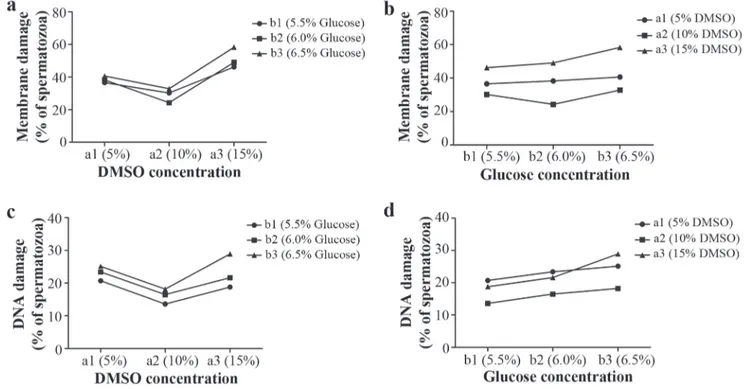
The results indicate that the membrane damage (D-Me) of bocachico frozen-thawed sperm varied according to DMSO and glucose concentration and that the two cryoprotectants had an interactive effect (P<0.05, ANOVA) (Table 1). On the contrary, there was no interactive effect between the two cryoprotectants upon DNA damage (F-DNA) during the cryopreservation process (P>0.05), however, it was evident that the DMSO as well as glucose concentrations can each generate damage independently (P<0.05) (Table 1).
Upon evaluating the principal effects of the glucose concentration factor (B) within each level of DMSO concentration (A) (Fig. 2a-c) (Table 2), it was found that the percentage of sperm with membrane damage was generated in a similar way by the three glucose concentrations (5.5%, 6%, o 6.5%) within the level of a1: 5% DMSO (P>0.05) (Fig. 2a). These damages were significantly reduced when the glucose level reached a concentration of 6% within the level of a2: 10% DMSO compared with whichever other concentration of glucose among the other levels of DMSO (Fig. 3). Nevertheless, the damages incremented in a highly significant form (P<0.0001) when the glucose reached a concentration of 6.5% within the maximum level of DMSO (15%) (Table 2) compared to the other glucose concentrations within the same level and other levels of DMSO (Fig. 3). Equally, the percentage of sperm with F-DNA (Fig. 2c) was originated in a statistically similar form (P>0.05) to each of the three concentrations of glucose, respectively, within each
Table 1. Analysis of variance for the experimental model in the cryopreservation process. D-Me: Membrane damage; F-DNA: DNA fragmentation. Significance level of 5%.
VARIABLE
“A” factor (DMSO concentration)
“B” factor (Glucose concentration)
A*B (interaction)
d.f. F-Value P-value d.f. F-value P-value d.f. F-value P-value
D-Me 2 157.29 <0.0001 2 18.20 <0.0001 4 4.51 0.0107
glucose concentration, factor (B) (Table 2) (Fig. 2b-d), it was found that within each level of glucose (b1: 5.5%, b2: 6%, and b3: 6.5%), the DMSO, in a concentration of 15%, contributed significantly the larger proportion of bocachico sperm cells with D-Me, especially when the DMSO reached the same concentration within the level b3: 6.5% glucose (Table 2) (Fig. 3). At the same time, the smallest damages were always generated when the DMSO concentration was 10%, except within the first level of glucose (b1), in which case 10% as much as 5% of DMSO generated low values and similar to the sperm with generated D-Me (P>0.05). Moreover, the percentage of sperm with F-DNA is contributed in a similar form by the concentrations of DMSO 15% and 5% (P>0.05) at any given level of glucose, whereas the smallest damages are generated when the DMSO reached, within all levels of glucose, a concentration of 10%, in comparison with DMSO 5% (P<0.05) (Fig. 4).
The cryopreservation significantly increased (P<0.05) the percentage of bocachico sperm cells displaying D-Me (24.3 ± 1.1% to 58.3 ± 2.8%) (Fig. 3) and F-DNA (13.6 ± 1.6% to 28.9 ± 3.3) (Fig. 4), considering that PFS maintained a significantly low percentage of cells with D-Me (2.3 ± 0.5%) and with F-DNA (6.7 ± 1.5%) compared to each treatment of cryopreserved semen.
Similarly, it was found that the smallest D-Me generated in the treatment DMSO 10% + glucose 6% (24.3 ± 1.1%), consecutive of DMSO 10% + glucose 5.5% (30.2 ± 2.2%), without significant difference between them (P>0.05). On the contrary, high percentages of cells with D-Me observed in
Fig. 2. Mean values of the percentage of bocachico Prochilodus magdalenae cryopreserved spermatozoa with (a, b) damaged membrane, and (c, d) DNA fragmented to the “A”: DMSO concentration (5%, 10%, and 15% levels) and “B”: glucose concentration (5.5%, 6.0%, and 6.5% levels) factors (n = 6).
Table 2. Analysis of main effects for “A” factor: DMSO concentration and “B” factor: glucose concentration (Reciprocal analysis of the levels of a factor within each specific level of the other). D-Me: Membrane damage; F-DNA: DNA fragmentation; A: DMSO concentration; B: Glucose concentration; a1: 5% DMSO; a2: 10% DMSO; a3: 15% DMSO; b1: 5.5% glucose; b2: 6% glucose; b3: 6.5% glucose. Significance level of 5%.
Variable Source of variation d.f. F-Value P-Value
D-Me
B within a1 2 1.81 0.1930
B within a2 2 8.25 0.0029
B within a3 2 17.16 <0.0001
A within b1 2 27.70 <0.0001
A within b2 2 65.84 <0.0001
A within b3 2 72.76 <0.0001
F-DNA
B within a1 2 2.90 0.0807
B within a2 2 3.08 0.0707
B within a3 2 15.88 0.0001
A within b1 2 7.79 0.0370
A within b2 2 7.46 0.0044
A within b3 2 17.32 <0.0001
level of DMSO (a1: 5% and a2: 10%) (Table 2). Nevertheless, these DNA damages grew significantly when the glucose reached a concentration of 6.5% among the level of a3: 15% DMSO (P=0.0001).
treatments such as DMSO 15% + glucose 6.5% (58.3 ± 2.8%), which was significantly different from the other treatments (P<0.05). Moreover, it was found that the treatment in which the highest rate of F-DNA was DMSO 15% + glucose 6.5% (28.9 ± 3.3%) (Fig. 5), with no significant difference between the treatments of DMSO 5% + glucose 6% or 6.5% (23.4 ± 3.4% and 25.1 ± 0.9%, respectively). Instead, the lowest rate of cells with DNA damage was found in the treatment DMSO 10% + glucose 5.5% (13.6 ± 1.6%) with no significant differentiation from the treatments of DMSO 10% + glucose 6% or 6.5% (P>0.05).
Correlation between F-DNA and D-Me
A positive correlation was determined (r = 0.7761) highly significant (P<0.0001) between F-DNA and D-Me in the cryopreservation process (Fig. 6).
Discussion
Some authors mention three possible factors which may cause DNA damage sperm during cryopreservation: i) direct damage of the sperm genome by the possible development of intracellular ice, which can act similarly to gamma radiation upon the DNA fragmentation (Miskolczi et al., 2005), ii) direct damage of the sperm genome by a type of oxygen reactant released by the dead cells or damaged during the freezing or thawing processes (Labbé et al., 2001) and, iii) induction of apoptotic processes on part of the cryopreservation (Cabrita et al., 2010). The factors (i) and (iii) possibly also involved in the damages to the sperm’s plasmatic membrane.
DNA fragmentation during cryopreservation and its relationship with membrane damage
In the present work, DMSO 5% and DMSO 15% produce the largest percentage of cells with DNA fragmentation, or in
other words, the extreme concentrations of cryoprotectant. Considering this, it is possible that in the case of the cells with DMSO 5%, the appearance of the cryoprotectant to the interior of the cell was minimal due in part to the low concentration gradient (Bolsover et al., 2004), not being sufficiently great enough to spread a high quantity into the interior. Consequently, it is possible that the high levels of F-DNA have generated due to the vulnerability that originated the low presence of the cryoprotectant within the interior of the cell; vulnerability to the supposed formation of free radicals or to the formation of ice crystals (when not reduced the point of freezing). During the cryopreservation of european sea bass sperm cells Dicentrarchuslabrax, when the cryoprotectant (DMSO 10%) was not added, the DNA fragmentation was duplicated in relation to treatments where DMSO was added (Zilli et al., 2003). Christensen & Tiersch (1997) also note that when the cryopreservation of rainbow trout sperm cells Oncorhynchus mykiss was carried out with only the extender (absent of the crioprotectant), almost 99% of the cells received damage.
Exposing DNA to oxidized stress originated more than 20 different types of base damage, producing oxidation and breaking of the same nitrogen rings (Slupphaug et al., 2003). The reactive oxygen species that are endogenously produced can attack lipids, proteins and nucleic acids simultaneously in the live cell. This way, in the nuclear and mitochondrial DNA, 8-hydroxydesoxyguanosine (8-OHdG), an oxidized DNA nucleoside, is the metabolite most frequently detected and studied in DNA lesions (Wu et al., 2004). In human sperm, the cryopreservation effect upon DNA fragmentation was evaluated, finding a significant increment in the 8-OHdG concentration of post-thaw semen compared with the same semen pre-freezing, which correlated significantly with the increase of DNA fragmentation (Thomson et al., 2009).
Although this metabolite has not been measured in the
Fig. 4. Effect of the interaction treatments between DMSO (5%, 10%, 15% v/v) and glucose concentration (5.5%, 6%, 10% w/v) on DNA fragmentation following cryopreservation of sperm from bocachico Prochilodus magdalenae. Between 400 and 500 cores for each sample within each treatment were recorded. Values are expressed as mean ± SD, (n = 6). Mean with different letters differ significantly. Significance level of 5%.
cryopreservation of fish, Ciereszko et al. (2005) demonstrate that even refrigeration at low temperatures induces DNA fragmentation sea lamprey sperm Petromyzon marinus which in turn leads to the conclusion that such damages can be originated by the DNA oxidation. Similar fragmentation results originated through oxidation of nucleic acids can be found in rainbow trout sperm (Dietrich et al., 2005).
In the case of a high DNA fragmentation in the treatment DMSO 15% + glucose 6.5%, one can predict that it could have been caused by the high osmotic force of the cryoprotectant (accentuated by the dehydration exerted by the glucose) originating high toxicities, which could have resulted in cellular infeasibility, and in turn inducing the processes of cellular death (apoptosis), consequently presenting the maximum grade of DNA damage. This assertion is supported when it is observed that in the same treatment the D-Me rises to almost 60%, accompanied by a significant correlation with F-DNA, suggesting a common origin. In fact, authors such as Khan et al.(2009), found that the apoptosis increases significantly after cryopreservation, and that this variable is positively correlated with the membrane damages, both at a significant rate after cryopreservation.
Apoptotic death can be triggered by intra or extracellular signals. Extracellular signals can be physical (temperature, radiation) and chemical (pharmaceutical, toxic) (Thompson, 1995). Among the principle changes made in the cell that later receive the apoptosis signal are the increase in the species of oxygen reactions (Kroemer et al., 1997), the activation of caspase enzymes, and the activation of nucleases called CAD (Caspase Activated Dnase), Dnases in charge of fragmenting the DNA (Enari, 1998). The caspases complete functions such as the inactivation of proteins that participate in DNA repair (Kothakota, 1997) and the destruction of the nuclear membrane (Goulet et al.,1998; Neamati et al.,1995).
In studies regarding the cryopreservation of human sperm, it was determined that the increase of concentration of positive caspases during this event was closely correlated with the increase of DNA fragmentation, and also, the DNA damages and the positive caspase concentration were much higher in semen post-thaw than in fresh semen (Thomson et al., 2009). The appearance of apoptotic characteristics was also evident
during the cryopreservation of bull semen (Martín et al.,2007). In paddlefish Polyodon spathula, three values of sucrose based osmolarity were tested(50mM, 75mM, and 100mM) in combination with three levels of methanol, finding that the highest rate of sperm DNA fragmentation was present in the treatments with the minimum and maximum tested concentration of methanol, and the higher tested concentration of sucrose (Li et al., 2008). DNA damage (by single- or double-strand breaks: comet assay) after sperm cryopreservation has also been confirmed in other marine and freshwater fishes as goldfish (Nathanailides et al., 2011), trout (Cabrita et al., 2005), sea bream (Cabrita et al., 2005) and loach (Kopeika et al., 2004).
Membrane damage during cryopreservation
Regarding membrane damages, they could have originated presumably due to the excessive (toxic) or deficient concentration of cryoprotectant in the interior of the cell (formation of ice crystals). The membrane damages show the highest values when the cryoprotectant reaches its maximum concentration (15%) or the minimum concentration (5%). With respect to this, it is possible to think that the percentage of bocachico sperm cells possesses both a maximum and minimum tolerance level to the cryoprotectant. It is possible to mention that the appearance of membrane damage in an identical percentage of cells beneath the same concentration of DMSO (5%) in great measure is due to the fact that the concentration of cryoprotectant could have been deficient in order to protect cells against nucleation with DMSO (5%) (Drain-Bennet & White, 1977; Drokin et al., 1998). In other words, it do not
Fig. 5. Sperm from bocachico Prochilodus magdalenae tested for DNA fragmentation (post-thawing), 40X. Interaction treatment between DMSO 15% and glucose 6.5%. (a): Sperm with fragmented DNA, (b): Sperm with non-fragmented DNA
decreased the freezing points (Kopeika & Kopeika, 2008), that permits the formation of ice with greater efficiency causes membrane damages during freezing or thawing (Chao & Liao, 2001). In a similar case, during the cryopreservation of striped bass Morone saxatilis, it was detected by a scanning electron microscope that with a minimum concentration of DMSO (2.5%), large damages in the plasmatic membrane occurred, suggesting that the damages were due to the low or nonexistent protection that the sperm received from the cryoprotectant, associating it with the formation of intracellular ice crystals (He & Woods, 2004).
It is clear that a high concentration of DMSO (e.g. 15%) is capable of producing greater membrane damage than DMSO 10% and 5%. In striped bass, when three levels of DMSO (2.5%, 5%, 10%) were tested, it was found that the greatest damage to the plasmatic membrane was generated under the maximum concentration, being statistically different (P<0.05) from the mean concentration (DMSO 5%, resulting in the least membrane damage) (He & Woods, 2004). In this respect, Kopeika & Kopeika (2008) signal that the movement rate of water through the cellular membrane is proportional to the difference in the concentration of the solution (e.g. DMSO and glucose) in both sides of the membrane. This, in turn adds that during the cryopreservation process the water abandons the cell and during thawing it is necessary to reverse this process. With this in mind, it would be easy to suggest that at the DMSO treatments of 15%, this concentration was too high for the bocachico sperm and, possibly due to its great osmotic force, the DMSO triggered rapid displacement of intracellular water and quickly spread throughout the membrane towards the interior of the cell replacing it, occurring again during thawing. It is probable that the membrane could not endure it, generating damages in two possible ways: 1) rupture of the membrane during freezing or thawing through the processes of dehydration or rehydration, whose excessive water friction when transporting through the bilayers exceeds the membrane’s diffusion capacity (Muldrew & McGann, 1990), 2) formation of pores in bilayers of palmitoyl-phosphatidylcholine, provoked by DMSO molecules (Notman et al., 2006).
Considering Kopeika & Kopeika (2008), an increase in external osmolarity also could have driven a larger contraction of the membrane or a greater dehydration, which could have resulted in lesions of the membrane in both cases. It is also possible that the concentration of glucose affects the membrane through changes in osmotic pressure, considering that the damages increased significantly when reaching levels of 6% (360 mOsm kg-1)
to a more hyperosmotic concentration of 6.5% (385 mOsm kg-1) respecting the seminal plasma of the species: 250
mOsm kg-1 (Martínez et al., 2011).
The results of the present study permit the conclusion that cryopreservation originates significant damage to the DNA and plasmatic membrane of sperm cells of bocachico whose magnitude will depend on the variation of the
concentration of the external cryoprotectant (glucose) as well as the internal (DMSO) and their interactions. These damages are lesser with the use of DMSO concentrations at 10%, in combination with glucose at concentrations of 5.5% or 6% utilizing 0.5 mL straws and under freezing and thawing conditions used in the present study.
The method of differential eosin-nigrosin tinting could suggest an indirect method of estimation about the genetic quality of the sperm of this species, above all for practical effects of rural producers and fishermen.
Acknowledgements
We thank Liliana Cardona, Víctor Arroyo and Jhonatan Vergara for technical support during this experiment and Alexis Rhyner for her translation of this manuscript. We also express gratitude to the infrastructure of Fishculture Research Center, University of Córdoba. This work was funded by the Research Division of the Universidad Nacional de Colombia, Sede Medellín (DIME) by source project: 20101007633 and the Ministry of Agriculture and Rural Development in Colombia through the project code: MADR-2007U7723-401.
Literature Cited
Atencio, G. V. J. 2001. Producción de alevinos de especies nativas. Revista de Medicina Veterinaria y Zootecnia-Córdoba, 6: 9-14. Bobe, J. & C. Labbé. 2010. Egg and sperm quality in fish. General
and Comparative Endocrinology, 165: 535-48.
Bolsover, R. S., J. S. Hyams, E. A. Shephard, H. A. White & C. G. Wiedemann. 2004. Cell Biology. New York, Wiley & Sons, 531p. Cabrita, E., C. Sarasquete, S. Martínez-Páramo, V. Robles, J. Beirao, S. Pérez-Cerezales & M. P. Herráez. 2010. Cryopreservation of sh sperm: applications and perspectives. Journal of Applied Ichthyology, 26: 623-635.
Cabrita E., V. Robles, L. Rebordinos, C. Sarasquete & M. P. Herráez. 2005. Evaluation of DNA damage in rainbow trout (Oncorhynchus mykiss) and gilthead sea bream (Sparus aurata) cryopreserved sperm. Cryobiology, 50: 144-153.
Christensen, J. M. & T. R. Tiersch. 1997. Cryopreservation of channel catfish spermatozoa: Effect of cryoprotectants, straw size, and formulation extender. Theriogenology, 47: 639-645. Ciereszko, A., T. D. Wolfe & K. Dabrowski. 2005. Analysis of
DNA damage in sea lamprey (Petromyzon marinus) spermatozoa by UV, hydrogen peroxide, and the toxicant bisazir. Aquatic Toxicology, 73: 128-38.
Cruz-Casallas, P. E., S. C. Pardo-Carrasco, J. A. Arias-Castellanos, P. E. Lombo-Castellanos, D. A. Lombo-Rodríguez & J. E. Pardo-Mariño. 2004. Cryopreservation of yamú Brycon siebenthalae milt. Journal of World Aquaculture Society, 35: 529-35.
Cruz-Casallas, P. E., V. M. Medina-Robles & Y. M. Velasco-Santamaría. 2006. Protocolo para la crioconservación de semen de yamú (Brycon amazonicus Spix and Agassiz 1829). Revista Colombiana de Ciencias Pecuarias, 19: 146-151.
Carolsfeld, J., H. P. Godinho, E. Zaniboni-Filho & B. J. Harvey. 2003. Cryopreservation of sperm in Brazilian migratory fish conservation. Journal of Fish Biology, 63: 472-489.
www.cci.org.co/cci/cci_x/datos/BoletinesIncoder/Publicaciones/ Informecompleto2009.pdf (Accessed 26 October, 2010) Chao, N. H., H. P. Chen & I. C. Liao. 1975. Study on cryogenic
preservation of grey mullet sperm. Aquaculture, 5: 389-406. Chao, N. H. & I. C. Liao. 2001. Cryopreservation on finfish and
shellfish gametes and embryos. Aquaculture, 197:161-189. Drain-Bennet, A. & I. G. White. 1977. Influence of the cholesterol
content of mammalian spermatozoa on susceptibility to cold shock. Journal of Cryobiology, 24: 466-470.
Dietrich, G. J., A. Szpyrka, M. Wajtczak, S. Dobosz, K. Goryczko, L. Zakowski & A. Ciereszko. 2005. Effects of UV irradiation and hydrogen peroxide on DNA fragmentation, motility, and fertilizing ability of rainbow truot (Oncorhynchus mykiss) spermatozoa. Theriogenology, 64: 1809-1822.
Drokin, S., H. Stein & H. Bartscherer. 1998. Effect of cryopreservation on the fine structure of spermatozoa of rainbow trout (Oncorhynchus mykiss) and Brown Trout (Salmo trutta F. Fario). Journal of Cryobiology, 37: 263-270.
Enari, M. 1998. A caspase-activated DNase that degrades DNA during apoptosis, and its inhibitor ICAD. Nature, 8: 365-372. Fernández, J. L., L. Muriel, M. T. Rivero, V. Goyanes, R. Vázquez & J. G. Alvarez. 2003. The sperm chromatin dispersion test: a simple method for the determination of sperm DNA fragmentation. International Journal of Andrology, 24: 59-66. Flesch, F. M. & B. M. Gadella. 2000. Dynamic of the mammalian
sperm plasma membrane in the process of fertilization. Biochemistry and Biophysics Acta, 1469: 197-235.
Goulet, D. I., J. C. Courvalin & B. Buendía. 1998. LBR, a chromatin and lamin binding protein from the inner nuclear membrane, is proteolyzed at the late stages of apoptosis. Journal of Cell Science, 111: 1441-1451.
Gwo, J. C., C. Y. Wu, W. S. Chang & H. Y. Cheng. 2003. Evaluation of damage in Pacific oyster (Crassostrea gigas) spermatozoa before and after cryopreservation using comet assay. Cryo Lett 24: 171-180.
He, S. & L. C. Woods. 2004. Changes in motility, ultrastructure, and fertilization capacity of striped bass Morone saxatilis spermatozoa following cryopreservation. Aquaculture, 236: 677- 686. Khan, D. R., N. Ahmad, M. Anzar & A. A. Channa. 2009. Apoptosis
in fresh and cryopreserved buffalo sperm. Theriogenology, 71: 872-876.
Kopeika, E. & J. Kopeika. 2008. Variability of sperm quality after cryopreservation in fish. Pp. 347-396. In: Alavi, S. M. H., J. J. Cosson, K. Coward, G. Rafiee (Eds.). Fish Spermatology. Alpha Science Inc, Oxford, 465p.
Kopeika, J., E. Kopeika, T. Zhang, D. M. Rawson & W. V. Holt. 2004. Effect of DNA repair inhibitor (3-aminobenzamide) on genetic stability of loach (Misgurnus fossilis) embryos derived from cryopreserved sperm. Theriogenology, 61: 1661-1673. Kothakota, S. 1997. Caspase-3-generated fragment of gelsolin: effector
of morphological change in apoptosis. Science, 278: 294-298. Kroemer, G., N. Zamzami & S. A. Susin. 1997. Mithochondrial
control of apoptosis. Immunology Today, 18: 44-51. Labbé, C., A. Martoriati, A. Devaux & G. Maisse. 2001. Effects of
sperm cryopreservation on sperm DNA stability and progeny development in rainbow trout. Molecular Reproduction and Development, 60: 397-404.
Li, P., Q. Wei & L. Liu. 2008. DNA integrity of Polyodon spathula
cryopreserved sperm. Journal of Applied Ichthyology, 24:121-125.
Lopez-Fernandez, C., M. J. G. Gage, F. Arroyo, A. Gosalbez, A. M. Larran, J. L. Fernandez & J. Gosalvez. 2009. Rapid rates of
sperm DNA damage after activation in tench (Tinca tinca: Teleostei, Cyprinidae) measured using a sperm chromatin dispersion test. Reproduction, 138: 257-266.
Martin, G., N. Cagnon, O. Sabido, B. Sio, G. Grizard, P. Durand & R. Levy. 2007. Kinetics of occurrence of some feature of apoptosis during the cryopreservation process of bovine spermatozoa. Human Reproduction, 22: 380-388.
Martínez, J. G., V. J. Atencio-García & S. C. Pardo-Carrasco. 2011. Effect of glucose concentration on sperm motility activation in bocachico Prochilodus magdalenae (Pisces, Characiformes). Revista de Medicina Veterinaria y Zootecnia-Córdoba, 16: 2554-2563.
Medina-Robles, V. M., Y. M. Velasco-Santamaría & P. E. Cruz-Casallas. 2007. Efecto del volumen de empaque sobre la tasa de congelación-descongelación y la fertilidad de semen crioconservado de yamú (Brycon amazonicus). Archivos de Medicina Veterinaria, 39: 229-237.
Miskolczi, E., S. Mihalffy, V. Pata-Kiné, B. Urbanyi & A. Horváth. 2005. Examination of larval malformation in African catfish (Clarias gariepinus) following fertilization with cryoconserved sperm. Aquaculture, 247: 119-125.
Mojica, J. I., C. Castellanos, S. Usma & R. Álvarez. 2002. Libro rojo de peces dulceacuícolas de Colombia. La serie Libros Rojos de Especies Amenazadas de Colombia. Instituto de Ciencias Naturales - Universidad Nacional de Colombia, Ministerio del Medio Ambiente, Bogotá, 288p.
Muldrew, K. & L. E. McGann. 1990. Mechanisms of intracellular ice formation. Biophysics Journal, 57: 525-532.
Nathanailides, C., T. Chanzaropoulos, A. Barbouti, C. Perdikaris & T. Zhang. 2011. DNA fragmentation, linear velocity and fertilizing ability of reactivated cryopreserved goldfish sperm using different cryoprotectants. Biotechnology, 10: 514-520. Neamati, N., A. Fernández, S. Wright, J. Kiefer & D. J. McConkey.
1995. Degradation of laminin B1 precedes oligonucleosomal DNA fragmentation in apoptotic thymocytes and isolated thymocyte nuclei. Journal of Immunology, 154: 3788-3795. Notman, R., M. Noro, B. O’Malley & J. Anwar. 2006. Molecular
Basis for Dimethylsulfoxide (DMSO) action on lipid membranes. Journal of the American Chemical Society, 128: 13982-13983.
Pan, J., S. Ding, J. Ge, W. Yan, C. Hao, J. Chen & Y. Huang. 2008. Development of cryopreservation for maintaining yellow catfish
Pelteobagrus fulvidraco sperm. Aquaculture, 279: 173-176. Pérez-Cerezales, S., S. Martínez-Páramo, J. Beirão & M. P. Herráez.
2010. Evaluation of DNA damage as a quality marker for rainbow trout sperm cryopreservation and use of LDL as cryoprotectant. Theriogenology, 74: 282-289.
Shen, H. M. & C. N. Ong. 2000. Detection of oxidative DNA damage in human sperm and its association with sperm function and male infertility. Free Radical Biology and Medicine 28: 529-536.
Singht, N. P., M. T. McCoy, R. R. Tice & E. L. Schneider. 1988. A simple technique for quantitation of low-levels of DNA damage in individual cells. Experimental Cell Research, 175: 184-191. Slupphaug, G., B. Kavli & H. E. Krokan. 2003. The interacting
pathways for prevention and repair of oxidative DNA damage. Mutation Research, 531: 231-251.
Thompson, C. B. 1995. Apoptosis in the pathogenesis and treatment of disease. Science, 267: 1456-1462.
oxidative stress rather than apoptosis. Human Reproduction, 24: 2061-2070.
Viveiros, A. T. M., L. H. Orfão, A. N. Maria & I. B. Allaman. 2009. A simple, inexpensive and successful freezing method for curimba Prochilodus lineatus (Characiformes) semen. Animal Reproduction Science, 112: 293-300.
Watson, P. F. & W. V. Holt. 2001. Cryobanking the Genetic Resource: Wildlife Conservation for the Future? Taylor and Francis, London, 463p.
Wildt, D. E. & C. Wemmer. 1999. Sex and wildlife: the role of reproductive science in conservation. Biodiversity Conservation, 8: 965-976.
Wu, L. L., C. C. Chiou, P. Y. Chang & J. T. Wu. 2004. Urinary 8-OHdG: a marker of oxidative stress to DNA and risk factor for cancer, atherosclerosis and diabetics. Clinica Chimica Acta, 339: 1-2.
Zilli, L., R. Schiavone, V. Zonno, C. Storelli & S. Vilella. 2003. Evaluation of DNA damage in Dicentrarchus labrax sperm following cryopreservation. Journal of Cryobiology, 47: 227-235.