ROLFSII (SACC.)
Sikandar Hayat1*, Christos Christias2
1
Post-Doctoral Fellow Alberta Research Council, Vegreville, Alberta, Canada; 2Professor of Microbiology University of Patras,
Greece.
Submitted: February 03, 2009; Returned to authors for corrections: April 26, 2009; Approved: August 23, 2009.
ABSTRACT
Sclerotium rolfsii (Sacc.) is a serious plant pathogenic fungus and lacks perfect (basidial) stage in
production. Protoplast fusion technology was employed to reconstruct fusants from this fungus. Two
strains designated as A and R were used. Maximum protoplast yields of 3.8x10⁵ /g mycelia and 2.8x10⁵ /g
mycelia were formed in strains A and R respectively. Osmotic stabilizer sucrose 1M gave maximum yield.
Lysing enzyme at the rate of 15mg/ml was found best for yield. Fusion of protoplasts from strains A and R
was carried out in fusion media containing PEG 4000 30% (w/v) with 0.2mM CaCl2. Four fusants F1, F2,
F3 and F4 were recovered. Morphological, physiological and pathogenic characters of fusants were
compared with parent strains on carrots, beans and tomato.
Key words: Protoplast, fusion, Sclerotium rolfsii, Pathogenicity and osmotic stabilizer.
INTRODUCTION
Protoplast fusion represents an approach to gene transfer
in microbial organisms including bacteria and fungi. Protoplast
Fusion Technology (PFT) has been used to bypass many
natural barriers to cross-breeding in fungi, as reported in
numerous species of Deuteromycetes, where no sexual cycle
exists (14, 17, 22, 29). Protoplast fusion allows investigators to
bypass mating type and incompatibility group barriers (7, 9) to
study mitochondrial genetics, (8, 21). Hyun-Choi et al (16)
performed inter-generic protoplast fusion in Saccharomycopsis
fibuligera and Saccharomyces cerevisiae. Salamiah et al (27)
applied protoplast fusion system in non-pathogenic Alternaria
alternata and a tomato pathotype Alternaria alternata using
electrofusion and PEG and studied pathogenicity in hybrid
strains. Couteaudier et al (6) fused protoplasts of two different
strains of entomopathogenic fungus Beauveria bassiana to get
fusants for more effective control against Ostrinia nubilalis.
Sclerotium rolfsii is a serious plant pathogen fungus. Its
perfect stage is very rare in nature. In addition the complete
absence of asexual spores in this fungus results inability of use
of traditional hybridization techniques for the development of
new strains. In view of serious economical losses caused by
this fungus, protoplast fusion technology being inexpensive
and simple technology was used in this study. This is the first
report of construction of new strains from the inter-strains
protoplast fusion from Sclerotium rolfsii.
*Corresponding Author. Mailing address: Post-Doctoral Fellow Alberta Research Council, Vegreville, Alberta, Canada.; Tel.: 780-632-8225.; E-mail:
MATERIALS AND METHODS
Materials
Two strains of Sclerotium rolfsii designated as A and R
were used through out the study. Strain A was isolated from
Myoporum sp. (5). Strain R a cotton isolate was obtained from
The National Institute of Biology, Athens, Greece. Osmotic
stabilizers including Sucrose, Sodium Chloride (NaCl),
Potassium Chloride (KCl), Ammonium Chloride (NH4Cl) were
purchased from Merck Germany. Magnesium sulphate
(MgSO4) was purchased from Reidel-de Hach AG Germany.
Lysing enzyme (Trichoderma harzianum), Cellulase
(Aspergillus niger), Driselease (Basidiomycetes), Chitinase
(Serretia marcescens) and Beta-Glucuronidase (Helix pomatia)
were purchased from Sigma Chemicals Co. (St Louis, USA).
Media and growth conditions
Potato Dextrose Agar (PDA) media was prepared boiling
200g of potato and adding 20g of glucose and 15g of agar in
total volume of 1000ml. Media was autoclaved at 121°C for 15
minutes and used for cultivation of fungal culture.
Preparation of semi-permeable cellophane membranes Semi-permeable cellophane membranes (9cm diameter)
were used to harvest clean and pure mycelia of the fungus, free
of any traces of agar from the Petri-dishes. Sterile cellophane
membranes were transferred on Petri-dishes and were
inoculated with 5mm mycelial disc of strain A and R and were
incubated at room temperature for 4-5 days.
Pre-treatment of mycelia and preparation of digestion mixtures
One gram of three days old mycelia from the both strains
was incubated in 20ml of NaCl 0.9M pH 5.8 for 20 minutes for
pre-treatment. Digestion mixtures were prepared using
different osmotic stabilizers including sucrose 1M, NaCl 1M,
KCl 0.9M, NH4Cl 1M and MgSO4 (0.4M, 0.6M, 0.8M and
1M) at pH 5.8-6.0 containing different lytic enzymes. Lysing
enzyme was used at the rate of 5mg, 10mg and 15mg/ml,
Cellulase 15mg/ml, Driselease 15mg/ml, Chitinase 1mg/ml and
Beta-glucuronidase 0.06mg/ml in the digestion mixture. The
digestion mixtures were autoclaved at 121C for 15 minutes
prior to the addition of lysing enzymes.
Protoplast formation
One gram of fresh 3 days old pre-treated mycelia was
mixed in digestion mixture in a total volume of 10ml using
each osmotic stabilizer and lysing enzyme in a 50 ml
Erlenmeyer flask in triplicates and was incubated in a rotary
shaker incubator at 75 rpm at 30C for 24 hours. Samples were
taken at 6, 12 and 24 hours to observe under the microscope for
cell wall digestion and protoplast formation.
Protoplast purification
The reaction mixture containing protoplasts and cell wall
debris was harvested after 24 hours and filtered through
sterilised cheese cloth to remove undigested materials under
aseptic conditions. The filtrates were centrifuged at 3000 rpm
for 15 minutes at 15C. The protoplasts were collected as pellet.
The protoplasts were washed in the same osmotic stabilizer to
remove lysing enzyme traces. The purified protoplasts were
re-suspended in the osmotic buffer. The size and number of
protoplasts was determined per ml per gram of fresh weight of
mycelia using haemocytometer. The average size of protoplasts
was expressed in micrometer (µ m).
Regeneration of protoplasts
Regeneration of protoplasts was carried out using 1 ml of
reaction mixture containing (1x10³) protoplasts in both solid
PDA and liquid PD media. The different phases of regeneration
of protoplasts into germ-tubes and hyphal cells were recorded
under the microscope.
Protoplast fusion
Protoplast fusion was performed according to a modified
method of (1, 2). A sample of 0.5 ml of the purified protoplast
suspension (1x10⁵) of strains A and R was mixed and
re-suspended in a fusion medium in a total volume of 1ml. The
fusion medium was prepared by adding poly-ethyl-glycol
(PEG) 4000, 30% (w/v) and 0.2 mM CaCl2 in 1M sucrose. The
fusion mixture was incubated for 30 minutes at 30C. Samples
were taken from fusion mixture and were spread on
Petri-dishes containing PDA regeneration media. The fusants were
picked on the basis of different pattern of mycelial growth
comparing with parent strains A and R.
Stability and growth rate of fusants
The stability of fusants was studied upto three generations
in PDA media. Growth rate of fusants was compared with
parent strains and was expressed in mm.
Study of properties of fusants in green house
A growth chamber experiment was carried out on three
crops namely tomato, carrots and beans to study
morphological, physiological and pathogenic parameters of
fusants comparing with the parent strains A and R.
A 550grams of garden soil was inoculated with homogenized
mycelia (slurry) in plastic bags with air pores. The soil bags
were kept at 25C for 10 days in order for fungal cultures to be
established and proliferate in the soil. Three-four days old
seedlings of each crop were transplanted into soil bags and kept
in green house.
Morphological parameters like plant height (cm), number
of leaves, leaf mid rib, fresh and dry weight of plants were
studied and the data was analysed. Physiological parameters
including chlorophyll fluorescence was measured by plant
efficiency analyzer (PEA) in beans only. Plants infected with
fusants, strains A and R and control were kept in dark
conditions for 1hour before measuring fluorescence. The data
in the form of initial fluorescence (F0), Maximum fluorescence
(FM), Fv variable difference between F0 and FM and ratio of
Fv/FM were recorded on PEA monitor. The ratio Fv/FM value
was used for chlorophyll fluorescence determination. This ratio
has been shown to be proportional to the quantum yield of the
photochemistry (3).
Estimation of chlorophyll a, b and carotenoids
The total chlorophyll amount and individual amounts of
chlorophyll a (Ca), chlorophyll b (Cb) and carotenoids in the
leaf samples were measured according to the method of
Lichtenthaler and Wellburn (20). Leaf samples were
homogenized in 5ml of 80% acetone and then centrifuged at
5000 rpm for 5 minutes. Supernatant was collected and volume
was made upto 5ml with 80% acetone and stored in darkness.
The measurements were taken using spectrophotometer
(Ultraspec 4050), LKB Biochem, England at three
wavelengths, 663, 646 and 470nm. The following formula was
used to calculate chlorophyll a, b and carotenoids. The
individual amount of chlorophyll a, b and carotenoids was
expressed in micrograms (µg) per ml of leaf extract.
Chlorophyll a, 12.21A663-2.81A646 Chlorophyll b 20.13A646-5.03A663 Carotenoids x+c = 1000A470-3.27Ca-104Cb/229
Statistical Analysis
All the data on growth and physiological parameters were
analyzed using one-way ANOVA and LSD test at P=0.05 level
by SPSS 9.0 and means were compared for significance.
RESULTS AND DISCUSSION
Optimization of protoplast yields from Sclerotium rolfsii Effect of osmotic stabilizers on protoplast yields
All osmotic stabilizers were used at 1M concentration
except KCl (0.9M). Strain A produced maximum protoplasts of
3.8x10⁵/gram fresh weight of mycelia in 1M sucrose and strain
R gave maximum protoplast yields of 2.8x10⁵ in the same
stabilizer (1M sucrose) after 24 hours incubation. Among
osmotic stabilizers, 1M sucrose was found to be optimum for
protoplast yields in both strains followed by 1M NaCl (Figure
1)
Effect of different lytic enzymes on protoplast yield
Five different commercial lytic enzymes including lysing
enzyme at the rate of 15mg/ml, cellulase (8.55units), driselease
15mg/ml, chitinase (0.072units) and beta-glucuronidase
except lysing enzyme did not produce protoplasts in both
strains A and R. Lysing enzyme at the concentration rate of
15mg/ml was found optimum for the isolation of protoplasts in
strains A and R (Figure 2).
0.0 6.0x10⁴ 1.2x10⁵ 1.8x10⁵ 2.4x10⁵ 3.0x10⁵ 3.6x10⁵
4.2x10⁵
A
R
MgSO4 1M KCl 0.9M
NH4Cl 1M NaCl 1M
Sucrose 1M
P
ro
to
p
la
s
t
y
ie
ld
s
/g
Figure 1. Effect of different osmotic stabilizers on protoplast yields of strains A and R of Sclerotium rolfsii, lysing enzyme 15mg/ml at pH 5.8 1g mycelia and incubation period 24 hours
5 10 15
0.0 7.0x10⁹ 1.4x10¹⁰ 2.1x10¹⁰ 2.8x10¹⁰ 3.5x10¹⁰ 4.2x10¹⁰
A R
P
ro
to
p
la
s
t
y
ie
ld
s
/g
Size of protoplast
The size of 150 protoplasts of both strains A and R was
measured randomly and the average size were found 15µ m.
Regeneration of protoplasts
The regeneration of protoplasts of both strains A and R
was started within 15 minutes after incubation in solid and
liquid media. The protoplasts of both strains formed bud like
structures, which developed into germ tubes. These germ tubes
later formed hyphal cells in 2-3 days. The protoplasts of both
strains formed sclerotia like the parent strains (Figure 3)
Fusion of protoplasts of strains A and R
Protoplast fusion experiment was conducted to recover
possible fusants. The results indicated that the fusion frequency
was very low as only four (4) fusants were recovered. It was
found under the microscope during the fusion, that both fusing
protoplasts after breaking down their cell membranes merged
their cytoplasmic contents (Figure 4). The four fusants
recovered were designated as F1, F2, F3 and F4. All four
fusants were found different from the parent strains and with
each other in morphology and sclerotia formation. F1 showed
thin and aerial growth of mycelia with whitish color. Few
sclerotia aggregates were also formed. F2 showed different
morphology from parent strains and other fusants. The mycelia
were thick and it did not produce sclerotia which was found a
rare character in this fungus. F3 exhibited different pattern of
sclerotial formation, it formed aggregates of smaller size
sclerotia along the periphery of the Petri-dishes. F4 derived
characteristics from both strains A and R. It formed sclerotia
small in size like strain A along the edge of Petri-dish unlike
strain A (Figure 5).
Stability experiments showed that out of 4 fusants, 3 were
found stable after three generations, while one fusant F3 lost its
acquired characters like mycelial morphology and sclerotial
formation.
A B C
D E
Figure 3. A. Purified protoplasts in media. Regeneration of protoplasts of
Sclerotium rolfsii. B. Onset of a
regenerating protoplast forming a bud
like structure. C. Germ tube formation
from the bud. D. Formation of a hypha
from germ tube. E. Elongation of hypha,
A B C
Figure 4. Fusion of protoplast of strains A and R of Sclerotium rolfsii, A. Close contact of two fusing protoplasts, B. rupture of membranes of fusing protoplasts, C. Complete rupture of membranes of fusing protoplasts and merging of cytoplasmic contents
(Magnification, 400 X)
Figure 5. Parent strains A and R of Sclerotium rolfsii and fusants F1, F2, F3 and F4. Strain A has thin mycelia, produces large number of small-sized sclerotia. The sclerotia are present all over the Petri dish. Strain R shows thick mycelia with large sized
sclerotia few in numbers. F1 shows aerial growth pattern of mycelia and whitish in color. The sclerotia are produced in irregular
manner in the Petri dish. F2 has thick mycelia with complete absence of sclerotia. F3 exhibits aggregation of small sized sclerotia,
black in color. The aggregations of sclerotia are present at the periphery of the Petri dish. F4 shows small size sclerotia in chains
present at the periphery of the Petri dish.
Assessment of pathogenicity of fusants and parent strains The pathogenic characteristics like plant wilting and
mortality percentage were investigated in fusants as compared
to the parent strains A and R on host crops like tomato, carrots
and beans. The growth parameters like plant height, number of
leaves, leaf mid rib length, fresh and dry weight of plants and
physiological parameters like chlorophyll a, b, carotenoids and
chlorophyll fluorescence was also determined.
Mortality %
The mortality was determined as percentage of dead
plants. The mortality rate was 0 in tomato plants infected with
fusants as well as parent strains A and R. Leaf discoloration
was found in plants infected with F1 and A strain. Mortality
rate of 40% was noted in two months old carrot plants infected
with F1 and 20% in F4.No mortality was reported in plants
showed that F1 was found to be the most virulent as compared
to other fusants and parent strains.
Highest mortality of 78% was noted in two months old
bean plants infected with strain A as compared to F1 the most
virulent fusant as well as strain R. The results indicated that
among host crops, beans were highly susceptible to pathogenic
strains A and R.
Growth parameters
The data on morphological parameters including plant
height, fresh and dry weight of plants in tomato plants infected
with fusants and parent strains A and R showed significant
results comparing fusants to the parent strains R and control in
plant height, fresh and dry weight of plants. Two months old
carrot plants infected with fusants and parent strains, plants
infected with F1 showed significant results in all morphology
parameters comparing within fusants and to parent strains and
control. In case of bean, plants infected with a parent strain A
had significant results when compared to fusants, parent strain
R and control.
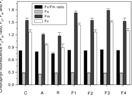
Physiological parameters
Chlorophyll fluorescence was measured as an indicator of
plant efficiency infected with pathogenic strains only in beans.
The Fv/FM ratio was measured and a maximum ratio of 0.8426
was noted in plants infected with F3 and results were found
non-significant when compared within fusants and with parent
strains (Figure 6).
0.0 0.3 0.6 0.9 1.2 1.5 1.8
F4 F3
F2 F1
R
A C
C
h
lo
ro
p
h
y
ll
fl
o
u
re
s
c
e
n
c
e
F
V
/FM
r
a
ti
o
,
FO
,
FM
a
n
d
FV
Fv/Fm ratio Fo Fm Fv
Figure 6. Chlorophyll fluorescence in beans leaf (two months old plants) infected with parent strains A and R and fusants F1, F2, F3 and F4 and control. The vertical bars represent standard errors
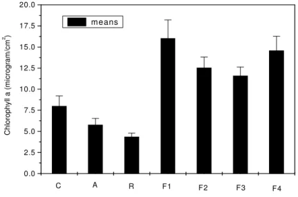
Estimation of chlorophyll a, b and carotenoids
Maximum chlorophyll (Ca) of 16.02 µg/cm2 was
determined in leaves of plants infected with F1 and minimum
amount of 4.36 µg/cm2 was found in the leaves infected with
the parent strain R. Highest amount of chlorophyll (Cb) of 3.55
µg/cm2 was recorded in the leaves of plants infected (Figures 7
and 8) with F1and maximum carotenoids content of 1.01
µg/cm2 were noted in the leaves of plants infected with F4 and
were found significantly different from the 0.293 µg/cm2 in the
0.0 2.5 5.0 7.5 10.0 12.5 15.0 17.5 20.0
F4 F3
F2 F1
R A
C
C
h
lo
ro
p
h
y
ll
a
(
m
ic
ro
g
ra
m
/c
m
2 )
means
Figure 7. Chlorophyll a in beans leaf (two months old plants) infected with parent strains A and R and fusants F1, F2, F3 and F4 and control. The vertical bars represent standard errors
Control A R F1 F2 F3 F4
0.0 0.7 1.4 2.1 2.8 3.5 4.2
C
h
lo
ro
p
h
y
ll
b
c
o
n
te
n
ts
(
m
ic
ro
g
ra
m
/c
m
2 )
means
Figure 8. Chlorophyll b in beans leaf (two months old plants) infected with parent strains A and R and fusants F1, F2, F3 and F4 and control. The vertical bars represent standard errors
In this research work, the aim was to isolate protoplasts
from the two morphologically different strains A and R of the
phytopathogenic fungus Sclerotium rolfsii and then to construct
fusants from these protoplasts. There is one report of isolation
of protoplasts from Sclerotium rolfsii by Deshpande et al. (10),
Kelkar et al. (18) and another report by Farina et al. (12) on the
formation and regeneration of protoplasts in Sclerotium rolfsii
ATCC 201126. They reported optimization of different culture
conditions for protoplast formation. Protoplasts were isolated
in 1M sucrose as osmotic stabilizer. The lysing enzyme from
Trichoderma harzianum was found to be the most effective
enzyme in our study with Sclerotium rolfsii. Our results are
supported by Farina et al (12) and Quigley et al. (26), when
they used commercial novozym-234 for the isolation of
protoplasts in Sclerotium rolfsii and Neurospora respectively.
To-date, there has been no rational explanation for the
superiority of one stabilizer over other. Our studies showed
sucrose and NaCl were the most effective stabilizers for
protoplasts yields. Van Diepeningen et al. (11) used 1M
sucrose for the isolation of protoplasts from Aspergillus
nidulans, whereas Farina et al (12) reported 0.6M MgSO4
osmotic stabilizer for protoplast formation. The regeneration of
protoplasts in sucrose solution showed that most of protoplasts
were found viable and our findings of protoplasts viability are
according to Cheng and Belanger (4). They reported 75%
viability of Pseudozyma flocculosa protoplasts regeneration in
0.8M sucrose medium. The size of protoplasts in both strains A
and R was measured 15µm, which was found larger as
compared to the size of protoplasts of Sclerotium rolfsii
reported by Deshpande et al (10). The reason for the different
protoplasts size can be due to the use of different isolates of
Sclerotium rolfsii and use of sucrose as stabilizing agent in
media in our studies. Protoplasts of both strains regenerated to
their normal vegetative cells both in liquid and solid media.
They formed bud like structures and increased its mass led to
the formation of germ tube which eventually gave rise hyphal
structures. The regeneration frequency of protoplasts was not
enough high in solid media. Peberdy (24) reported 0.1-50%
regeneration frequency in fungal protoplasts. Protoplast fusion
technology provides an alternative approach to obtain genetic
recombination in imperfect fungi (Hashiba and Yamada, (15);
Peberdy, (23) and Typas, (30). In Sclerotium rolfsii, there is no
report of intra-specific protoplast fusion so far. Therefore,
protoplast fusion technology was employed to construct fusants
from two distinct strains of this fungus. The results showed
fusion frequency was not enough high as compared to fusion
frequencies in other fungi species. Only four fusants were
recovered and designated as F1, F2, F3 and F4. Kirimura et al.
(19) reported fusion frequency of 3.6% in Aspergillus niger. As
there is no report of protoplast fusion in Sclerotium rolfsii it is
difficult to compare the fusion frequency of Sclerotium rolfsii
with other fungi genus. Ferenczy et al. (13) described PEG as
the most important factor affecting protoplast fusion frequency.
Punja and Sun (25) investigated genetic diversity among
species of Sclerotium rolfsii and reported difficulty in genetic
exchange and proposed construction of genetic markers for
further exploration. Four fusants F1, F2, F3 and F4 were
recovered on the basis of morphology and sclerotial formation
from the strains A and R. Three fusants F1, F2 and F4 were
found stable and F4 was found unstable after three generations
and showed stability percentage of 75%, while Salamiah et al
(28) reported 5.4% in Alternaria alternata. There are only two
reports on the pathogenicity of fusants derived by protoplast
fusion, one by Yang et al. (31) in Rhizoctonia solani and other
by Salamiah et al (28) in Alternaria alternata. In our study on
pathogenicity of fusants on three host crops, tomato, carrots
and beans, growth parameters and physiological parameters
like chlorophyll fluorescence and chlorophyll a, b and
carotenoids contents were studied as indicators. The data
showed F1 was found hyper-virulent among fusants and as
compare to strains A and R. Among host crops, the beans
showed totally opposite behaviour to fusants and showed
growth promotion unlike in other two host crops. This growth
promotion behaviour of beans was also supported by the data
on chlorophyll florescence and chlorophyll contents. This
selective expression of plant growth promoting in one crop and
pathogenic behaviour in another crop might be due to species
specificity or synthesis of growth promoting substances
triggered by certain genes.
In conclusion, during our study protoplast fusants were
constructed successfully using protoplast fusion technology.
The pathogenicity of fusants derived from protoplast fusion of
strains A and R of Sclerotium rolfsii provided value able
information about different host-pathogen relationships.
Further research is required to carry out complete genetic
analysis of fusants and parent strains to correlate physiological
ACKNOWLEDGEMENTS
This study was funded by a grant from (IKY) State
Scholarship Foundation, Athens, Republic of Greece.
REFERENCES
1. Anne, J. (1982). Comparison of penicillins produced by inter-species hybrids from Pencillium chrysogenum. European Journal of Microbial
Biotechnology, 15: 41-46.
2. Anne, J.; Peberdy, J.F. (1975). Conditions for induced fusion of fungal protoplasts in polyethylene glycol solutions. Arch. Microbiol., 105: 201-205.
3. Butler, W.L.; Kitajima, M. (1975). A tripartiate model for chloroplast fluorescence. In: Proceeding of the 3rd International Congress on
Photosynthesis. Edited by Avron, M. 13-24, Elsevier, Amsterdam. 4. Cheng, Y.; Belanger, R.R. (2000). Protoplast preparation and
regeneration from the spores of the bio-control fungus Pseudozyma
flocculosa. FEMS Microbiol. Lett., 190, 287-291.
5. Christias, C. (1975). Specific inhibition of sclerotium formation by 2-mercaptoethanol and related sulfhydryl compounds in Sclerotium rolfsii.
Can. J. Microbiol., 21: 1541-1547.
6. Couteaudier, Y.; Viaud, M.; Riba, G. (1996). Genetic nature, stability and improved virulence of hybrids from protoplast fusion in Beauveria.
Microb. Ecol., 32: 1-10.
7. Croft, J.H. (1985). Protoplast fusion and incompatibility in Aspergillus. In: Fungal protoplasts: Applications in Biochemistry and Genetics J.F. Peberdy and L. Ferenczy, Marcel Dekker, New York, PP.225-240. 8. Croft, J.H.; Dales, R.B.G.; Turner, C.; Earl, A. (1980). The transfer of
mitochondria between species of Aspergillus. In Advances in Protoplasts Research Ferenczy, L and G.L. Farkas, Akademiai Kiado, Budapest, Pergamon Press Oxford, pp. 85-92.
9. Dales, R.B.G.; Croft, J.H. (1977). Protoplast fusion and isolation of heterokaryons and diploids from vegetatively incompatible strains of
Aspergillus nidulans. FEMS Microbiol. Lett., 1: 201-204.
10. Deshpande, M.V.; Balkrishnan, H.; Ranjekar, P.K.; Shanker, V. (1987). Isolation and immobilization of Sclerotium rolfsii protoplasts.
Biotechnol. Lett., Vol.9, (1), 49-52.
11. Dipeningen, A.D. van; Debets, A.J.M.; Hoekstra, R.F. (1998). Intra and inter-specific virus transfer in Aspergilli via protoplast fusion. Fungal
Genetic and Biology, 25, 171-180.
12. Farina, J.I..; Molina, O.E.; Figueroa, L.I.C. (2003). Formation and regeneration of protoplasts in Sclerotium rolfsii ATCC 201126. J. Appl.
Microbiol., Vol.96, (2) 254-262.
13. Ferenczy, L.; Kevei, F.; Szegedi, M.; Franko, A.; Rojik, I. (1976). Factors affecting high frequency fungal protoplast fusion. Experientia, 32: 1156-1158.
14. Fournier, P.; Provost, A.; Bourguignon, C.; Heslot, H. (1977). Recombination after protoplast fusion in the yeast Candida tropicalis.
Arch. Microbiol., 115: 143-149.
15. Hashiba, T.; Yamada, M. (1984). Intra-specific protoplast fusion between auxotrophic mutants of Rhizoctonia solani. Phytopathology, vol. 74, No.4 398-401.
16. Hyun-Choi, S.; Sung, C.; Oh, M.; Kim, C. (1997). Inter-generic protoplast fusion in Saccharomyces fibuligera and Saccharomyces
cerevisiae. J. Ferment. Bioeng., 84: 158-161.
17. Kakar, S.N.; Partridge, R.M.; Magee, P.T. (1983). A genetic analysis of
Candida albicans: Isolation of a wide variety of auxotrophs and
demonstration of linkage and complementation. Genetics, 104: 241-255. 18. Kelkar, H.S.; Shankar, V.; Deshpande, M.V. (1990). Rapid isolation and
regeneration of Sclerotium rolfsii and their potential application for starch hydrolysis. Enzyme Microb. Technol. 12, 510-514.
19. Kirimura, K.; Yanuchi, T.; Usami, S. (1986). Intra-specific protoplast fusion of citric acid producing strains of Aspergillus niger. Journal of
Fermentation Technology, 64: 473-479.
20. Lichtenthaler, H.K.; Wallburn, A.R. (1983). Determination of total carotenoids and chlorophyll a and b of leaf extracts in different solvents.
Biochem. Soc. Trans. 11, 591-592.
21. Maraz, A.; Subik, J. (1981). Transmission and recombination of mitochondrial genes in Saccharomyces cerevisiae after protoplast fusion.
Mol. Gen. Genet., 181: 131-133.
22. Peberdy, J.F. (1980). Protoplast fusion: a tool for genetic manipulation and breeding of industrial microorganisms. Enzyme Microb. Technol., 2: 23-29.
23. Peberdy, J.F. (1989). Presidential address: Fungi without coats- protoplast as tool for mycological research. Mycol. Res., 93: 1-20. 24. Peberdy, J.F. (1991). Fungal protoplasts. In : More gene Manipulation in
Fungi by Bennett, J.W.; Lasure, L.L. pp. 307-318, Academic Press, California.
25. Punja, Z.A.; Sun, L. (2001). Genetic diversity among mycelial compatibility groups of Sclerotium rolfsii (telemoroph Athelia rolfsii)
and Sclerotium delephini. Mycol. Res. 105: 537-546.
26. Quigley, D.R.; Taft, C.S.; Stark, T.; Selitrennikoff, C.P. (1987). Optimal conditions for release of protoplasts of Neurospora crassa using novozm 234. Exp. Mycol., 11, 236-240.
27. Salamiah, A.H.; Fukumassa-nakai, Y.; Otani, H.; Kodama, M. (2000). Genetic analysis of pathogenicity and host-specific toxin production of Alternaria alternate tomato pathotype by protoplast fusion. J. Gen. Plant
Pathol., 67: 7-14.
28. Salamiah, A.H.; Fukumassa-nakai, Y.; Otani, H.; Kodama, M. (2001). Construction and genetic analysis of hybrid strains between apple and tomato pathotypes of Alternaria alternate by protoplast fusion. J. Gen.
Plant Pathol., 67: 97-105.
30. Types, M.A. (1983). Heterokaryon incompatibility and inter-specific hybridization between Verticillium alboatrum and Verticillium dahliae
following protoplast fusion and microinjection. J. Gen. Microbiol., 129: 3043-3056.