online | memorias.ioc.fiocruz.br
Studying nanotoxic effects of CdTe quantum dots in
Trypanosoma cruzi
Cecilia Stahl Vieira1, Diogo Burigo Almeida2,André Alexandre de Thomaz2, Rubem Figueredo Sadok Menna-Barreto3, Jacenir Reis dos Santos-Mallet1,
Carlos Lenz Cesar2, Suzete Araujo Oliveira Gomes1, 4, Denise Feder4/+
1Laboratório de Transmissores de Leishmanioses, Setor de Entomologia Médica e Forense
3Laboratório de Biologia Celular, Instituto Oswaldo Cruz-Fiocruz, Rio de Janeiro, RJ, Brasil 2Laboratório de Aplicações Biomédicas de
Lasers, Instituto de Física Gleb Wataghin, Universidade Estadual de Campinas, Campinas, SP, Brasil 4Laboratório de Biologia de Insetos,
Departamento de Biologia Geral, Instituto de Biologia, Universidade Federal Fluminense, Niterói, RJ, Brasil
Semiconductor nanoparticles, such as quantum dots (QDs), were used to carry out experiments in vivo and ex vivo with Trypanosoma cruzi. However, questions have been raised regarding the nanotoxicity of QDs in living cells, microorganisms, tissues and whole animals. The objective of this paper was to conduct a QD nanotoxicity study on living T. cruzi protozoa using analytical methods. This was accomplished using in vitro experiments to test the interference of the QDs on parasite development, morphology and viability. Our results show that after 72 h, a 200 µM cadmium telluride (CdTe) QD solution induced important morphological alterations in T. cruzi, such as DNA damage, plasma membrane blebbing and mitochondrial swelling. Flow cytometry assays showed no damage to the plasma membrane when incubated with 200 µM CdTe QDs for up to 72 h (propidium iodide cells), giving no evi-dence of classical necrosis. Parasites incubated with 2 µM CdTe QDs still proliferated after seven days. In summary, a low concentration of CdTe QDs (2 µM) is optimal for bioimaging, whereas a high concentration (200 µM CdTe) could be toxic to cells. Taken together, our data indicate that 2 µM QD can be used for the successful long-term study of the parasite-vector interaction in real time.
Key words: CdTe quantum dots - Trypanosoma cruzi - nanotoxicity
Trypanosoma cruzi (family Trypanosomatidae, or-der Kinetoplastida), the etiologic agent of Chagas dis-ease (Chagas 1909), is transmitted to humans by either blood sucking triatomine insect vectors, blood transfu-sion, organ transplantation or congenital transmission. Chagas disease is present in 18 tropical and subtropical countries and has affected 17 million individuals (Mon-cayo & Silveira 2009). Various fluorescent methods have been developed and utilized for T. cruzi studies. Previ-ous investigations have demonstrated that normal bio-logical function is maintained in the long term by living protozoan parasites such as T. cruzi after labelling with cadmium telluride (CdTe) quantum dots (QDs) (Feder et al. 2009). Therefore, QDs are considered to be safe biomarkers to study the parasite-vector interaction (T. cruzi-Rhodnius prolixus). However, detailed investiga-tions of the toxicity of QDs in parasites or parasite-cell interactions are still scarce.
Nanotoxicology is an emerging discipline that at-tempts to characterize and categorize the effects of en-gineered nanomaterials and to determine relationships between nanoparticles and toxicity (Marquis et al. 2009) that can be used to formulate rules for the design of safer
Financial support: FAPERJ (E-26/171.111/2005), IOC-FIOCRUZ, CEPOF, INFABIC, CNPq (479074/2008-9), FAPESP
+ Corresponding author: [email protected] Received 20 August 2010
Accepted 26 January 2011
nanomaterials. QDs are semiconductor nanocrystals used in the electronics industry and biomedical imaging. The main advantage of QDs in biomedical imaging is their high photostability, which allows long-term real-time study of in vitro, in vivo and ex vivocellprocesses (Feder et al. 2009, Kasemets et al. 2009, Marquis et al. 2009). However, this advantage would be overridden if QDs’ toxicity to cells can change their functionality and cause artefacts. For example, Cadmium (Cd) and Tel-lurium (Te), two widely used constituent metals in QD core metalloid complexes, are known to cause acute and chronic toxicity in vertebrates (Kondoh et al. 2002, Ha-milton 2004). Cd, a probable carcinogen, has a biologi-cal half-life of 15-20 years in humans, bioaccumulates, is known to cross the blood-brain barrier and placenta, and is systemically distributed to all bodily tissues, with liver and kidney being target organs of toxicity (Hard-man 2006). If Cd atoms dissociate from QDs, they would be toxic to the organism. QDs can be coated with inert materials, such as silica, to prevent dissociation, to make QDs less toxic than conventional organic dyes and to cre-ate electron-dense structures (Hoshino et al. 2004).
Several studies have suggested that the cytotoxic mechanism of QDs involve photolysis or oxidation. Un-der oxidative and photolytic conditions, the coatings of QDs are labile and are subject to degradation, which ex-poses potentially toxic “capping” material or intact core metalloid complexes or results in the dissolution of the core complex into the QDs’ core metal components [e.g., Cd, Selenium (Se)] (Derfus et al. 2004, Kirchner et al. 2005, Hardman 2006). It had been suggested that certain types of QDs may be cytotoxic; CdTe QDs coated with mercaptopropionic acid and cysteamine were cytotoxic to rat pheochromocytoma (PC12) cell cultures at concentra-tions of 10 µg/mL. Uncoated CdTe QDs were cytotoxic at 1 µg/mL (Lovrić et al. 2005a, Hardman 2006).
Previous reports have described the cytotoxicity of CdTe QDs in mammals, bacteria and protozoan cells (Jaiswal et al. 2003, Dumas et al. 2009). Some of the observed cellular changes suggest necrotic or apoptotic phenotypes characterized by chromatin condensation and membrane blebbing. Moreover, the major mecha-nism for QD toxicity has been determined to be oxida-tive stress or DNA damage (Lovrić et al. 2005a, Cho et al. 2007). DNA damage may be associated with apop-tosis and oxidative stress because the nanoparticles are in direct contact with the cell nucleus (Hardman 2006, Marquis et al. 2009). However, previous studies have clearly showed that cells containing QDs could main-tain cell function. Cells labelled with QDs remained vi-able for study over several days and generations (Parak et al. 2005). In one study, mammalian (HeLa) cells or Dictyostelium discoideum cells did not present any al-terations and continued the endocytic process of QDs after 20 days (Jaiswal et al. 2003). Similar experiments performed by our group have demonstrated that insects fed QDs presented no significant apparent dysfunction or toxicity for up to 10 days (Feder et al. 2009).
In this context, the goal of the current study was to evaluate the toxicity of our CdTe QDs on T. cruzi as a func-tion of QD concentrafunc-tion and time exposure and to investi-gate the parasite’s viability and nanoparticle localization.
MATERIALS AND METHODS
Parasites - T. cruzi epimastigotes (Dm28 clone), classified as TcI (Anonymous 1999), were grown at 28ºC in liver infusion and tryptose with hemin and folic acid supplemented with 10% bovine foetal serum (Chiari & Camargo 1984, Jaffe et al. 1984). All parasites were col-lected during an exponential growth phase.
QDs preparation-To obtain a green-emitting sample of QDs, we diluted 3.8 mg of Te powder (approximately 40 mesh) in 40 mL of Milli-Q water. This solution was placed in a three-neck flask under argon atmosphere and stirred vigorously for 15 min to purge dissolved oxygen. Next, 35 mg of NaBH4 in 1 mL of water was added to the solution to reduce metallic Te to Te2-.
The Te reduction process was performed at 80ºC for approximately 2 h. A secondary solution was produced by mixing 0.52 mL of a 0.1 mol/L solution of Cd perchlorate with 0.24 mL of a 4.7% of mercaptoacetic acid (MAA) solution. The pH of this secondary solution was adjusted
to 11 and injected into the primary solution resulting in a colloidal CdTe QD solution. CdTe QDs were built from the adaptation of two methods: Zhang et al. (2000) and Gaponik et al. (2002). For details, refer to the Materials and Methods section in the study by Feder et al. (2009).
Parasites labelling-For the in vitro assay, 5 μL of a T. cruzi (1X10⁷) suspension was incubated with 50 μL of diluted yellow-emitting CdSe QDs (10 μL CdSe to 90 μL of a 0.14 M PBS/0.01 M NaCl phosphate buffer, pH 7.2) and stabilized with MAA, for 60 min at 28°C (Feder et al. 2009). The parasites were then observed by confocal microscopy.
Cell proliferation -To determine the effect of QDs on live parasite cells, we constructed a dose-response le-thality curve using several concentrations. On the fifth day of culture, parasites were harvested in sterile tubes, centrifuged at 1,500 g at 4ºC for 10 min and washed twice with the cold buffer solution [phosphate buffered saline (PBS)] described above. The cell density was estimated using a haemocytometric chamber and the growth curve was initiated with 1 x 10⁶ cells followed by seven days of
incubation with different CdTe concentrations: 0.2, 2, 20 and 200 µM. Cell proliferation was verified at 0, 24, 48, 72, 96 and 168 h after incubation with the nanoparticles.
Flow cytometry-For flow cytometry analysis, para-sites (3 x 10⁶ cells in 300 μL) were incubated with QDs
for 72 h. To evaluate cell viability, parasites were incu-bated with 30 µg/mL of propidium iodide (PI) (Sigma-Aldrich, Saint Louis, USA) for 15 min at 28ºC. For cell-cycle analysis, parasites were permeabilized with 0.1% saponin for 15 min before incubation with 30 µg/mL PI to improve PI labelling (Menna-Barreto et al. 2007). Data collection and the analysis were conducted using the FACScalibur flow cytometer (Becton-Dickinson, San Jose, USA). A total of 10,000 events were acquired in the regions previously established for T. cruzi epimas-tigotes. The comparison between control and QD groups were made using the Mann-Whitney test. Differences were considered statistically significant with p ≤ 0.05.
Transmission electron microscopy - Epimastigotes incubated with different concentrations of CdTe QDs were fixed in 2.5% glutaraldehyde diluted in a 0.1 M ca-codylate buffer, pH 7.2, for 2 h at room temperature and postfixed in a 0.1 M cacodylate buffer containing 1% OsO4, 5 mM calcium chloride and 0.8% potassium fer-ricyanide for 1 h. The cells were dehydrated in a graded series of acetone and embedded in epoxy resin. Ultrathin sections were stained with uranyl acetate and lead citrate for observation under a Jeol JEM 1011 transmission elec-tron microscope (Tokyo, Japan).
RESULTS
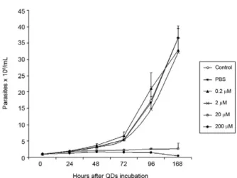
QD dose-response curve in live parasites - Based on a time-scale growth curve, we determined the doses of CdTe for which T. cruzi maintained normal (i) integ-rity for at least seven days, (ii) proliferation and (iii) motility. The results showed that the effects are dose- dependent but not time-dependent. Parasites incubated with 20 and 200 µM CdTe QDs could not proliferate at similar rates as the control.
Fig. 2 shows the exponential growth curve of these parasites over time, with no significant differences be-tween the control, 2 and 0.2 µM of CdTe groups until after 168 h (7 days). Treatments with 20 and 200 µM hin-dered the growth of parasites when compared with the control group. The integrity and motility of these para-sites remained intact up to 96 h. Cellular viability was assessed before and after incubation using the motility and Trypan blue cell dye exclusion methods (Gomes et al. 2006). Fig. 2 also shows that after 72 h, the 0.2 and 2.0 µM groups exhibit significant differences in growth. A significant increase in parasite death was observed for the 20 and 200 µM CdTe QD groups.
Viability of T. cruzi epimastigotes-Parasite viability was checked after 72 h of incubation. The negative control consisted of parasites without QDs and PI. The positive
control consisted of parasites that were permeabilised with 0.1% saponin and PI, as described in Materials and Meth-ods. After 72 h, the incubation with QDs (0.2-200 µM) did not cause plasma membrane disruption. The positive con-trol with saponin-permeabilized parasites showed a high percentage of PI+ cells (about 75%) (Fig. 3).
Cell cycle of T. cruzi epimastigotes - The genetic content of T. cruzi was evaluated by flow cytometry via observation of the following: (i) the percentage of fragmented DNA, (ii) interphasic DNA, which indicates that the parasites are not dividing and (iii) duplicated DNA, which indicates that the parasites are undergoing mitosis. According to our results shown in Fig. 4, we observed a significant increase in DNA fragmentation with CdTe QDs that was dose-dependent in parasites in-cubated with 20 and 200 µM (p < 0.011 and p < 0.006, respectively). No significant differences were observed
Fig. 1: confocal analysis of alive quantum dots (QDs)-labelled
Trypanosoma cruzi epimastigotes. A: 2 µM cadmium telluride (CdTe) QDs with excitation laser of 473 nm; B: 2 µM CdTe QDs with excita-tion laser of 405 nm. Confocal images from Olympus FV-1000, 60x oil immersion lens.
Fig. 2: growth curve of Trypanosoma cruzi epimastigotes (106 parasites/
mL) at different concentrations of cadmium telluride quantum dots (QDs) in a time scale. PBS: phosphate buffered saline. Bars represent mean ± standard deviation of a typical experiment performed in triplicate.
Fig. 3: viability analysis of Trypanosoma cruzi epimastigotes incu-bated with quantum dots (QDs). The incubation with different con-centrations of cadmium telluride QDs led to no important increase in % propidium iodide parasites. Negative control: epimastigotes in liver infusion and tryptose medium; positive control: parasites per-meabilized with 0.1% saponin for 15 min. Bars indicate the standard deviation of five independent experiments.
Fig. 4: cell cycle analysis of Trypanosoma cruzi epimastigotes in-cubated with quantum dots (QDs). Saponin-permeablized parasites were labeled with propidium iodide and the DNA content evaluated by flow cytometry. Black bars: fragmented DNA; grey bars: inter-phasic DNA; white bars: duplicated DNA. Asterisks mean significant
differences compared to control (p ≤ 0.05). Bars indicate the standard
for interphasic DNA. However, the incubation with 2, 20 and 200 µM of CdTe QDs showed less duplicated DNA, indicating at least a partial arrest of T. cruzi mitosis (p < 0.015; p < 0.012 and p < 0.004, respectively).
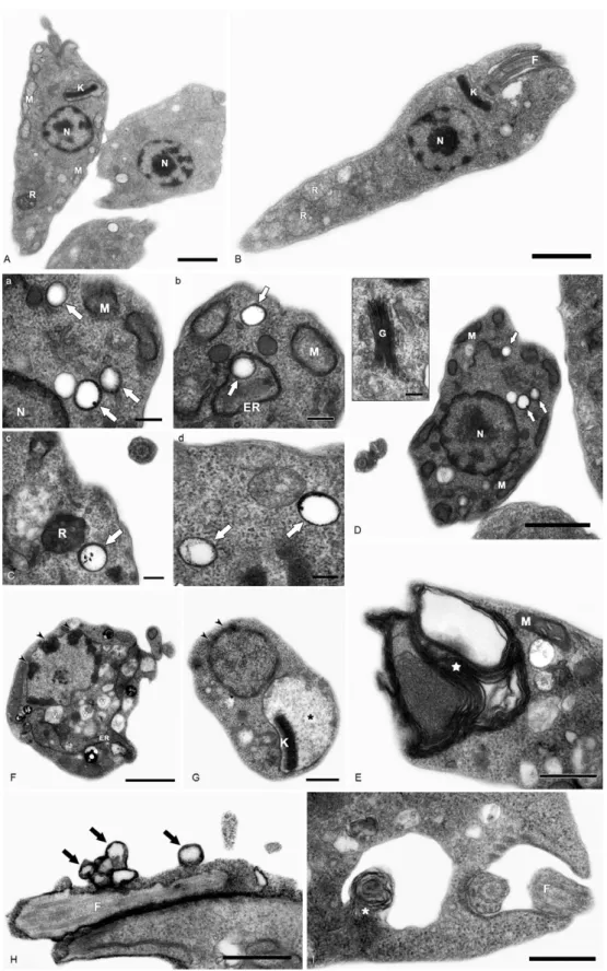
Transmission electron microscopy -Ultrastructural analysis of epimastigotes incubated with 2 μM CdTe QDs showed the integrity of cellular organelles such as mitochondria, nucleus, Golgi complex and plasma mem-brane (Fig. 5A, B). We observed QDs in vesicles scat-tered in the cytoplasm and near organelles, including reservosomes (Fig. 5Ca-d).
Parasites incubated with 200 μM CdTe QDs showed several alterations. Fig. 5E-G shows evidence of abnor-mal chromatin condensation, suggesting DNA damage and intense cytosolic vacuolisation. Endoplasmic reticu-lum profiles surrounding subcellular structures and my-elin-like figures were indicative of autophagic processes (Fig. 5F). This concentration led to mitochondrial swell-ing (Fig. 5G) with loss of its matrix and, occasionally, total disruption of the organelle. We also observed QDs in membrane vesicles close to the flagellar region (Fig. 5H) and extensive changes in the plasma membrane near the flagellar pocket (Fig. 5I), suggesting the occurrence of membrane shedding. It is important to mention that no change was observed in the flagellum and sub-pellicular microtubule layers of these parasites (Fig. 5H, I).
DISCUSSION
QD toxicity has been extensively studied in various cellular prokaryote and eukaryote cellular models (Der-fus et al. 2004, Pelley et al. 2009). The potential effects of CdTe in T. cruzi epimastigotes were evaluated in this work. This analysis involved parasite proliferation un-der different concentrations of CdTe QDs. High doses of QDs (20 and 200 µM) led to a decrease in T. cruzi growth patterns. However, lower concentrations presented simi-lar proliferative rates to the control. Derfus et al. (2004) noted the toxicity of CdSe functionalized with MAA in primary hepatocytes. The authors showed that CdSe at low concentrations (0.25 mg/mL), which is equivalent to more than 20 µM CdTe used in this study, was not toxic to cells and caused no deleterious effects on hepa-tocellular function over seven days. The concentrations used here to label T. cruzi and intestinal epithelial cells of R. prolixus (2 and 10 µM of CdTe and CdSe) were lower than those previous employed (Derfus et al. 2004), encouraging further study into the interaction between parasites and vertebrate host cells. Lovrić et al. (2005b) reported CdTe toxicity in murine microglia cells, dem-onstrating that high concentrations of QDs with green emission (10-100 μg/mL) induced cellular changes such as chromatin condensation, disorder of the nuclear mem-brane and a decrease in cellular metabolism. Our cell vi-ability data showed that T. cruzi incubated with 200 µM CdTe do not die by necrosis within 72 h (Fig. 3).
As previously described (Menna-Barreto et al. 2007), PI labelling was employed to evaluate the cell cycle of the parasite. PI is a well-known DNA marker; exten-sive studies relating the labelling intensity and different amounts of nucleic acids have been published. Intense labelling represented parasites with duplicated DNA;
low labelling (half the intensity of the high-labelled samples) showed parasites in interphase and no label-ling represented debris and/or G0 cells. In this work, the cell-cycle analysis showed that high concentrations (200 μM) of QDs decreased the percentage of duplicat -ed-DNA parasites (14.5%) and increased the percentage of fragmented-DNA parasites (36.7%, as compared with control groups (36.6% and 11.9%, respectively) (Fig. 4). These results suggest that high concentrations of CdTe QDs lead to DNA changes in T. cruzi, compromising the proliferation of the parasite (Fig. 2). Some authors have observed alterations in DNA caused by nanoparticles, including CdTe aggregation in the nucleus (Weis 2003, Lovrić et al. 2005b). It has already been shown that QDs capped with urea and acetate have affinity for the nuclei of mouse fibroblast cells (Bruchez et al. 1998).
Nanoparticle size is also an essential factor in the analysis of toxicity because smaller CdTe QDs with green emission (2 nm) seem to be more toxic to the cells than those with red emission (6 nm) (Lovrić et al. 2005b, Zhang et al. 2007). Our ultrastructural data showed that 2 µM QDs induced no alteration in T. cruzi morphol-ogy (Figs 3, 4). However, several important injuries were observed when parasites were incubated with 200 µM QDs. The extensive mitochondrial swelling observed was verified by Cho et al. (2007) who reported a similar effect when human breast cancer cells were incubated 10 mg/mL CdTe-Cys (about 100 µM CdTe). The authors also observed abnormal chromatin condensation (Cho et al. 2007). Morphological data, such as endoplasmic reticulum profiles surrounding the subcellular structure and myelin-like structures, led us to suggest that au-tophagy is occurring.
As reviewed by Pelley et al. (2009), the toxicity of QDs is associated with their physicochemical properties. Due to the large diversity of nanoparticles (CdTe, CdSe, indium phosphide) and different capping techniques (MAA, zinc sulfide, MAS) employed, it is not possible to elucidate all possible toxicity mechanisms. The concen-trations used in each model vary greatly. Several attempts have been made to reduce particle size by (i) selecting the capping of the nanoparticles, (ii) using minimal dos-es and (iii) modulating nanoparticle size (Bruchez et al. 1998, Hardman 2006, Cho et al. 2007, Tang et al. 2008). All of these factors are summarized in Table.
N
an
o
to
xici
ty o
f q
ua
n
tu
m d
o
ts i
n
T. c
ru
z
i
•
C
e
ci
lia V
ie
ira S
ta
h
l e
t a
l.
16
3
Comparison of quantum dots (QDs) toxicity on different cells
QDs Capping layer Celullar model
QDs concentration
Cellular
localization Toxicity References
CdSe/ZnS Dihydrolipoic acid Mammalian cells or
Dictyostelium discoideum cells
400-600 nM Vesicles and specific
membranes proteins
ND Jaiswal et al.
(2003)
CdTe MAA MCF-7 5 and 10 µg/mL Plasma membrane,
mitochondrion and nucleus
Damage to the plasma membrane, mitochondrion and nucleus
Lovrić et al.
(2005a)
CdTe MPA PC12 and murine
microglial cell line cells
10 μg/mL Cell membrane, cytoplasm
and nucleus
Chromatin condensation and membrane blebbing
Lovrić et al.
(2005b)
Red-emitting CdSe/ZnS QDs
1-ethyl-3-(3-dimethyla-minopropyl) carbodiimide
hydrochloride
PC12 cells 1 μM Membrane surface, endocitic
vesicles and mitochondria
ND Khatchadourian et
al. (2007)
CdTe-CdS-glucose
MAA Living yeast cells (Hebron) ND Membrane transport system ND Farias et al.
(2007)
CdTe and CdSe/ ZnS
MPA, cysteamine, or N-acetylcysteine conjugated to cysteamine
MCF-7 30-150 nM Intracellular compartments
and lysosomes
Cytotoxicity: lysosomal damage and reactive oxygen
species formation
Cho et al. (2007)
CdSe/ZnS Methoxy-PEG-5000 Mice cells 20 μL of a 2-μM Blood and tissue ND Yang et al.
(2007)
CdSe/ZnS Poly-L-lysine,
methoxy-terminated PEG350-thiol
Zebrafish larvae 0.2-200 μM Zebrafish body ND King-Heiden et al.
(2009)
Corroborating these results, transmission electron microscopy results demonstrated the presence of QDs in cytoplasmic vesicles, dispersed in the cytoplasm and near reservosomes. These results suggest that nanoparticles are being endocytosed via an unknown mechanism. We did not observe the presence of QDs within the reservo-somes, showing that they are probably not stored in this organelle as suggested by confocal microscopy results. In addition, we verified the presence of QDs around lipid droplets and dispersed in the cytoplasm (data not shown), indicating that QDs not conjugated to specific molecules may be located in different compartments. Some stud-ies have shown that the size of the nanoparticle influ-ences its subcellular distribution. Smaller QDs tend to accumulate in the nucleus whereas larger QDs remain in the cell cytoplasm (Lovrić et al. 2005b, Hardman 2006). Studies on the ejection of QDs by cells are rare and are usually conducted in mammalian models (Chen et al. 2008, Lin et al. 2008); studies on the elimination of QDs by microorganisms are very scarce. Our results indicate a possible T. cruzi pathway for the elimination of QDs; their presence in vesicles and the plasma membrane shedding data suggest that QDs could be externalized by these mechanisms. Vesicles labelled with QDs were also observed surrounded by profiles of the endoplasmic reticulum, indicating an autophagic process that may be an attempt to eliminate the QDs.
In the present study we demonstrated that T. cruzi labelled with low concentrations of CdTe QDs did not affect (i) parasite integrity up to seven days, (ii) parasite cell division or (iii) parasite motility. This fact was rein-forced by the low level of QD cytotoxicity in the para-sites. In summary, our results show that T. cruzi labelled with 2 µM of QDs do not experience toxic effects and that these QDs are suitable for in vivo cellular studies.
REFERENCES
Anonymous 1999. Recommendations from a satellite meeting. Mem Inst Oswaldo Cruz 94 (Suppl. I): 429-432.
Bruchez M Jr, Moronne M, Gin P, Weiss S, Alivisatos AP 1998. Semi-conductor nanocrystals as fluorescent biological labels. Science 281: 2013-2016.
Chagas C 1909. Nova tripanozomiaze humana: estudos sobre a mor-folojia e o ciclo evolutivo do Schizotrypanum cruzi n. gen., n. sp., ajente etiolojico de nova entidade morbida do homem. Mem Inst Oswaldo Cruz 1: 159-218.
Chen Z, Chen H, Meng H, xing G, Gao x, Sun B, Shi x, Yuan H, Zhang C, Liu R, Zhao F, Zhao Y, Fang x 2008. Bio-distribution and metabolic paths of silica coated CdSeS quantum dots. Toxicol Appl Pharmacol 230: 364-371.
Chiari E, Camargo EP 1984. Culturing and cloning of Trypanoso-ma cruzi, In Morel CM (ed.), Genes and antigens of parasites. A laboratory manual, 2nd ed., Fundação Oswaldo Cruz, Rio de Janeiro, p. 23-26.
Cho SJ, Maysinger D, Jain M, Röder B, Hackbarth S, Winnik FM 2007. Long-term exposure to CdTe quantum dots causes func-tional impairments in live cells. Langmuir 23: 1974-1980. Derfus AM, Chan WCW, Bhatia SN 2004. Probing the cytotoxicity of
semiconductor quantum dots. Nano Lett 4:2163-2169.
Dumas EM, Ozenne V, Mielke RE, Nadeau JL 2009. Toxicity of CdTe quantum dots in bacterial strains. IEEE Trans Nanobio-science 8: 58-64.
Farias PMA, Santos BS, Menezes FD, Brasil JRAG, Ferreira R, Motta MA, Castro-Neto AG, Vieira AAS, Fontes A, César CL 2007. Highly fluorescent semiconductor core-shell CdTe-CdS nanocrystals for monitoring living yeast cell activity. Appl Phys A 89: 957-961.
Feder D 2009a. Motilidade e divisão do parasita: mostra motilidade e parasitos intactos marcados com pontos quânticos CdSe que emitem cor amarela. Available from: www.fiocruz.br/chagas/cgi/ cgilua.exe/sys/start.htm?sid=135.
Feder D 2009b. Ensaio de interação in vitro: mostra a aderência do
T. cruzi ao epitélio do intestino médio de R. prolixus marcado por pontos quânticos que emitem fluorescência verde CdTe (fluo-rescência confocal de 3 quadros por segundo de uma célula viva de T. cruzi). Available from: www.fiocruz.br/chagas/cgi/cgilua. exe/sys/start.htm?sid=135.
Feder D, Gomes SA, de Thomaz AA, Almeida DB, Faustino WM, Fontes A, Stahl CV, Santos-Mallet JR, Cesar CL 2009. In vitro
and in vivo documentation of quantum dots labeled Trypanosoma cruzi-Rhodnius prolixus interaction using confocal microscopy.
Parasitol Res 106: 85-93.
Gaponik N, Dmitri VT, Rogach AL, Hoppe K, Shevchenko EV, Kor-nowski A, Eychmüller A, Weller H 2002. Thiol-capping of CdTe nanocrystals: an alternative to organometallic synthetic routes.
J Phys Chem B 106: 7177-7185.
Gomes SA, Fonseca de Souza AL, Silva BA, Kiffer-Moreira T, San-tos-Mallet JR, Santos AL, Meyer-Fernandes JR 2006. Trypano-soma rangeli: differential expression of cell surface polypeptides and ecto-phosphatase activity in short and long epimastigote forms. Exp Parasitol 112: 253-262.
Hamilton SJ 2004. Review of selenium toxicity in the aquatic food chain. Sci Total Environ326:1-31.
Hardman R 2006. A toxicologic review of quantum dots: toxicity de-pends on physicochemical and environmental factors. Environ Health Perspect 114: 165-172.
Hoshino A, Hanaki K, Suzuki K, Yamamoto K 2004. Applications of T-lymphoma labeled with fluorescent quantum dots to cell tracing markers in mouse body. Biochem Biophys Res Commun 314: 46-53.
Jaffe CL, Grimaldi G, McMahon-Pratt D 1984. The cultivation and cloning of Leishmania. In Morel CM (ed.), Genes and antigens of parasites. A laboratory manual, 2nd ed. Fundação Oswaldo Cruz, Rio de Janeiro, p. 48-91.
Jaiswal JK, Mattoussi H, Mauro JM, Simon SM 2003. Long-term multiple color imaging of live cells using quantum dot bioconju-gates. Nat Biotechnol 21: 47-51.
Kasemets K, Ivask A, Dubourguier HC, Kahru A 2009.Toxicity of nanoparticles of ZnO, CuO and TiO2 to yeast Saccharomyces cerevisiae. Toxicol In Vitro 23: 1116-1122.
Khatchadourian R, Bachir A, Clarke SJ, Heyes CD, Wiseman PW, Nadeau JL 2007. Fluorescence intensity and intermittency as tools for following dopamine bioconjugate processing in living cells. J Biomed Biotechnol2007: 70145-70155.
King-Heiden TC, Wiecinski PN, Mangham AN, Metz KM, Nesbit D, Pedersen JA, Hamers RJ, Heideman W, Peterson RE 2009. Quan-tum dot nanotoxicity assessment using the zebrafish embryo. En-viron Sci Technol43: 1605-1611.
Kondoh M, Araragi S, Sato K, Higashimoto M, Takiguchi M, Sato M 2002. Cadmium induces apoptosis partly via caspase-9 activation in HL-60 cells. Toxicology 170:111-117.
Lin P, Chen JW, Chang LW, Wu JP, Redding L, Chang H, Yeh TK, Yang CS, Tsai MH, Wang HJ, Kuo YC, Yang RS 2008. Computa-tional and ultrastructural toxicology of a nanoparticle, quantum dot 705, in mice. Environ Sci Technol 42: 6264-6270.
Lovrić J, Bazzi HS, Cuie Y, Fortin GR, Winnik FM, Maysinger D
2005a. Differences in subcellular distribution and toxicity of green and red emitting CdTe quantum dots. J Mol Med 83: 377-385.
Lovrić J, Cho SJ, Winnik FM, Maysinger D 2005b. Unmodified cad -mium telluride quantum dots induce reactive oxygen species formation leading to multiple organelle damage and cell death.
Chem Biol 12: 1227-1234.
Marquis BJ, Love SA, Braun KL, Haynes CL 2009. Analytical meth-ods to assess nanoparticle toxicity. Analyst 134: 425-439. Menna-Barreto RF, Corrêa JR, Pinto AV, Soares MJ, de Castro SL
2007. Mitochondrial disruption and DNA fragmentation in Try-panosoma cruzi induced by naphthoimidazoles synthesized from beta-lapachone. Parasitol Res101: 895-905.
Moncayo A, Silveira AC 2009. Current epidemiological trends for Chagas disease in Latin America and future challenges in epide-miology, surveillance and health policy. Mem Inst Oswaldo Cruz 104 (Suppl. I): 17-30.
Parak WJ, Pellegrino T, Plank C 2005. Labelling of cells with quan-tum dots. Nanotechnol 16: R9-R21.
Pelley JL, Daar AS,Saner MA 2009. State of academic knowledge on toxicity and biological fate ofquantumdots. Toxicol Sci 112: 276-296.
Tang M , xing T, Zeng J, Wang H, Li C, Yin S, Yan D, Deng H, Liu J, Wang M, Chen J, Ruan DY 2008. Unmodified CdSe quantum dots induce elevation of cytoplasmatic calcium levels and impairment of functional properties of sodium channels in rat primary cultured hippocampal neurons. Environ Health Perspect116: 915-922. Weis K 2003. Regulating access to the genome: nucleocytoplasmic
transport throughout the cell cycle. Cell 112: 441-451.
Yang RS, Chang LW, Wu JP, Tsai MH, Wang HJ, Kuo YC, Yeh TK, Yang CS, Lin P 2007. Persistent tissue kinetics and redistribution of nanoparticles, quantum dot 705, in mice: ICP-MS quantitative assessment. Environ Health Perspect115: 1339-1343.
Zhang W, Zhang L, Cheng Y, Hui Z, Zhang x, Yixie, Qian Y 2000. Synthesis of nanocrystalline lead chalcogenides PbE (E = S, Se, or Te) from alkaline aqueous solutions. Mat Res Bull 35: 2009-2015. Zhang Y, Chen W, Zhang J, Liu J, Chen G, Pope C 2007. In vitro and