Stamen morphoanatomy of
Dyckia
Schult.f. (Bromeliaceae,
Pitcairnioideae) species: new data for taxonomic use
Jordano Dorval Tavares de Carvalho1, João Marcelo Santos de Oliveira1*, Cesar Carvalho de Freitas1 and Meriélem Saldanha Martins1
Received: April 4, 2016 Accepted: June 13, 2016
ABSTRACT
Th is study presents a morphoanatomical analysis of Dyckia ibicuiensis, D. polyclada and D. racinae stamens. Flowers at anthesis were desiccated and their stamens were processed using common microtechniques for light microscopic analysis. Morphologically, the androecium in these species is diff erentiated by the disposition of the stamens around the gynoecium and by general anther characteristics. Included stamens, with antesepalous fi laments diff erent from those of the antepetalous, anthers in a radial disposition, always curved, and sporangia facing the gynoecium characterize D. ibicuiensis and D. racinae. Th is confi guration results from the fi lament connation, as well as special anatomical characteristics of the anthers, such as the connective with thickenings. In addition, these species are diff erentiated by the U-shaped thickening in the endothecium. D. polyclada is characterized by its small, free and exserted stamens, with a very short common tube, its anthers, that are not curved or organized around the gynoecium, and its divergent sporangia. Th e anatomical and morphological characteristics identifi ed here are important for characterization of these species. Considering that the androecium is important in the delimitation of Dyckia, the increased use of these data should be equally important for other species and infrageneric groupings.
Keywords. anatomy, androecium, anther, common tube, connective, Dyckia,endothecial thickening, stamen vasculature
Introduction
Dyckia is a very diverse genus of Pitcairnioideae (Bromeliaceae), with over 160 terricolous or saxicolous species, with adaptations to xerophytism, distributed across South America, especially in Brazil (Smith & Downs 1974; Leme et al. 2012; Forzza et al. 2015; Gouda et al. 2015). Phylogenetic hypotheses based on morphological (Forzza 2001) and molecular (Krapp et al. 2014) traits have confi rmed the monophyly of the genus. However, there are still diffi culties regarding both the circumscription of some species and the proposition of infrageneric groupings
(Krapp et al. 2014). Th is genus has a large number of endemic or rare species (Martinelli et al. 2008), and is considered problematic from a taxonomic point of view, either because of insuffi cient records of specimens, or due to overlaps in key analytical characteristics (Smith 1967; Versieux & Wendt 2007). For this reason, Dyckia has been of interest in morphoanatomical studies of reproductive organs in order to defi ne the species more consistently (Fagundes & Mariath 2010; Dorneles et al. 2014; Martins et al. 2015; Oliveira et al. 2015; Santos-Silva et al. 2015) and to serve as a basis for understanding infrageneric relations and making phylogenetic inferences, which are rare and hampered by
1 Departamento de Biologia, Centro de Ciências Naturais e Exatas, Universidade Federal de Santa Maria Av. Roraima 1000, 97105-900, Santa Maria,
RS, Brazil
the homogeneity of traits in this group, due to its recent radiation (Krapp et al. 2014).
Characteristics of floral morphology, such as the presence of extranuptial nectaries and, mainly, fusion of the stamens, are important in the definition of the genus, which is phylogenetically and morphologically quite similar to Encholirium. However, the androecium is usually little pronounced in the original descriptions of Dyckia species, with emphasis in some species on the connation and relative size. Nevertheless, the importance of the stamens is not restricted to the delimitation of the genus, having been referred to in epithets, such as in Dyckia choristaminea (“dyckia of separated stamens”), D. tubifilamentosa (“dyckia of filaments in tube”), D. exserta (“dyckia of exserted stamens”), demonstrating the plasticity of this organ in the group, whose floral characteristics are considered quite homogeneous.
Anatomical analysis of reproductive organs has been used for taxonomic and phylogenetic purposes in Bromeliaceae and Dyckia (Sajo et al. 2005; Fagundes & Mariath 2010; Nogueira et al. 2015; Santos-Silva et al. 2015), due to not only the descriptive and diagnostic potential but also to the value of this kind of analysis in evolutionary considerations or the relative homogeneity of some often used morphological traits. In Dyckia, in addition to general anatomical traits of the flower (Dorneles et al. 2014; Martins et al. 2015), stamen anatomy, especially of the anther, has been investigated in some works from ontogenetic (Sajo et al. 2005; Mendes et al. 2012), taxonomic (Dorneles et al. 2014; Martins et al. 2015) and histochemical (Oliveira et al. 2015) points of view, especially in endemic or rare species. This type of analysis has contributed to a more precise characterization and delimitation of the respective species of Dyckia. The original descriptions of the species analyzed in the present study, Dyckia racinae, D. ibicuiensis and D. polyclada, presented only general aspects of flowers (Smith 1988; 1989; Strehl 1997) and few specific elements. The difficulties in identification not only exacerbate the condition of ‘critically endangered’ of D. ibicuiensis, but also hamper the classification of D. racinae and D. polyclada into the accurate category of endangerment, due to insufficient data and a lack of knowledge about the distribution and/or their respective population states (DOE RS 2014).
Considering the lack of information about the stamen in Dyckia, more detailed analyses of this organ are important in research on traits with potential phylogenetic and taxonomic use. Thus, the present study aims to characterize morphoanatomically the androecium of D. racinae, D. ibicuiensis and D. polyclada, highlight relevant characteristics of the stamens and anthers for each species and compare them using these characteristics.
Materials and methods
The processing and analysis of floral samples was carried out at the Laboratório de Botânica Estrutural (LABOTE) of
the Departamento de Biologia at the Universidade Federal de Santa Maria (UFSM). Inflorescences of six specimens of D. ibicuiensis Strehl, ten specimens of D. racinae L.B. Sm. and ten specimens of D. polyclada L.B. Sm. were collected from natural populations in: Morro Itaquatiá, São Pedro do Sul, RS, Brazil (29°42.941’S/ 53°43.087’ W), São Pedro do Sul, RS, Brazil (29°35’22.2’’S/ 54°49’49.4’’W) and Distrito de Santo Antão, Santa Maria, RS, Brazil (29°42.911’S/ 53°43.093’W), respectively. The plant material was deposited at the herbarium of UFSM, under the registration numbers SMDB 13840, SMDB 15923 and SMDB 15924.
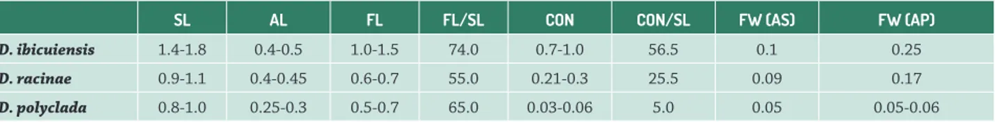
Morphometric data were obtained from random individuals, using stamens of 10 to 20 flowers at anthesis for each species, at different heights of inflorescence. The total size of the stamen, the length of the anther and the filament, the ratio between filament length and total length of the stamen, the height of connation, the height of connation relative to the total size of the stamen and the width of the filament were measured. The terminology used is in accordance with Smith & Downs (1974).
For the light microscopy, the botanical material was fixed in a solution containing 1% glutaraldehyde and 4% formaldehyde (McDowell & Trump 1976) in 0.1 M sodium phosphate buffer, pH 7.2 (Gabriel 1982), followed by washing in the same buffer, washing in distilled water and finally dehydration in an ethyl alcohol series (O’Brien & McCully 1981). The material was pre-infiltrated in a 2-hydroxyethyl methacrylate (HEMA) and pure ethanol solution (1:1) during 12h, followed by infiltration in HEMA for approximately 4h and embedding in a Teflon holder until polymerization was completed (Gerrits & Smid 1983). The anthers and filaments were sectioned to 3μm in thickness using a Leica RM2245 rotary microtome. Toluidine blue O in 0.05% sodium benzoate buffer, pH 4.4 was used to stain the histological slides (Sidman et al. 1961).
For analysis of fibrous thickening, portions of anthers were macerated using a 3:1 solution of hydrogen peroxide and glacial acetic acid, with subsequent temporary preparation in alcoholic glycerine (Johansen 1940) and analyzed using Differential Interference Contrast (DIC).
For analysis of vascularization, fixed anthers and filaments were clarified using 3% sodium hypochlorite (Johansen 1940).
Photographic documentation and analysis were performed using a Leica DM2000 microscope® and Stereomicroscope Leica M80 with DFC295 image capture system and Zeiss AxioImager A2 with a Zeiss Axiocam RMC digital photographic system.
PhotoshopTM was used to process the images.
Results and discussion
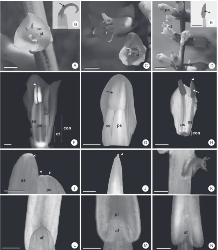
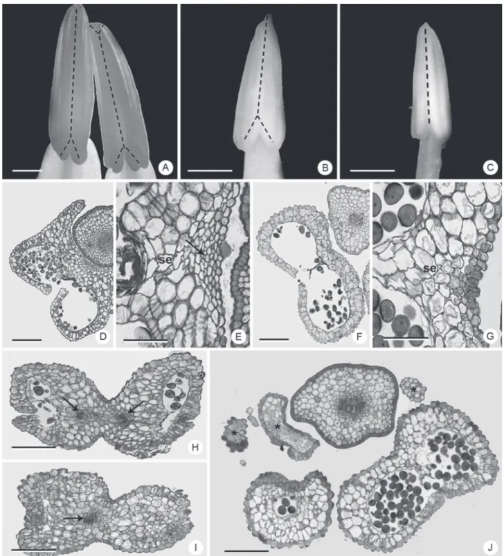
Figure 1. General aspects of the stamens of Dyckia species. A, B, F, I, L: D. ibicuiensis. C, G, J, M: D. racinae. D, E, H, K, N: D. polyclada. A, C, D: General appearance of the flower in vivo. B, E: detail of dehiscent anther. F, G, H: general appearance of antepetalous and antesepalous stamens after chemical fixation. G: cross section of filaments occurred near the distal region of the common tube. I, J: rostra. K: projection of tissue at the base of the anther of D. polyclada. L, M, N: detail of the region of filament insertion. Arrows: region of dehiscence; arrow heads: rostra; af: apex of the filament; con: connation; ct: common tube with the petals; pa: flower at pre-anthesis; pr: projection around the apex of the filament; ps: antepetalous stamen; ss: antesepalous stamen; st: stamen. Bars: A, C, D: 5 mm; B, E, F-H: 2 mm; I-N: 500 μm.
common tube with the petals (Smith & Downs 1974). Thus, there are two stamen whorls, which can be connate
and exserted in D. polyclada (Fig. 1A-H). D. racinae and D. ibicuiensis present an evident common tube with the petals (Fig. 1F); in D. polyclada this tube is short or imperceptible (Fig. 1H). The anthers are triangular, dorsifixed, introrse and bithecous, with longitudinal dehiscence (Fig. 1F-H), and present a pointed rostrum (Fig. 1 H-J), which is bilobed in the antepetalous stamens of D. ibicuiensis (Fig. 1I). The general characteristics of the androecium, in the three species analyzed, corroborate previous data for Dyckia (Smith & Downs 1974; Reitz 1983) in large taxonomic studies of the genus. However, the original descriptions of the species analyzed here (Smith 1988; 1989; Strehl 1997), merely reported the height relative to the corolla and the occurrence of connation.
Morphometrically, the stamens differ by total size, size relative to the corolla, extent of connation and length and width of the filaments (Tab. 1). D. ibicuiensis and D. racinae present the largest stamens, with antepetalous filaments wider than the antesepalous filaments and with a greater extent of connation (Tab. 1). The filament in these two species represent 74% and 55%, respectively, of the total size of the stamen (Tab. 1). D. polyclada presents the smallest stamens and smallest extent of connation. In addition, the filaments are thin and the antepetalous and antesepalous filaments are similar (Tab. 1). The filament comprise more than 60% of the total size of the stamen (Tab. 1). Based on interpretation of the data, it is possible to infer that despite these ratios being unique for each species, there is no direct relationship between relative size of the filament and length relative to the corolla, which may be related to the length and curvature of the petals. Nevertheless, it is observed that the filaments continue to lengthen after anthesis in D. polyclada, while the petals cease to grow in this period (Fig. 1D), a common phenomenon in several angiosperms according to Greyson (1994). This characteristic could be investigated in future studies, associated to temporal development patterns of flower organs, during anthesis. Similarly, it is not possible to evaluate the relationship between the longer common tube and stamens, with greater extent of connation, and the relative sizes observed which differentiate D. racinae and D. ibicuiensis from D. polyclada, since the common tube with petals may also lengthen late, without leading to exposition of the stamens. It is important to note that relative measurements are rarely used in descriptions of species, despite generally being
robust, as they do not present overlaps and they provide good comparisons.
At anthesis, the stamens of D. ibicuiensis and D. racinae are regularly positioned around the carpels, giving the anthers a radial organization, with zones of dehiscence facing toward the gynoecium (Fig. 1A-C). This radial disposition is made possible by the greater extension of connation of the stamens, and by the greater opening of the corolla in these species, due to the dorsal curvature of the petals in D. racinae and D. ibicuiensis (Fig. 1A, C). In contrast, D. polyclada presents an irregular stamen distribution (Fig. 1D), mainly due to the smaller opening of the corolla and by the short connation. In addition, D. ibicuiensis and D. racinae, in vivo, show a marked dorsal curvature of the anthers (Fig. 1A-C), while in D. polyclada, there is only a slight curvature (Fig. 1D-E). After fixation, or when the anthers are rehydrated, there is a return to the rectilinear shape observed prior to dehiscence (Fig. 1F-G). This curvature does not appear to be a result of differential growth processes, but rather a result of typical dehydration of the anther during dehiscence, in association with its mature tissues. This consideration is supported by the fact that after rehydration, the anthers resume the typical pre-anthesis form. Changes in morphology, as well as position of the anther during anthesis have been observed in other plant groups (Dettke & Santos 2011), including Dyckia (Rogalski et al. 2009) and are related mainly to strategies of pollen grain dispersal and reproductive systems adopted by the species. As indicated by Rogalski et al. (2009), in addition to the radial arrangement of the anthers, which turn the sporangia toward the gynoecium, this curvature also characterizes a self-pollination system commonly observed in other Bromeliaceae genera, including Dyckia (Smith & Downs 1974; Benzing 2000).
Irregular projections of tissue were observed from the base of the theca of some stamens in D. polyclada (Fig. 1 K). This structure possesses variable and asymmetric forms. Since the presence of these projections is not constant, its taxonomic utilization is not viable, but nevertheless is a new record for the species and genus.
The insertion point of the filament is different in each species (Fig. 1L-N). In D. ibicuiensis and D. polyclada the apical region of the filament is exposed (Fig. 1L, N), unlike D. racinae, where this region is covered by the projection of the connective and sporangia around the apex of the
Table 1. Morphometry of the stamens of Dyckia ibicuiensis, D. racinae and D. polyclada. SL: stamen length. AL: anther length. FL: filament length (including the common tube with the petals). FL/SL: approximate mean ratio of filament length relative to total stamen length CON: connation height (including the common tube with petals). CON/SL: mean ratio between connation height and total stamen length. FW: filament width immediately above the connation point. FW (AS): filament width of antesepalous stamens. FW (AP): filament width of antepetalous stamens. SL, AL, FL, CON and FW measurements in cm; CON/SL and FL/SL: percentage values.
SL AL FL FL/SL CON CON/SL FW (AS) FW (AP)
D. ibicuiensis 1.4-1.8 0.4-0.5 1.0-1.5 74.0 0.7-1.0 56.5 0.1 0.25
D. racinae 0.9-1.1 0.4-0.45 0.6-0.7 55.0 0.21-0.3 25.5 0.09 0.17
filament, similar to a groove (Fig. 1M). These species also differ by the shape of the filament apex, both antesepalous and antepetalous, which are obtuse in D. ibicuiensis and D. racinae (Fig. 1L-M) and acute in D. polyclada (Fig. 1N). In all the species, there is a gradual narrowing of the filament towards the apex (Fig. 1L-N). The insertion point of the filament is often associated with the movement of the anther in the reproductive period (D’Arcy 1996; Dettke & Santos 2011). The morphology of this region suggests constraint of lateral and dorso-ventral movements of the anther in D. ibicuiensis and D. racinae, regardless of its curvature. In D. polyclada, on the other hand, there seems to be articulation of the anthers, which, combined with the free exserted stamens, the rectilinearity of the anthers and the divergent sporangia, may be evidence of variations in the pollination process.
Another important feature is the differential morphology between the antepetalous and the antesepalous stamens of D. ibicuiensis and D. racinae (Fig. 1F-G) (Tab. 1). Differences between stamen whorls, as indicated by Greyson (1994), are related to initiation time and differential spacing between the antepetalous and antesepalous stamens during the early stages of floral development. These different widths of filaments in each whorl may demonstrate different patterns of floral development between these species and D. polyclada, whose filaments presented only a slight dimorphism, making their use in future taxonomic analyses relevant for both revisions of species of genus and for new descriptions. In the anthers, it is noteworthy that there is a forked rostrum only in the antepetalous stamens in D. ibicuiensis, which may be used as a diagnostic trait.
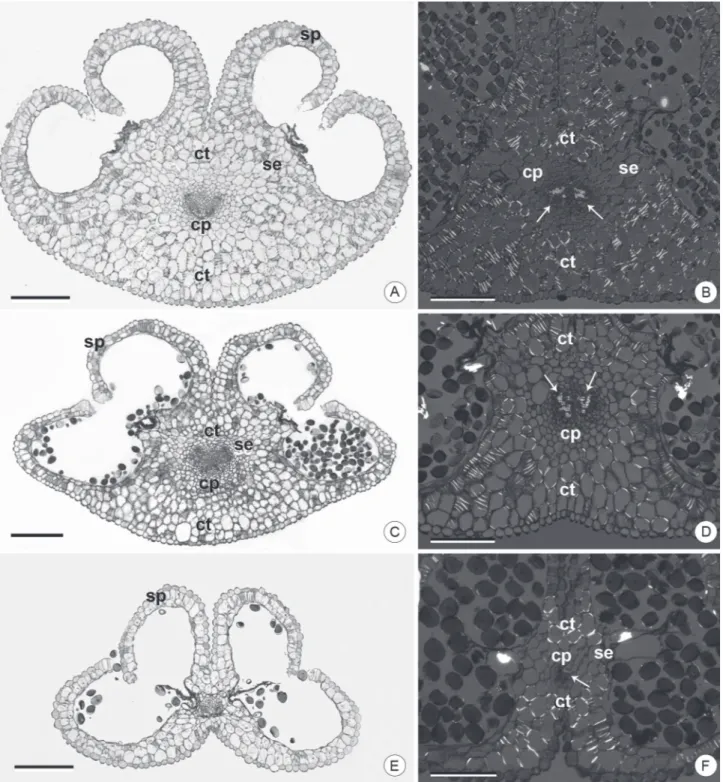
The anatomy and histology of the mature anthers in a cross-section of the species analyzed here was in line with typical expectations for Dyckia and other Bromeliaceae (Johri et al. 1992; Sajo et al. 2005; Mendes et al. 2012), with regard to the presence of a connective with central vascular bundle and four sporangia with parietal layers consisting of epidermis, endothecium and middle layer (Fig. 2A-F). Details of the distribution and constitution of these tissues, however, showed important anatomical and histological differences among the species.
In the cross section, the middle portion of the anthers presents typical histological sites, the connective and the sporangial site (Fig. 2A-F). The connective in D. ibicuiensis and D. racinae (Fig. 2A-D) presents a relatively more developed mid-dorsal region of the anther, unlike that in D. polyclada (Fig. 2E-F). The sporangial site comprises four sporangia facing the gynoecium in D. ibicuiensis and D. racinae (Fig. 2A, C), where in D. polyclada the sporangia assume a divergent position (Fig. 2E).
The connective, in a broad sense, is a tissue that comprises the entire area inside the anther, except for the sporangial tissue (D’Arcy 1996). In addition, it is the site of vascular bundle differentiation and, thus, sporangial nutrition. It also provides support for the anther and assists
in its dehiscence, in addition to being a site for storage and mobilization of metabolites (Clément et al. 1994; Oliveira et al. 2015).
In the present study, the presence of fibrous thickenings allowed for differentiation between two regions in the connective (Fig. 2A-F). The region without fibrous thickenings is central and made up of a collateral vascular bundle, with two parallel xylem poles, surrounded by parenchymatous tissue, similar to a sheath, with four to six cell layers extending to the septal region (Fig. 2A-F). The sheath, and its extensions to the septal sporangia are probably associated with the transfer of nutrients to the sporangia. In addition, they possibly aid in anther support, especially in D. polyclada, where this tissue forms almost the entirety of the connective (Fig. 2E-F). The connective dorsal and ventral regions in D. ibicuiensis and D. racinae are bulky and have cells with fibrous thickenings (Fig. 2B, D), unlike in D. polyclada (Fig. 2E), where this region is limited to a few thick cell layers (Fig. 2E-F). Considering the anthers in cross-section, the dorsal region in D. ibicuiensis and D. racinae is protruding (Fig. 2A, D), and in D. polyclada it is always concave (Fig. 2E), due to the connective bulk. Apparently, the greater accumulation of mechanical tissue in the dorsal and ventral region of the connective is important for the conformation of the anther in the anthesis period. Hufford & Endress (1989) and Dettke & Santos (2011) described the dehiscence of the anthers in different species as a result of the association between mechanical tissues of the connective and the endothecium, which likely occurs in the present study. The mechanical function is associated with the forces generated by differential dehydration of cell walls of mechanical tissues, as described by D’Arcy (1996). Thus, the dorsal curvature observed in the anthers after dehydration was interpreted as a result of deformation of the connective in D. racinae and D. ibicuiensis. In D. polyclada, the presence of few cells in the connective region and a more limited distribution of thickenings allow for this interpretation. Another result of the volume of the connective in the species analyzed in this work is related to the convergent position of the sporangia, facing the ventral region, especially in D. ibicuiensis and D. racinae, where this tissue orientates the growth and direction of the sporangia. In D. polyclada, whose connective is basically limited to the parenchymal region surrounding the vascular bundle, there is no mechanical restriction on sporangial development, which takes a lateralized position. From a taxonomic point of view, it is important to check this anatomical trait in future descriptions or infrageneric considerations, given its importance in the final structuring of anthers and its constancy in different flowers and different individuals.
(1996) have interpreted these portions of the connective as an endothecial-type of hypodermis or as a tabular configuration of the endothecium, due to the fact that the presence of fibrous thickenings is also associated with the opening of the anthers.
As for the sporangial region (Fig. 3), in the mature
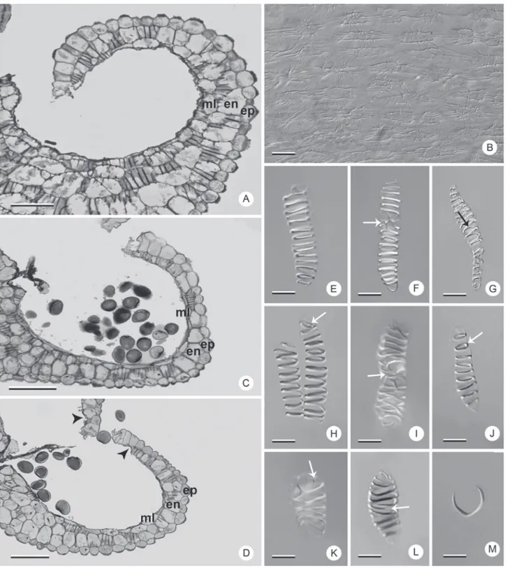
anther, there only remains cells from the epidermis, fibrous endothecium and some cells from the middle layer, despite there being an increase of cell layers in regions of transition from the sporangia to the connective (Fig. 3A, C, D).
When comparing the three species, the epidermis is striated, with thickened and cutinized external periclinal
Figure 3.Sporangia and fibrous thickenings in Dyckia species. A, B, E-G, M: D. ibicuiensis. C, H-J: D. racinae. D, K, L: D. polyclada. A, C, D: sporangia in median cross section under bright field microscopy. B, E-M: Differential Interference Contrast microscopy (DIC). B: sporangial surface detail. E-M: endothecium cells showing different patterns of fibrous thickening. Arrows: bifurcations or rings; arrow heads: region with absent endothecium; en: endothecium; ep: epidermis; ml: middle layer. Bars: A, C, D: 100 μm; B, E-M: 10 μm. walls, making the organ surface irregular (Fig. 3B). In
addition, it presents cells with anticlinal elongation in the region adjacent to the stomium (Fig. 3A, C, D). Lobo et al. (2013) consider these characteristics of Dyckia epidermis to reflect a xeromorphic condition common to the genus,
being that the taxonomic use of this layer is limited due to its structural simplification in the stamens.
thickenings present in this layer, which generate differential tensions with typical dehydration during anthesis, aiding to open the sporangia (Keijzer 1987), by means of hygroscopic mechanisms. The greater structural diversity of the endothecium cells and the conservation of these types in different groups of plants (Manning 1996) justify its utilization in phylogenetic and taxonomic analyses. In the present study, in the same anther, different types of fibrous thickenings in endothecial cells were observed, in addition to types exclusive to the species (Fig. 3E-M).
In the species analyzed here, the endothecium consists of a layer of cells that is continuous until the stomial region, with the exception of D. polyclada, in which the endothecium cells do not occur in that region (Fig. 3D). In all the species, there is a predominance of helical thickening, combined or not with rings at the extremities or with bifurcations (Fig. 3E-I, K-L). In addition, cells with annular thickenings may also be observed (Fig. 3J). D. ibicuiensis and D. racinae also present U-shaped thickenings (Fig. 3M), not observed in D. polyclada. The predominant types in the three species are variations of the helical type described by Manning (1996), which, according to the author, is one of the most important types from a phylogenetic point of view, in addition to the U-shapes. Manning & Linder (1990) consider U-shaped thickenings as a derived type in Poales families. According to them, this type emerged from the helical type in this group, a hypothesis that may justify future studies in Dyckia, due to the variability in fibrous thickenings found in the three species analyzed. Besides the absence of U-shaped thickenings, D. polyclada is also characterized by the absence of the endothecium near the stomial region.
The middle layer, in the three species, consists of a non-uniform layer of cells that also presents fibrous thickenings (Fig. 3A, C-D). This parietal layer is conspicuous only in D. ibicuiensis (Fig. 3A). The structure of the middle layer in the mature anther proved to be of interest in the comparison among the species since, by remaining in the anthers, its cells also developed fibrous thickenings. The permanence of the middle layer with thickenings in the mature anter is common and has been reported in other monocot families, such as Rapateaceae (Oriani & Scatena 2013) and Commelinaceae (Hardy et al. 2000), despite being unidentified in Dyckia and other Bromeliaceae. It probably aids in anther dehiscence together with the endothecium, due to the presence of fibrous thickening.
Some studies investigating the anther anatomy in Dyckia have shown the location and types of vascular bundles (Sajo et al. 2005; Mendes et al. 2012; Dorneles et al. 2014). Likewise, a number of studies have recognized the phylogenetic and taxonomic importance of anther vasculature in other taxonomic groups (Hufford 1980; French 1986; Dettke & Santos 2011). In this study, D. ibicuiensis and D. racinae present a bifurcation of the vascular bundle in the anter, extending from the insertion point of the filament to the basal region of both theca (Fig. 4A-B), unlike D.polyclada, which presents vascular bundles
without ramifications (Fig. 4C). D. ibicuiensis presents a small bifurcation at the apex of the antepetalous stamens, corresponding to the rostral lobes (Fig. 4A), which does not occur in the other two species. The vascularization patterns described are easily observed anatomically along the anther (Fig. 4 D-I), due to the occurrence not only of conducting elements, but also the vascular sheaths. In D. polyclada this tissue is also present along the entire length of the anther, although in the basal region only the vascular sheaths occur (Fig. 4F-G). In the basal region of the theca of some anthers of this species there are projections of non-vascular tissue (Fig. 1K), with epidermis and parenchymatous content (Fig. 4J). Relationships between anther volume and vascular branching in the connective have been proposed previously (Dettke & Santos 2011) and in the present study, it is possible to observe a simplification of the vascular tissues in D. polyclada, possibly associated with the smaller size of the anther. In the other species, the vascular branching possibly occurs due to the larger size of the anthers and to the distance from the site of entry of vascularization in the connective.
Figure 4.Vascularization of the anthers in Dyckia species. A, H, I: D. ibicuiensis. B, D, E: D. racinae. C, F, G, J: D. polyclada. A-C: images of stereomicroscopy. D-J: bright field microscopy. A-C: dashed line indicates vascularization of the anthers. D-G, J: basal portion of the anther. H, I: apices of anthers of antepetalous and antesepalous stamens, respectively; Arrows: vascular bundles; asterisk: projections of non-vascular parenchymatous tissue; se: bundle sheath extension. Bars: A-C: 1 mm; D, F, J: 200 μm. E, G, H, I: 50 μm.
flowers remains relatively well-conserved, being important for evolutionary inferences, especially related to fusions and structural losses. Therefore, the variations found in D. racinae are an important record of the variability in stamen vasculature, demonstrating the need for studies
of flower development in order to verify the origin of these configurations and their possible utilization in species characterization.
Figure 5.Vascularization and histology of filaments in Dyckia species. A, D: D. ibicuiensis. B, G-H: D. racinae. C, E-F: D. polyclada. A-C: filaments clarified under stereomicroscopy. D-H: median cross-section of filaments under bright field microscopy. C, E, G: filaments of antesepalous stamens. F, H: filaments of antepetalous stamens. Arrow: eccentric vascular bundle; arrow heads: xylem poles; pf: antepetalous filament; sf: antesepalous filament; vb: vascular bundle. Bars: A-C: 500 μm; D-H: 200 μm.
“maritima complex” (Strehl & Beheregaray 2006), a group that has been neglected, but which shares various floral characteristics, including the morphology of the androecium observed in this study. According to HM Büneker et al. (unpubl. res.) several species of Dyckia from southern Brazil are part of this group, including D. myriostachya, D. retroflexa, D. rigida, D. maritima, D. selloa, D. alba, D. agudensis, D. nigrospinulata, D. tomentosa, D. domfelicianensis, D. hebdingii, D. delicata, as well as D. polyclada. Apparently, the androecium in all these species is quite similar, with slightly connate or free stamens, often exserted, with a short common tube with the petals, similar and narrow antesepalous and antepetalous filaments and small anthers not distributed radially around the gynoecium, in contrast
to D. racinae and D. ibicuiensis, which are likely related to other dyckias of a second group (Strehl & Beheregaray 2006), apparently heterogeneous. It is important to note that there are significant overlaps in taxonomic descriptions, especially in floral characterization of the “maritima complex” species, for example, the demarcation between D. polyclada and D. maritima (Smith & Downs 1974; Reitz 1983), whose few diagnostic characteristics overlap markedly. This points to the need for an analysis that includes a greater number of floral features, especially those related to the androecium, which, according to the data of this study, may be relevant.
unpublished data for future studies of the genus. For comparative purposes, a number of characteristics can be highlighted, including the connective with thickened cells, the curvature of the anthers, the types of fibrous thickenings found in the endothecium, the particularities of each stamen whorl and stamen vasculature. Finally, the stamen morphanatomy in Dyckia was found to be important in the characterization of three rare and endangered species of this diverse genus. We emphasize that the stamen features present taxonomic potential both for revisions of already described species and for characterization of new Dyckia species.
Acknowledgements
We thank Henrique Mallmann Büneker for the correct identification of the species and collection of biological material.
References
Benzing DH. 2000. Bromeliaceae: profile of an adaptive radiation. Cambridge, Cambridge University Press.
Clément C, Chavant L, Burrus M, Audran JC. 1994. Anther starch variations in Lilium during pollen development. Sexual Plant Reproduction 7: 347-356.
D’Arcy WG. 1996. Anthers and stamens and what they do. In: D’Arcy WG, Keating RC. (eds.) The anther: form, function and phylogeny. Cambridge, Cambridge University Press. p. 1-24.
Dettke GA, Santos RP. 2011. Morfologia externa, anatomia e histoquímica da antera e grãos de pólen de Passifloraceae do Rio Grande do Sul, Brasil. Revista Brasileira de Biociências 9: 48-74.
DOE RS. 2014. Decreto 51.109 de 19 de dezembro de 2014. <http://www. fzb.rs.gov.br/conteudo/4809/?Homologada_a_nova_Lista_da_Flora_ Ga C3%BAcha_Amea%C3%A7ada_de_Extin%C3%A7%C3%A3o.> 29 May 2016.
Dorneles MP, Oliveira JMS, Canto-Dorow TS. 2014. Dyckia racinae L.B. Sm. (Bromeliaceae): morphological description emphasizing the reproductive structures. Iheringia Serie Botânica 69: 397-404. Fagundes NF, Mariath JEA. 2010. Morphoanatomy and ontogeny of fruit
in Bromeliaceae species. Acta Botanica Brasilica 24: 765-779. Forzza RC. 2001. Filogenia da tribo Puyeae Wittm. e revisão taxonômica
do gênero Encholirium Mart. ex Schult. & Schult.f. (Pitcarnioideae-Bromeliaceae). PhD Thesis, Universidade de São Paulo, Brazil. Forzza RC, Costa A, Siqueira Filho JA, et al. 2015. Bromeliaceae in Lista de
Espécies da Flora do Brasil, Jardim Botânico do Rio de Janeiro. <http:// reflora.jbrj.gov.br/jabot/floradobrasil/FB34334>. 10 Oct. 2015. French JC. 1986. Patterns of stamen vasculature in the Araceae. American
Journal of Botany 73: 434-449.
Gabriel BL. 1982. Biological Electron Microscopy. New York, Van Nostrand Reinhold Company.
Gerrits PO, Smid L. 1983. A new less toxic polymerization system for the embedding of soft tissue in glycol methacrylate and subsequent preparing of serial sections. Journal of Microscopy 132: 81-85. Gouda EJ, Butcher D, Gouda K. 2015. [continuously updated]. Encyclopaedia
of Bromeliads Version 3.1.<http://botu07.bio.uu.nl/bcg/encyclopedia/ brome/>. 10 Oct. 2015.
Greyson RI. 1994. The Development of Flowers. Oxford, Oxford University Press.
Hardy CR, Stevenson DW, Kiss HG. 2000. Development of the gametophytes, flower, and floral vasculature in Dichorisandra thyrsiflora
(Commelinaceae). American Journal of Botany 87: 1228-1239.
Hufford LD. 1980. Staminal vascular architecture in five dicotyledonous angiosperms. Proceedings of the Iowa Academy of Science 87: 96-102. Hufford LD, Endress PK.1989. The diversity of anther structures and
dehiscence patterns among Hamamelididae. Botanical Journal of the Linnean Society 99: 301-346.
Johansen DA. 1940. Plant microtechnique. New York, MacGraw-Hill. Johri BM, Ambegaokar KB, Srivastava OS. 1992. Comparative Embryology
of Angiosperms. Berlin, Springer.
Keijzer CJ. 1987. The processes of anther dehiscence and pollen dispersal. New Phytologist 105: 487-498.
Krapp F, Pinangé DSB, Benko-Iseppon AM, Leme EM, Weising K. 2014. Phylogeny and evolution of Dyckia (Bromeliaceae) inferred from chloroplast and nuclear sequences. Plant Systematics and Evolution 300: 1591-1614.
Leme EMC, Ribeiro OBDC, Miranda ZDJG. 2012. New species of Dyckia
(Bromeliaceae) from Brazil. Phytotaxa 67: 9-37.
Lobo GM, Souza TV, Voltolini CH, Reis A, Santos M. 2013. Leaf Epidermis of the Rheophyte Dyckia brevifolia Baker (Bromeliaceae). The Scientific World Journal 2013: 1-7.
Manning JC. 1996. Diversity of endothecial patterns in the angiosperms. In: D’Arcy WG, Keating RC. (eds.) The anther: form, function and phylogeny. Cambridge, Cambridge University Press. p. 136-158. Manning JC, Linder HP. 1990. Cladistic analysis of patterns of endothecial
thickenings in the Poales/Restionales. American Journal of Botany 77: 196-210.
Martinelli G, Vieira CM, Gonzalez M, Leitman P, Piratininga A, da Costa AF, Forzza, RC. 2008. Bromeliaceae da Mata Atlântica brasileira: lista de espécies, distribuição e conservação. Rodriguésia 59: 209-258. Martins MS, Dorneles MP, Oliveira JMS, Nicoloso FT. 2015. Morphological
characterization of the ovarian wall, stigma, stylus and ovules in two species of Dyckia (Bromeliaceae). Rodriguésia 66: A32.
McDowell EM, Trump BF. 1976. Histologic fixatives suitable for diagnostic light and electron microscopy. Archives of Pathology & Laboratory Medicine 100: 405-414.
Mendes SP, Costa CG, Toni KLG. 2012. Androecium development in the bromeliad Dyckia pseudococcinea L.B.Sm. (Pitcairnioideae - Bromeliaceae), an endangered species endemic to Brazil: implications for conservation. Flora 207: 622-627.
Nogueira FM, Fagundes NF, Kuhn SA, Fregonezi JN, Mariath, JEA. 2015. Ovary and ovule anatomy in the nidularioid complex and its taxonomic utility (Bromelioideae: Bromeliaceae). Botanical Journal of the Linnean Society 177: 66-77.
O’Brien TP, McCully ME. 1981. The study of plant structure principles and selected methods. Melbourne, Termarcarphi Pty Ltd.
Oliveira JMS, Martins MS, Dorneles MP, Freitas CC. 2015. Starch distribution in anthers, microspores and pollen grains in Aechmea recurvata (Klotzsch) L.B. Sm., Dyckia racinae L.B. Sm. and Tillandsia aeranthos (Loisel.) L.B. Sm. (Bromeliaceae). Acta Botanica Brasilica 29: 103-112.
Oriani A, Scatena VL. 2013. The taxonomic value of floral characters in Rapateaceae (Poales-Monocotyledons). Plant Systematics and Evolution 299: 291-303.
Puri V. 1951. The role of floral anatomy in the solution of morphological problems. The Botanical Review 17: 471-553.
Reitz R. 1983. Bromeliáceas e a malária-bromélia endêmica. In: Reitz R. (ed.). Flora Ilustrada Catarinense. Itajaí, Herbário Barbosa Rodrigues p. 1-808.
Rogalski JM, Reis A, Reis MSD, Hmeljevski KV. 2009. Reproductive biology of the rheophyte Dyckia brevifolia Baker (Bromeliaceae), on the Itajaí-Açu River, Santa Catarina, Brazil. Brazilian Journal of Botany 32: 691-702.
Sajo MG, Rudall PJ, Prychid CJ. 2004. Floral anatomy of Bromeliaceae, with particular reference to the evolution of epigyny and septal nectaries in commelinid monocots. Plant Systematics and Evolution 247: 215-231. Sajo MG, Furness CA, Prychid CJ, Rudall PJ. 2005. Microsporogenesis and
anther development in Bromeliaceae. Grana 44: 65-74.
Sidman RL, Mottla PA, Feder N. 1961. Improved polyester wax embedding for histology. Stain Technology 36: 279-284.
Smith LB. 1967. Notes on Bromeliaceae. Phytologia 14: 457-491. Smith LB. 1988. A giant Dyckia mystery. Journal of the Bromeliad Society
38: 248-249.
Smith LB. 1989. Another giant Dyckia mystery. Journal of the Bromeliad Society 39: 206.
Smith LB, Downs RJ. 1974. Pitcairnioideae (Bromeliaceae). In:Flora Neotropica Monography 14, part. 1. New York, The New York Botanical Garden.
Strehl T. 1997. Novas bromélias da flora gaúcha - I, Dyckia ibicuiensis Strehl sp. nov. Bromélia - Revista da Sociedade Brasileira de Bromélias 4: 14-16.
Strehl T, Beheregaray RCP. 2006. Morfologia de sementes do gênero
Dyckia, subfamília Pitcairnioideae (Bromeliaceae). Pesquisas Botânicas 57: 103-120.