AR
TI
GO
O
RI
GI
NA
L
/ O
RI
GI
NA
L
AR
TI
CL
RECURRENT AND
DE NOVO
NON-ALCOHOLIC STEATOHEPATITIS
FOLLOWING ORTHOTOPIC
LIVER TRANSPLANTATION
Raquel F. Liermann GARCIA
1, Eugenia MORALES
2,
Christian Evangelista GARCIA
3, Sushma SAKSENA
4,
Stefan G. HÜBSCHER
5and Elwin ELIAS
6ABSTRACT – Background – Non-alcoholic steatohepatitis was coined in 1980 to describe pathological and clinical features
of non-alcoholic disease associated with pathological features, commonly seen in alcoholic-liver disease itself. It is now a well-recognised cause of end-stage liver disease and a rare cause of orthotopic liver transplantation. A small number of cases with recurrent non-alcoholic steatohepatitis following liver transplantation have been reported, however de novo alcoholic steatohepatitis in the liver allograft is not well recognised. Aims/Results - We report four cases of non-alcoholic steatohepatitis following orthotopic liver transplantation describing the factors related with the pathology. The recurrence of fatty infiltration occurred within 21 months and transition from mild steatosis to non-alcoholic steatohepatitis and early fibrosis was observed within 60 months post transplant in all four patients. All four cases had association with one or multiples risk factors (obesity, type 2 diabetes and/or hyperlipidemia). Conclusions - Management of this risk factors may play a therapeutic role in the prevention of recurrent and de novo non-alcoholic steatohepatitis following orthotopic liver transplantation.
HEADINGS – Fatty liver. Hepatitis. Liver transplantation. Diabetes mellitus. Recurrence.
Liver Unit, Queen Elizabeth Hospital, Birmingham, B15 2TH, UK.
Presented at the 35th Annual Meeting of the European Associaton for Study of the Liver (Rotterdam 2000), as poster.
1 Former Honorary Registrar in Clinical Hepatology at the Liver Unit - Queen Elizabeth Hospital, Birmingham, UK. Médica Assistente da Equipe de Clínica Médica do
Hospital Professor Edmundo Vasconcelos, São Paulo, SP.
2 Former Honorary Registrar in Clinical Hepatology at the Liver Unit - Queen Elizabeth Hospital, Birmingham, UK. Consultant in Internal Medicine at University of Santiago, Chile.
3 Former Honorary Registrar in Liver Transplantation at the Liver Unit - Queen Elizabeth Hospital, Birmingham, UK. Médico Assistente em Cirurgia Hepatobiliar e
Transplante de Fígado no Centro Terapêutico Especializado em Fígado da Real e Benemérita Sociedade de Beneficência Portuguesa de São Paulo, SP.
4 Former Clinical Hepatology Fellow at the Liver Unit - Queen Elizabeth Hospital, Birmingham, UK. 5 Senior Lecture in Pathology at the Liver Unit - Queen Elizabeth Hospital, Birmingam, UK.
6 Professor in Hepatology at Liver Unit - Queen Elizabeth Hospital, Birmingham, UK.
INTRODUCTION
Nonalcoholic steatohepatitis (NASH) is a clinicopathological entity characterised by the development of histological changes in the liver that are nearly identical to those induced by excessive alcohol
intake, but in the absence of alcohol abuse(7). NASH is a disease of
emerging identity and importance, and is now considered as one of the commonest liver diseases in western countries. Establishing a diagnosis requires the secure exclusion of alcohol abuse and viral hepatitis as an alternate cause of liver injury. The gold standard of diagnosis is liver biopsy.
The cause remains unclear but it is frequently associated with severe obesity; especially abdominal adiposity. A substantial weight
loss following gastroplasty(4) is accompanied by a marked reduction
in the prevalence and the severity of the various biological abnormalities of the metabolic syndrome and, concomitantly, by an important regression of liver steatosis in most obese patients. However, in some patients, this rapid and drastic weight loss may result in a mild increase in inflammatory lesions (hepatitis), which might result from the rapid mobilization of fatty acids or cytokines from adipose tissue. Non-insulin-dependent diabetes mellitus (NIDDM) is reported to occur in up to 50% to 75% of patients with NASH, although as few as 20% have had NIDDM in some series. Hyperlipidaemia is associated with NASH in up to 20% to 80% of patients, sometimes with concurrent
diabetes mellitus(11). Other conditions implicated include short bowel
syndrome, long-term administration of parenteral hyperalimentation, abetalipoproteinemia or hypobetalipoproteinemia and Weber-Christian disease.
Unfortunately, NASH, although a common entity, is without a satisfactory treatment. Weight reduction, optimum blood glucose control, and elimination of medications implicated in causing the disorder are sensible first steps in treatment. Drug therapy should not be used unless there is histologic evidence of necro-inflammatory injury and/or fibrosis. Ursodeoxycholic acid (UDCA) has been reported to be of
benefit in non-controlled trials and anecdotal case reports(15).
The natural history of the disease remains unknown, it is clear that some patients may follow an indolent course whereas some, 20% or more, may develop cirrhosis. A small number of patients progress to end stage liver disease and require orthotopic liver transplantation (OLT).
There is recent evidence to suggest that some cases of cirrhosis previously classified as cryptogenic may have developed as a
consequence of NASH(5). A small number of cases with recurrent
NASH following liver transplantation have been reported(12), however
de novo NASH in the liver allograft is not well recognised.
We report four cases of NASH following OLT, where two patients had previous diagnosis of cryptogenic cirrhosis, one primary biliary cirrhosis and one with alpha-1 anti-trypsin deficiency and iron overload.
MATERIALS AND METHODS
Medical records of all four patients were reviewed. Transplan-tation for all cases was performed between May 1992 and December 1995. Follow up was between 5 and 7 years. Six hundred seventeen orthotopic liver transplantation was performed at the liver unit from May1992 to December 1995.
A complete pre and post transplant evaluation including laboratory and histological examinations had been undertaken in each patient. Medical records were reviewed with regard to patient demographics, height, weight, biochemical parameters, graft histology and major clinical events.
The diagnosis of NASH was unequivocally establish in all patients based on the following criteria:
a) persistent abnormal liver biochemistry for 3 months; b) liver histology: histological features relating to steatohepatitis
(fatty change, Mallory's hyaline, parenchymal neutrophils, and pericellular fibrosis) were assessed and graded semi quantitatively on a scale of 0-3: 0 (absent), 1 (mild), 2 (moderate), 3 (severe).
For fatty change:
1 = fatty droplets <25% of hepatocytes 2 = fatty droplets in 25% to 50% of hepatocytes 3 = fatty droplets in >50% of hepatocytes For fibrosis:
1 = zonal fibrosis
2 = bridging fibrosis (central-central or central-portal) 3 = nodule formation (cirrhosis)
c) appropriate exclusion of other liver disease such as alcoholic liver disease, viral hepatitis, autoimmune hepatitis, drug induced liver disease and rejection.
Individuals with a body mass index (BMI) greater than 30, have been recognised to be obese. BMI was calculated using the formula:
weight in kilograms/(height in meters)2.
Immunosuppression therapy was based in tacrolimus (with azathioprine in one case and mycophenolate mofetil in another) in two cases and cyclosporin (neoral) plus azathioprine in other two cases. Glucocorticoid therapy was discontinued after 3 months according to the unit protocol.
RESULTS
Patients characteristics pre-transplantation
patients at the time of initial pre-transplant evaluation are summarised in Table 1. The presence of NASH was known before transplantation in one patient (case 4). Two patients (case 1 and 4) were obese (BMI >30). Two patients were diabetic previous OLT (case 2 and 4), and cases 2, 3 and 4 had mild to moderate hyperlipidaemia. Median time for the development steatohepatitis was 43.7 months.
Outcome of liver transplantation
The features post-OLT in all four patients is summarised in Table 2. All the patients are alive with no graft failure after being followed for up to 5 years (5 to 7 years).
With regard to the biochemical parameters, no characteristic pattern was identified for the development of recurrent or de novo NASH in the graft. The liver enzymes were abnormal in all patients at the time of the biopsy, with AST/ALT ratio >1 in 50% of them. Iron studies
were abnormal in the patient (case 1) with previous diagnosis of alpha-1 antitrypsin and iron overload (HFE negative), needing venosection after transplantation.
Three patients (cases 2, 3, 4) maintained high levels of cholesterol with one developing associated hypertriglyceridemia. Patient 1 developed high levels of triglyceride isolated. The two patients previously obese (1 and 4) maintained their body weight above ideal after OLT.
Diagnostic criteria for steatohepatitis post-OLT
In all cases, there was development of moderate to severe macrovesicular steatosis (Fig. 1) accompanied by ballooned hepatocytes and Mallory's hyaline (Fig. 2). All biopsies are summarised in Table 3. In some cases special techniques (Ubiquitin immunostaining) were required to demonstrate Mallory's hyaline (Fig. 3).
TABLE 1 – Patients characteristion pre-transplantation
Patient S e x Age Indication for OLT Pre OLT risk Pre OLT histological
factors for NASH evidence of NASH
1 Male 45 α-1 antitrypsin deficiency Obese No
and iron overload
2 Male 45 Cryptogenic cirrhosis Diabetic No
3 Female 66 Primary biliary cirrhosis Diabetic Yes
4 Female 60 Cryptogenic cirrhosis Morbid obesity hyperlipidemic
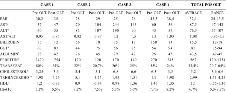
TABLE 2 – Biochemistry pre and post-transplantation: post-transplant data at the time of the biopsy with NASH
CASE 1 CASE 2 CASE 3 CASE 4 TOTAL POS OLT
Pre OLT Post OLT Pre OLT Post OLT Pre OLT Post OLT Pre OLT Post OLT AVERAGE RANGE
BMI1 30,2 35 28 29 25 26 43,5 38,4 32,1 25-43,5
AST2 57 47 70 104 244 143 60 56 87,5 47-143
ALT3 60 55 85 107 190 90 45 54 76,5 55-107
AST/ALT 0,95 0,85 0,82 0,97 1,2 1,5 1,3 1,03 1,08 0,85-1,5
BILIRUBIN5 75 12 56 18 75 18 158 14 15,5 12-18
GGT6 60 87 44 75 56 85 54 94 85 75-94
ALBUMIN7 28 42 26 45 29 42 25 45 43,5 42-45
FERRITIN8 2420 1754 170 120 170 149 270 245 567 120-1754
TRANSF.SAT 80% 64% 22% 20,7% 26% 25% 35% 24% 33,4% 20,7-64%
CHOLESTEROL9 3,25 3,6 5,4 5,1 6,8 6,6 6,3 5,5 5,2 3,6-6,6
TRIGLYCERIDES10 1,98 4,25 5,1 4,25 1,95 1,51 1,9 1,98 2,99 1,51-4,25
HDL11 1,2 0,97 0,9 0,74 0,98 1,36 1,46 1,35 1,1 0,75-1,36
HbA1c12 5,2% 5,5% 7,2% 7,5% 5,3% 5,6% 7,7% 8,2% 6,7% 5,5-8,2%
1BMI (Body Mass Index): normal BMI = 18.5 to 24.9 kg/m2, overweight = 25.0 to 29,9 kg/m2, obesity = 30,0 to 39,9 kg/m2, and morbid obesity > 40 kg/m2
2AST = 5 – 43 U/L, 3ALT = 5-35 U/L, 5Bilirubin = 1 – 22 mmol/L, 6GGT = 9 – 50 U/L, 7Albumin = 34 – 51 g/L, 8Ferritin = 10 – 300 mmol/L, 9Cholesterol < 5 mmol/L, 10Triglycerides
FIGURE 1 – Severe macronodular steatosis in a biopsy 22 months post-OLT (patient 1)
FIGURE 2 – Steatohepatitis in a biopsy 64 months post-OLT (patient 2). Balloned hepatocytes contain Mallory’s hyaline. There is an inflammatory infiltrate including neutrophils
TABLE 3 – Histological findings Post-OLT
Patient Months post-OLT Fatty change Mallory’s Neutrophils Fibrosis Main diagnosis
hyaline
1 22 3 1 1 1 1) Steatohepatitis
49 3 1 0 1 1) Steatohepatitis
2 12 2 0 0 1 1) Fatty change? Cause
24 2 0 0 1 1) Fatty change? Cause
37 2 0 0 1 1) Fatty change? Cause
64 3 2 1 2 1) Steatohepatitis/Fibrosis
54 2 1 1 2-3 1) Steatohepatitis/Fibrosis with early cirrohosis
3 1 2 0 0 0 1) Moderate acute rejection
4 1 0 0 0 1) Mild acute rejection
24 3 0 0 1 1) Mild chronic hepatitis
2) Severe fatty change? Cause
27 2 0 0 1 1) Moderate chronic hepatitis
2) Fatty change? Cause
45 1 2 1 1 1) Mild chronic hepatitis
2) Steatohepatitis/Fibrosis
4 29 3 0 0 1 1) Fatty change? Cause
44 2 1 1 2-3 1) Steatohepatitis/Fibrosis with early cirrohosis
70 2 1 1 2-3 1) Steatohepatitis/Fibrosis with early cirrohosis
Development of fibrosis
In all biopsies there were features of pericellular fibrosis (Fig. 4). Pericellular fibrosis was also noted in some biopsies showing fatty changes without other diagnostic feature of steatohepatitis.
In two cases (patients 1 and 3) fibrosis persisted as a mild lesion confined to perivenular lesions (acinar zone 3).
FIGURE 4 – Pericellular fibrosis in a biopsy 44 months post-OLT. Haematoxylin Van Gienson
FIGURE 3 – Steatohepatitis in biopsy 64 months post-OLT (same biopsy as Figure 2) Ubiquitin immunostaining reveals abundant Mallory’s hyaline like material in perivenular hepatocytes (zone 3)
FIGURE 5 – Micronodular cirrhosis in a biopsy 44 months post-OLT (same biopsy as Figure 4). Haematoxylin Van Gienson
DISCUSSION
A small number of cases with recurrent NASH following liver
transplantation have been reported(12), however de novo NASH in the
liver allograft has not been, so far described. Recurrences after liver transplantation suggest persistence of the pathogenic mechanisms responsible for the original disease or introduction of new mechanisms that produce the same histologic pattern.
According to CZAJA(6), two hypotheses of pathogenesis are
pertinent: the metabolic and free radical hypothesis. The metabolic hypothesis presumes that there is decreased tissue sensitivity to
insulin(3, 17) reducing tissue responsiveness, most commonly occurring
in conjunction with diabetes and/or obesity. This lack of responsiveness results in relative hyperinsulinism, which in turn
impairs beta-oxidation of fatty acids and promotes hepatic steatosis(16).
Free fatty acids (especially the unsaturated variety) can impair hepatocytic membrane integrity, depress enzyme activity, directly injure blood vessels, cause mithocondrial swelling, and increase
lysosomal fragility(1).
The free radical hypothesis presumes that endotoxemia is the basis for disturbances in hepatic lipid metabolism. Decreased hepatic clearance of endotoxin by the newly implanted liver and/or excessive endotoxin production from occult infection or intestinal bacterial overgrowth are unproven requisites for the hypothesis. Endotoxin can produce peroxisomal dysfunction, stimulate super oxide and hydrogen peroxide production, and release tumor necrosis factor from
monocytes(8, 10). These consequences can result in hepatic steatosis
and liver cell necrosis. Indeed, depletions of glutathione, vitamin E,
and superoxide dismutase occur commonly in systemic illness(8, 10),
and similar deficiencies of intrinsic antioxidants may be present before and after liver transplantation.
In this small series we report four patients with NASH post-transplantation, where in two cases NASH could be the cause of end-stage liver disease and in the other two NASH was a new event in the graft.
All four patients are alive with no graft failure after follow-up up to 5 years. The recurrence of fatty infiltration occurred within 21 months and transition from mild steatosis to NASH and early fibrosis was observed within 60 months post transplant in all four patients. This appears far more rapid and aggressive than the progression in the native liver reported in the literature, although the rate of
progression of NASH is debated(2). Glucocorticoids are frequently
implicated in the pathogenesis of steatosis and steatohepatitis; however
there is little evidence to support this association(14) and all four
patients were withdrawn from steroid within 3 months post-transplantation, following the unit policy.
and its inherent complications. The reason for different course of patients suffering apparently the same condition suggest that a less aggressive mechanism of liver injury operates in some patients whereas, in others, NASH seems to be potentiate by aggravating factors leading to increased fibrosis and cirrhosis. In a large series
published by ANGULO et al.(2) shown that some of these factors
include older age, greater BMI (obesity) and presence of diabetes mellitus. In all four patients described, risk factors pre and post-OLT for developing NASH were identified. Two patients (cases 2 and 4) were diabetic pre-OLT, on insulin control. None of the patients has developed new-onset diabetes post-transplant.
The relation of body weight and recurrence or development of steatohepatitis suggests that the persistence of obesity and/or weight gain play a role in the pathogenesis of post-transplant NASH. Again, obesity was observed in two of four patients.
Hyperlipidaemia occurs frequently in the transplant setting and is attributed to the immunosuppression. All patients developed hyperlipidaemia post-OLT, three with hypercholesterolemia, one associated hypertriglyceridemia and one patient (case 1) developed hight level of triglyceride alone.
Liver function tests were abnormal in all patients, at the time of the liver biopsy. With regard to biochemical parameters, no characteristic pattern was identified. The AST/ALT ratio less than 1 was found in two patients and more than 1 in the other two. The two patients presented with early cirrhosis had AST/ALT more and less
than 1 respectively, not reflecting some findings that change from <1.0 in uncomplicated NASH to > than 1 when cirrhosis is present.
Iron studies were abnormal in one patient (with α-1 antitrypsin
deficiency and iron overload).
In conclusion, all four cases of NASH post-transplantation demonstrate that recurrence and new onset of steatohepatitis in the liver allograft is possible. These cases-reports can suggest the hypothesis of perpetuation or creation of a pathogenic process that is specific for the host. Obesity, diabetes, drugs or alcohol abuse, chronic viral infection and bulimia are examples of host-dependent disorders that could afflict the native and implanted liver in a similar fashion.
The time of recurrence may be shorter than in the native liver and some patients develop hepatic fibrosis within 2 years. Indeed, NASH must be considered the cause of end-stage liver disease for which OLT was indicated in two cases. In those who were transplanted for other indications, the association with risk factors, such as obesity, diabetes and hyperlipidaemia, probably relates for the development of de novo NASH.
Although biochemical tests are not diagnostic, steatosis/ steatohepatitis can be a cause for sustained abnormalities of liver function tests in patients post-transplantation. Management of obesity, diabetes mellitus and hyperlipidaemia may play a therapeutic role in the prevention of recurrent and de novo NASH following OLT.
Garcia RFL, Morales E, Garcia CE, Saksena S, Hübscher SG, Elias E. Recurrência e "de novo" esteatohepatite não-alcoólica após transplante ortotópico de fígado. Arq Gastroenterol 2001;38(4):247-253.
RESUMO – Racional – O termo NASH (esteatohepatite não-alcoólica) foi introduzida em 1980 para descrever "características patológicas
e clínicas de doença não-alcoólica observadas comumente na própria doença alcoólica". Atualmente é causa reconhecida de doença hepática crônica e rara indicação de transplante hepático. Pequeno número de casos de recurrência de NASH pós-transplante foram descritos na literatura; entretanto, de novo NASH no enxerto jamais foi relatado. Objetivos/Resultados - Reportam-se quatro casos de NASH pós-transplante, descrevendo fatores associados a esta patologia. A média de recurrência da infiltração gordurosa foi de 21 meses com transição para esteatohepatite/fibrose aos 60 meses pós-transplante. Os quatro casos possuiam associação com um ou vários fatores de risco (obesidade, diabetes tipo 2 e/ou hiperlipidemia) no período que se seguiu ao transplante. Conclusão - Manejo destes fatores provavelmente exercem papel terapêutico na prevenção da recurrência e no aparecimento de NASH no pós-transplante.
DESCRITORES – Fígado gorduroso. Hepatite. Transplante de fígado. Diabetes mellitus. Recidiva.
REFERENCES
1. Acosta D, Wenzel DG. Injury produced by free fatty acids to lysosomes and mitochondria in cultured heart muscle and endothelial cells. Atherosclerosis 1974;20:417-26.
2. Angulo P, Keach JC, Batts KP, Lindor KD. Independent predictors of liver fibrosis in patients with nonalcoholic steatohepatitis. Hepatology 1999;30:1356-62. 3. Braillon A, Capron JP, Herve MA, Degott C, Quenum C. Liver in obesity. Gut
1985;26:133-9.
4. Burke GW, Cirocco R, Hensley G, Ranjan D, Reddy R, Jeffers L, Schiff E, Miller J. Liver transplantation for cirrhosis following jejuno-ileal bypass – regional cytokine differences associated with pathological changes in the transplant liver. Transplantation 1992;54:374-7.
5. Caldwell SH, Oelsner DH, Iezzoni JC, Hespenheide EE, Battle EH, Driscoll CJ. Cryptogenic cirrhosis: clinical characterization and risk factors for underlying disease. Hepatology 1999;29:664-9.
7. Diehl AM, Goodman Z, Ishak KG. Alcohollike liver disease in nonalcoholics. A clinical and histologic comparison with alcohol-induced liver injury. Gastroenterology 1988;95:1056-62.
8. Doherty JF, Golden MH, Brooks SE. Peroxisomes and the fatty liver of
malnutrition: an hypothesis. Am J Clin Nutr 1991;54:674-7.
9. Dray X, Tainturier MH, De La Lande P, Marty O, Mallet L. Cirrhose avec
stéatohépatite non alcoolique: rôle du tamoxifene.. Gastroenterol Clin Biol 2000;24:1122-3.
10. Golden MH, Ramdath D. Free radicals in the pathogenesis of kwashiorkor. Proc Nutr Soc 1987;46:53-68.
11. James O, Day C. Non-alcoholic steatohepatitis: another disease of affluence. Lancet 1999;353:1634-6.
12. Kim WR, Poterucha JJ, Porayko MK, Dickson ER, Steers JL, Wiesner RH.
Recurrence of nonalcoholic steatohepatitis following liver transplantation. Transplantation 1996;62:1802-5.
13. Ludwig J, Viggiano TR, McGill DB, Oh BJ. Nonalcoholic steatohepatitis: Mayo Clinic experiences with a hitherto unnamed disease. Mayo Clin Proc 1980;55:434-8.
14. Manton ND, Lipsett J, Moore DJ, Davidson GP, Bourne AJ, Couper RT. Non-alcoholic steatohepatitis in children and adolescents. Med J Aust 2000;173:476-9. 15. Sorrell MF, Mukherjee S. Non-alcoholic steatohepatitis (NASH). Curr Treat
Options Gastroenterol 1999;2:447-50.
16. Tankurt E, Biberoglu S, Ellidokuz E, Hekimsoy Z, Akpinar H, Cümlekçi A, Okan A, Sagol O. Hyperinsulinemia and insulin resistance in non-alcoholic steatohepatitis. J Hepatol 1999;31:963.
17. Wanless IR, Lentz JS. Fatty liver hepatitis (steatohepatitis) and obesity: an autopsy study with analysis of risk factors. Hepatology 1990;12:1106-10.