NON-ALCOHOLIC FATTY LIVER DISEASE -
CHANGING THE PREVALENCE OF LIVER CANCER?
Benedetta Campana,
1,2David Semela,
3Markus Heim,
1,2*Christine Bernsmeier
1,3,41. Department of Biomedicine, University of Basel, Basel, Switzerland
2. Division of Gastroenterology and Hepatology, University Hospital Basel, Basel, Switzerland 3. Division of Gastroenterology and Hepatology, Cantonal Hospital St. Gallen, St. Gallen, Switzerland
4. Institute of Liver Sciences, King’s College London, London, UK *Correspondence to [email protected]
Disclosure: No potential conflict of interest.
Received: 31.10.14 Accepted: 03.12.14
Citation: EMJ Hepatol. 2015;3[1]:12-18.
ABSTRACT
Due to its increasing prevalence, exceeding 25% of the Western population, non-alcoholic fatty liver disease (NAFLD) merits recognition as one of the most frequent chronic liver diseases (CLD) and requires consideration of the associated disease-related complications and their consequences for the surveillance and treatment of patients and the socio-economy worldwide. Along with the increasing incidence of NAFLD-related cirrhosis and end-stage liver disease, the frequency of NAFLD-related hepatocellular carcinoma (HCC) is rising and expected to surpass HCC related to chronic hepatitis C in the upcoming future. These epidemiologic changes will impact on the overall mortality of CLD and the requirement of organs for transplantation. Although the risk of HCC in NAFLD, similar to other CLD, is related to fibrosis (advanced fibrosis increases the risk of HCC 25-fold), there are reports suggesting a considerable rate of HCC also developing in simple hepatic steatosis. Moreover, HCC is nowadays the leading cause of obesity-related cancer mortality; cancers of other origin such as colorectal cancer are more prevalent in patients with NAFLD and obesity. The pathophysiology of HCC has mainly been studied in models of viral hepatitis. Given the expected raise in NAFLD-related HCC, a better understanding of the pathophysiology of carcinogenesis in NAFLD and obesity is desired in order to better define chemopreventive strategies. Here we review the epidemiology, aetiology, and pathogenesis of HCC on the background of NAFLD and deduce potential consequences for the management of patients in respect to the NAFLD epidemic.
EDITOR’S PICK
With the introduction of effective curative treatment for hepatitis C, the incidence of the hepatitis C virus (HCV)-associated hepatocellular carcinoma (HCC) will be gradually declining within the next 20 years, at least in Western countries which will have wider access to these drugs within the next few years. Since chronic hepatitis B is also fairly well controlled in patients with known infection, alcoholic and non-alcoholic fatty liver disease are the two remaining big aetiologies of chronic liver disease in need of better treatments. Amongst the two, it is non-alcoholic fatty liver disease (NAFLD) that has shown the biggest increase in prevalence globally, mostly in developed countries but also increasingly in developing countries. Despite the fact that severe liver damage occurs only in a small fraction of patients with NAFLD, the proinflammatory environment constitutes an important precancerous condition, making liver cancer the most important obesity-related malignancy, particularly in men. Understanding this means putting a major effort into preventing NAFLD, into identifying what drives the
inflammation in the liver, and ultimately into finding a cure for it.
NON-ALCOHOLIC FATTY LIVER DISEASE
(NAFLD)
NAFLD is one of the rising epidemics of the 21st
century in developed countries given its close association with obesity and the metabolic syndrome (MetS). The disorder was first described and named ‘non-alcoholic steatohepatitis (NASH)’ in 1980 by Ludwig et al.1 It is defined as triglyceride
accumulation of >5% in hepatocytes.2,3 Histological
features of NASH are indistinguishable from those that hallmark alcoholic steatohepatitis (fatty vacuoles, lobular hepatitis, mixed inflammatory infiltrate, presence of Mallory bodies, and fibrosis in some cases) but patients present without a personal history of alcohol consumption. For the diagnosis of NAFLD a maximal alcohol intake of 20 g/day of alcohol for women and 40 g/day for men is tolerated. Nowadays NAFL (non-alcoholic fatty liver) and NASH are classified using the NAFLD activity score (NAS) composed of 14 histological features.2 Recently, the group of
Bedossa4,5 developed and validated the
SAF-Score (S: steatosis; A: activity; F: fibrosis for the histopathological classification of liver lesions in patients with morbid obesity. The SAF score allows a better distinction between NAFL and NASH, and high interobserver agreement. To date, the prevalence of NAFLD exceeds 25% in Western countries6 and requires clear guidelines as to how
patients need to be stratified and surveyed.
Epidemiology
Clinical studies estimate the prevalence of NAFLD and NASH in Western countries at 20-30% and 3-5%, respectively.7 The studies are based on
different diagnostic criteria including ultrasound (US),8 liver biopsies performed before planned liver
transplantation, and autopsies. Numbers are even higher in autopsy-based studies including obese patients.9 The prevalence of NAFLD in normal
weight individuals (body mass index [BMI] ≤25 kg/ m2) in the NHANES III study was 4-times lower than
in obese patients (7% versus 28%).7
Aetiology
NAFLD is associated with obesity, impaired glucose tolerance, Type 2 diabetes mellitus (T2DM) and arterial hypertension – and regarded as the hepatic manifestation of the MetS. Pathophysiologically,
there is evidence that the accumulation of intracellular lipids in hepatocytes reduces insulin clearance and promotes insulin resistance (IR), respectively.10 The presence of IR is regarded as the
main predisposing factor for metabolic diseases, independently of the BMI and the visceral fat deposit.11 However, whether NAFLD is the cause
or consequence of metabolic disorders, needs to be clarified. Fortunately NAFL, otherwise termed ‘simple steatosis’, is supposed to follow a benign course, generally without the risk of complications or life-threatening consequences, although reports of cirrhosis in 4% of patients with simple steatosis have been published.12
About 25% of all patients with fatty liver develop inflammation and fibrosis termed NASH.13 NASH is
considered to be the more severe form of NAFLD, bearing the risk of hepatic and extrahepatic complications. In about one-third of patients, NASH leads to cirrhosis, which in turn may lead to liver related death, including the development of hepatocellular carcinoma (HCC).14 To investigate
the natural history, NAFLD patients were re- biopsied at least 3 years after their first histologic assessment in a non-interventional study. Indeed, progression to cirrhosis differs between NAFL and NASH patients: none of the patients diagnosed with NAFL at the time of the first histology showed fibrosis in the repeated biopsy, but one-third of patients with initial diagnosis of NASH had a progression of the degree of fibrosis.15 NAFLD/
NASH is expected to represent the leading indication for liver transplantation within the next few years.16
Besides this, the cardiovascular risk is markedly increased in patients with MetS and NAFLD. Patients with NAFLD have increased intima thickness of carotid artery, and also higher incidence of coronary, periphery, and cerebrovascular diseases.17 While a cohort study showed no impact
on survival after a follow-up of 13 years in patients with NAFL, survival was significantly reduced in patients with NASH.18 A different cohort revealed
a higher liver-related mortality in patients with NASH. Overall, mortality was higher if patients were older at diagnosis, had T2DM, or a low level of albumin.19
Pathophysiology
The sequence of progression from simple steatosis (NAFL) to inflammation and fibrosis (NASH) remains poorly understood. The mechanisms contributing to the accumulation of fat in hepatocytes and successive induction of a local inflammatory response in the liver need further elucidation. There is a link between hepatic steatosis and increased hepatic activity of the nuclear factor
κ
B (NFκ
B). The upregulation of NFκ
B related pro-inflammatory cytokines (interleukin 6 [IL-6], tumour necrosis factor alpha [TNF-α
], interleukin 1 beta [IL-1β
]) activates Kupffer cells, contributing to inflammation and cell injury.20There is substantial evidence for a genetic component in the development of NAFLD. Observational studies detected lower prevalence in African Americans than in Hispanic Americans.21
A number of genes have subsequently been associated with the phenotype of NAFLD. One documented genetic association is a missense variant in PNPLA3. PNPLA3 (Patatin like phospholipase domain-containing protein 3) is encoding a lipase protein. A single nucleotide polymorphism (rs738409, encoding I148M) was associated with hepatic fat accumulation.22
Patients that are homozygous for this allele accumulated twice as much intrahepatic fat, compared to those who didn’t carry the mutation. Moreover, differences in the composition of the gut microbiota of obese and NAFLD patients have been observed. Modifications of the microbiota can promote gut permeability, allowing the translocation of endotoxins and other bacterial products and of luminal antigens, which may enhance hepatic inflammation.23
HCC IN NAFLD
The epidemic of NAFLD elicits a rising number of NAFLD associated advanced fibrosis and increases the burden of HCC. The overall 5-year survival is as low as 15%, given that only 13% of patients diagnosed with HCC are eligible for therapeutic interventions.24 HCC screening strategies for
patients with NAFLD need to be defined in order to detect HCC early and enable treatment within the means of healthcare resources. Effective chemopreventive strategies for patients at increased risk for HCC are desirable.16 Numbers
for the incidence of HCC in NAFLD vary upon the population studied. A single-centre cohort study recently reported development of HCC in >12%
of patients with NASH cirrhosis over a median observation period of 3.2 years, consistent with a yearly HCC incidence of 2.6% after the diagnosis of cirrhosis.25 Another Danish cohort had reported
a yearly incidence of 4.6%.26 As additional risk
factors for the development of HCC, older age, higher BMI, and tobacco and alcohol consumption were detected.27,28
The incidence of HCC increased over the last decades, representing 90% of all liver cancers and the third cause of cancer-related mortality worldwide.29 Known risk factors for developing
HCC are chronic hepatitis B (HBV) and chronic hepatitis C virus (HCV) infection, but also chronic alcohol consumption. The incidence of HCC in patients with chronic hepatitis C and advanced fibrosis is higher compared to the incidence in patients with NASH (7% versus 2%, respectively).25
Because of the high prevalence, cases of NAFLD-associated disease have become one of the leading causes of HCC in the United States. The reported proportion of HCC attributed to the diagnosis of NAFLD ranges between 4-22%.30 Marrero et al.28
found underlying ‘cryptogenic’ cirrhosis in 29% of the investigated cases with HCC; among these, 50% displayed clinical and histological features of NASH.
Although advanced fibrosis increased the risk of HCC in NAFLD about 25-fold,31,32 there is evidence
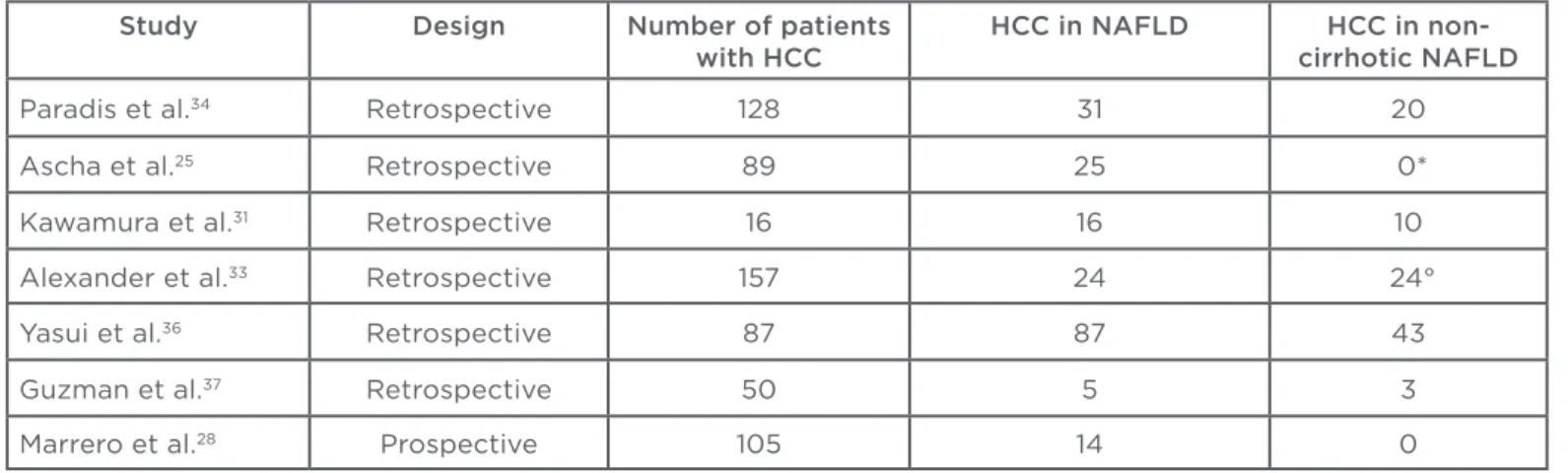
suggesting that HCC can also develop in a simple steatotic liver or in NASH without the presence of cirrhosis.33,34 In a retrospective histopathological
study, including 128 cases of HCC that had undergone resection, Paradis et al.34 identified
that 65% of HCC, in the group of patients with underlying MetS-related liver disease, developed in livers free of significant fibrosis (F0-F2), versus only 26% in the group of patients with other underlying chronic liver diseases (CLD). A similar study in >800 patients diagnosed with HCC, revealed that 42.6% of tumours developed in a non-cirrhotic liver regardless of the diagnosis.35 A Japanese
study reported a rate of complete cirrhosis in liver tissue surrounding the HCC in only 51% of patients with histologically proven NASH. Moreover, the prevalence of cirrhosis was lower in male compared to female individuals (39% versus 70%).36 Another
cohort found HCC in NAFL without features of inflammation, fibrosis or cirrhosis in 3 out of 50 patients (see Table 1).37
with and without cirrhosis. The association between NAFLD and risk of HCC seemed to be limited to individuals showing features of cirrhosis.38 In
summary, cirrhosis is apparently not required for the development of HCC in chronic NAFLD-related liver disease. However, the relatively low risk of developing HCC in a non-cirrhotic NAFLD liver does not justify HCC screening of the entire NAFLD population. This is an evolving topic. The differential morphological features and pathophysiological mechanisms of hepatocarcinogenesis, in respect to the underlying liver diseases, i.e. NAFLD versus other CLD, and the individual contributions of obesity, T2DM, steatosis, NASH, and degree of fibrosis need detailed elucidation.
Pathophysiology
As detailed for other malignancies, HCC emerges mostly in patients with CLD and develops over years following a dysplasia-carcinoma sequence.39 The
pathophysiological mechanism of HCC development has mainly been studied in models of chronic viral infection with HBV and HCV. In HCC developing in the livers of patients with NAFLD, hepatic oncogenic drivers such as steatosis and inflammation as well as systemic factors, i.e. obesity and T2DM, have been identified. Hepatic steatosis can cause inflammation with subsequent up-regulation of pro-inflammatory cytokines, e.g. IL-1
β
, IL-6, and TNFα
.40 In accordance, activation of hepatic Kupffercells has been described in the liver of obese patients, as well as induced fibrosis.41 Vice-versa,
oxidative stress and inflammatory responses are
reduced in obese persons after fasting for 48 hours42 or during a low calorie (1,000 kcal/day)
diet over 4 days.43 In a murine model, NASH and
HCC have been induced after (6-) 12 months of American Lifestyle-Induced Obesity Syndrome (ALIOS) diet and sedentary lifestyle. Mice developed typical metabolic changes of NAFLD, such as higher transaminases and higher triglyceride. These histological alterations reflected the characteristics found in the liver of patients with NAFLD: steatosis, ballooned hepatocytes, Mallory bodies, lobular inflammation, peri- sinusoidal fibrosis, and activation of stellate cells. Conclusively, a higher NAS score was observed after 12 months in mice on the ALIOS diet and sedentary lifestyle, compared to mice on a control diet. Surprisingly, 6 out of 10 mice developed hepatocellular neoplasms after 12 months.44
Another study published by the same group explored the effect of 5-alpha-reductase (5
α
R).45 5α
R is needed to convert cortisolto inactive metabolites, and testosterone to dihydrotestosterone. Both steroids are involved in the regulation of lipid metabolism. The expression level of 5
α
R-isoforms (Type 1 and Type 2) was similar in NAFLD and healthy control livers. The authors suggest a potential protective role for reduced expression of 5α
R Type 1, since 5α
R-/- knock-out mice (Type 1, only present inmice) displayed enhanced hepatic steatosis, but developed fewer hepatocellular lesions in response to the ALIOS diet. This mechanism needs to be verified in humans.
Table 1: Epidemiologic studies evaluating the development of hepatocellular carcinoma (HCC) in non-alcoholic fatty liver disease (NAFLD).
Study Design Number of patients with HCC
HCC in NAFLD HCC in non-cirrhotic NAFLD
Paradis et al.34 Retrospective 128 31 20
Ascha et al.25 Retrospective 89 25 0*
Kawamura et al.31 Retrospective 16 16 10
Alexander et al.33 Retrospective 157 24 24°
Yasui et al.36 Retrospective 87 87 43
Guzman et al.37 Retrospective 50 5 3
Marrero et al.28 Prospective 105 14 0
As discussed above, a genome-wide association study identified a variant in the PNPLA3 gene was associated with susceptibility to NAFLD. The same gene was independently associated with a higher risk of progressive liver fibrosis and HCC. This homozygous mutation is often found in HCC with lower differentiation, multiple foci at diagnosis, and reduced survival for these patients.46,47 Various
studies suggested a higher incidence of neoplastic lesions other than HCC, such as colorectal adenoma and high-grade dysplastic neoplasms of the colon in patients with NAFLD.48,49 On the
other hand, in a meta-analysis of 26 studies, El- Serag et al.50 found a higher incidence of HCC in
patients with diabetes mellitus, regardless of the presence of NAFLD. Also obesity alone has been linked to a higher incidence of malignancies of diverse organs (i.e. colon, endometrium, breast, intestine, liver).51
Vaccine and antiviral strategies against HBV and therapy for chronic HCV are effective in lowering the incidence of HCC in these conditions. A recent review by Singh at al.16 systematically discussed
chemopreventive strategies against HCC. The use of statins seemed to reduce the risk of HCC in epidemiological studies, but has not been demonstrated in double-blind placebo-controlled trials. These trials had mostly been designed to evaluate cardiovascular end points and addressed the incidence of HCC as a post-hoc analysis only. Other observational studies in diabetic patients detected a reduced risk of HCC in those treated with metformin. The effect may be promoted through inhibition of cell growth and downregulation of c-Myc. Aspirin, bearing an anti-inflammatory effect, had some antineoplastic properties. However, epidemiological studies might be biased and overestimate the actual effect. Dietary changes have also been evaluated: coffee, known to reduce the risk of fibrosis and cirrhosis, seemed to be effective in reduction of HCC regardless of the chronic underlying liver disease.16
RECOMMENDATIONS FOR DIAGNOSTICS,
TREATMENT, AND FOLLOW-UP OF
PATIENTS WITH NAFLD
In medical practice, patients with persistently elevated transaminases should be referred to a specialised hepatology centre for work up of the underlying condition. Hepatic steatosis can be detected using non-invasive imaging techniques, such as US and magnetic resonance imaging
(MRI), but these do not allow for the assessment of hepatic fibrosis. Non-invasive methods to identify fibrosis in NAFLD include NAFLD Fibrosis Score, levels of cytokeratin 18 (CK18), and measuring transient elastography of the liver.52 The NAFLD
Fibrosis score is a calculated score based on age, BMI, albumin, aspartate aminotransferase/alanine transaminase ratio, hyperglycaemia, and platelet count.53 CK18 is significantly increased in patients
with NASH compared to NAFL or absence of steatosis.54 The assessment of liver stiffness by
transient elastography (Fibroscan) in individuals with biopsy proven NAFLD showed a high negative predictive value to exclude advanced fibrosis, but often failed in patients with high BMI.55
Although an invasive procedure bearing the risk of complications, liver biopsy is still the diagnostic gold standard and the only method allowing the detection and quantification of fibrosis and inflammation in the steatotic liver. Also, it is important to screen for other CLD since steatosis can be detected in hepatitis C, Wilson’s and coeliac disease, or as a consequence of the toxic effects of alcohol or prescription drugs. Since, depending on the population studied, >25% of the population in Western countries are predicted to carry the diagnosis of NAFLD, a general follow-up of each and every patient in a specialised centre cannot be recommended. However, patients with biopsy proven NASH need counselling regarding the risk of disease progression towards cirrhosis and possible life-threatening complications, such as acute-on-chronic liver failure or HCC. Furthermore, potential extrahepatic complications such as malignancies other than HCC and the risk of cardiovascular disease need to be evaluated and treated. Regular follow-up of patients with NASH is recommended in order to monitor disease progression. The recommended treatment for NAFLD is lifestyle modification with the aim to reduce weight but also IR, the major target of current therapeutic approaches.56 Until
now, no effective pharmaceutical therapy has been approved.52
A number of clinical trials evaluating novel targets are ongoing. Promising effects were shown in the FLINT trial57 evaluating obeticholic acid (OCA)
versus placebo in patients with NASH. Treatment with OCA for 72 weeks improved histological findings. However pruritus occurring in 23% of patients on OCA requires further clarification of drug-safety.57 Another multicentre randomised
REFERENCES
1. Ludwig J et al. Nonalcoholic steatohepatitis: Mayo Clinic experiences with a hitherto unnamed disease. Mayo Clin Proc. 1980;55(7):434-8.
2. Kleiner DE et al; Nonalcoholic Steatohepatitis Clinical Research Network. Design and validation of a histological scoring system for nonalcoholic fatty liver disease. Hepatology. 2005;41(6):1313-21. 3. Brunt EM. Nonalcoholic fatty liver disease: what the pathologist can tell the clinician. Dig Dis. 2012;30 Suppl 1:61-8. 4. Bedossa P et al. Histopathological algorithm and scoring system for evaluation of liver lesions in morbidly obese patients. Hepatology. 2012;56(5):1751-9.
5. Bedossa P; FLIP Pathology Consortium. Utility and appropriateness of the fatty liver inhibition of progression (FLIP) algorithm and steatosis, activity, and fibrosis (SAF) score in the evaluation of biopsies of nonalcoholic fatty liver disease. Hepatology. 2014;60(2):565-75. 6. Lazo M et al. Prevalence of nonalcoholic fatty liver disease in the United States: the Third National Health and Nutrition Examination Survey, 1988-1994. Am J Epidemiol. 2013;178(1):38-45.
7. Armstrong MJ et al. Presence and severity of non-alcoholic fatty liver disease in a large prospective primary care cohort. J Hepatol. 2012;56(1):234-40. 8. Musso G et al. Meta-analysis: natural history of non-alcoholic fatty liver disease (NAFLD) and diagnostic accuracy of non-invasive tests for liver disease severity. Ann Med. 2011;43(8):617-49.
9. Zois CD et al. Steatosis and steatohepatitis in postmortem material
from Northwestern Greece. World J Gastroenterol. 2010;16(31):3944-9.
10. Tamura Y et al. Effects of diet and exercise on muscle and liver intracellular lipid contents and insulin sensitivity in type 2 diabetic patients. J Clin Endocrinol Metab. 2005;90(6):3191-6.
11. Fabbrini E et al. Obesity and nonalcoholic fatty liver disease: biochemical, metabolic, and clinical implications. Hepatology. 2010;51(2): 679-89.
12. Matteoni CA et al. Nonalcoholic fatty liver disease: a spectrum of clinical and pathological severity. Gastroenterology. 1999;116(6):1413-9.
13. McCullough AJ. The clinical features, diagnosis and natural history of nonalcoholic fatty liver disease. Clin Liver Dis. 2004;8(3):521-33, viii.
14. Powell EE et al. The natural history of nonalcoholic steatohepatitis: a follow-up study of forty-two patients for up to 21 years. Hepatology. 1990;11(1):74-80.
15. Fassio E et al. Natural history of nonalcoholic steatohepatitis: a longitudinal study of repeat liver biopsies. Hepatology. 2004;40(4):820-6.
16. Singh S et al. Chemopreventive strategies in hepatocellular carcinoma. Nat Rev Gastroenterol Hepatol. 2014;11(1):45-54.
17. Targher G et al. Risk of cardiovascular disease in patients with nonalcoholic fatty liver disease. N Engl J Med. 2010;363(14):1341-50.
18. Ekstedt M et al. Long-term follow-up of patients with NAFLD and elevated liver enzymes. Hepatology. 2006;44(4):
865-73.
19. Rafiq N et al. Long-term follow-up of patients with nonalcoholic fatty liver. Clin Gastroenterol Hepatol. 2009;7(2):234-8. 20. Day CP. Natural history of NAFLD: remarkably benign in the absence of cirrhosis. Gastroenterology. 2005;129(1):375-8.
21. Schwimmer JB et al. Heritability of nonalcoholic fatty liver disease. Gastroenterology. 2009;136(5):1585-92. 22. Romeo S et al. Genetic variation in PNPLA3 confers susceptibility to nonalcoholic fatty liver disease. Nat Genet. 2008;40(12):1461-5.
23. Miura K, Ohnishi H. Role of gut microbiota and Toll-like receptors in nonalcoholic fatty liver disease. World J Gastroenterol. 2014;20(23):7381-91. 24. El-Serag HB et al. Treatment and outcomes of treating of hepatocellular carcinoma among Medicare recipients in the United States: a population-based study. J Hepatol. 2006;44(1):158-66. 25. Ascha MS et al. The incidence and risk factors of hepatocellular carcinoma in patients with nonalcoholic steatohepatitis. Hepatology. 2010;51(6):1972-8.
26. Sørensen HT et al. Risk of cancer in patients hospitalized with fatty liver: a Danish cohort study. J Clin Gastroenterol. 2003;36(4):356-9.
27. Dyson J et al. Hepatocellular cancer: the impact of obesity, type 2 diabetes and a multidisciplinary team. J Hepatol. 2014;60(1):110-7.
28. Marrero JA et al. Alcohol, tobacco and obesity are synergistic risk factors for hepatocellular carcinoma. J Hepatol.
dehydrogenase Type 1 (11
β
-HSD1) versus placebo in patients with in MRI-detected NAFLD. 11β
-HSD1 reduced hepatic fat content by 2% while there was no difference in the placebo group.58 Inour NAFLD/NASH cohort, we documented a significantly lower secretion of glucose induced glucagon-like peptide-1 (GLP-1) along with a reduced glucose-lowering effect in NAFLD and NASH patients, endorsing ongoing clinical studies evaluating the use of GLP-1-analogs for the treatment of NAFLD.59
Gastric bypass or sleeve gastrectomy decrease the BMI and improve extrahepatic diseases related to MetS.60 However, the beneficial effect on hepatic
fibrosis or cirrhosis in patients with NASH could not be proven in cohort studies.61 Comorbidities
of MetS and a multidisciplinary team evaluation,
involving endocrinologists, surgeons, and gastroenterologists/hepatologists, clearly need to justify the indication for and the risks of this incisive surgical procedure. A recent study highlights the significantly increased peri and postoperative morbidity and mortality in cirrhotic patients showing 10% of postoperative mortality, 32% of major complications, and 43% deterioration of liver function after major surgery.62 All patients
2005;42(2):218-24.
29. European Association For The Study Of The Liver; European Organisation For Research And Treatment Of Cancer. EASL-EORTC clinical practice guidelines: management of hepatocellular carcinoma. J Hepatol. 2012;56(4):908-43.
30. Michelotti GA et al. NAFLD, NASH and liver cancer. Nat Rev Gastroenterol Hepatol. 2013;10(11):656-65.
31. Kawamura Y et al. Large-scale long-term follow-up study of Japanese patients with non-alcoholic fatty liver disease for the onset of hepatocellular carcinoma. Am J Gastroenterol. 2012;107(2):253-61. 32. Hashimoto E, Farrell GC. Will non-invasive markers replace liver biopsy for diagnosing and staging fibrosis in non-alcoholic steatohepatitis? J Gastroenterol Hepatol. 2009;24(4):501-3.
33. Alexander J et al. Non-alcoholic fatty liver disease contributes to hepatocarcinogenesis in non-cirrhotic liver: a clinical and pathological study. J Gastroenterol Hepatol. 2013;28(5): 848-54.
34. Paradis V et al. Hepatocellular carcinomas in patients with metabolic syndrome often develop without significant liver fibrosis: a pathological analysis. Hepatology. 2009;49(3):851-9. 35. Nzeako UC et al. Hepatocellular carcinoma in cirrhotic and noncirrhotic livers. A clinico-histopathologic study of 804 North American patients. Am J Clin Pathol. 1996;105(1):65-75.
36. Yasui K et al; Japan NASH Study Group, Ministry of Health, Labour, and Welfare of Japan. Characteristics of patients with nonalcoholic steatohepatitis who develop hepatocellular carcinoma. Clin Gastroenterol Hepatol. 2011;9(5):428-33; quiz e50.
37. Guzman G et al. Does nonalcoholic fatty liver disease predispose patients to hepatocellular carcinoma in the absence of cirrhosis? Arch Pathol Lab Med. 2008;132(11):1761-6.
38. White DL et al. Association between nonalcoholic fatty liver disease and risk for hepatocellular cancer, based on systematic review. Clin Gastroenterol Hepatol. 2012;10(12):1342-1359.e2.
39. Kojiro M, Nakashima O. Histopathologic evaluation of hepatocellular carcinoma with special reference to small early stage tumors. Semin Liver Dis. 1999;19(3): 287-96.
40. Shoelson SE et al. Obesity, inflammation, and insulin resistance. Gastroenterology. 2007;132(6):2169-80. 41. Farrell GC et al. NASH is an inflammatory disorder: pathogenic, prognostic and therapeutic implications. Gut Liver. 2012;6(2):149-71.
42. Dandona P et al. The suppressive effect of dietary restriction and weight loss in the obese on the generation of reactive oxygen species by leukocytes, lipid peroxidation, and protein carbonylation. J Clin Endocrinol Metab. 2001;86(1):355-62. 43. Dandona P et al. Inhibitory effect of a two day fast on reactive oxygen species (ROS) generation by leucocytes and plasma ortho-tyrosine and meta-tyrosine concentrations. J Clin Endocrinol Metab. 2001;86(6):2899-902.
44. Dowman JK et al. Development of hepatocellular carcinoma in a murine model of nonalcoholic steatohepatitis induced by use of a high-fat/fructose diet and sedentary lifestyle. Am J Pathol. 2014;184(5):1550-61.
45. Dowman JK et al. Loss of 5α-reductase type 1 accelerates the development of hepatic steatosis but protects against hepatocellular carcinoma in male mice. Endocrinology. 2013;154(12):4536-47. 46. Valenti L et al. PNPLA3 I148M polymorphism, clinical presentation, and survival in patients with hepatocellular carcinoma. PLoS One. 2013;8(10):e75982. 47. Hassan MM et al. Genetic variation in the PNPLA3 gene and hepatocellular carcinoma in USA: risk and prognosis prediction. Mol Carcinog. 2013;52 Suppl 1:E139-47.
48. Huang KW et al. Patients with nonalcoholic fatty liver disease have higher risk of colorectal adenoma after negative baseline colonoscopy. Colorectal Dis. 2013;15(7):830-5.
49. Wong VW et al. High prevalence of colorectal neoplasm in patients with non-alcoholic steatohepatitis. Gut. 2011;60(6):829-36.
50. El-Serag HB et al. The association between diabetes and hepatocellular carcinoma: a systematic review of epidemiologic evidence. Clin Gastroenterol Hepatol. 2006;4(3):369-80.
51. Wolk A et al. A prospective study of obesity and cancer risk (Sweden). Cancer Causes Control. 2001;12(1):13-21.
52. Chalasani N et al. The diagnosis and
management of non-alcoholic fatty liver disease: practice guideline by the American Association for the Study of Liver Diseases, American College of Gastroenterology, and the American Gastroenterological Association. Hepatology. 2012;55(6):2005-23.
53. Angulo P et al. The NAFLD fibrosis score: a noninvasive system that identifies liver fibrosis in patients with NAFLD. Hepatology. 2007;45(4):846-54.
54. Wieckowska A et al. In vivo assessment of liver cell apoptosis as a novel biomarker of disease severity in nonalcoholic fatty liver disease. Hepatology. 2006;44(1):27-33.
55. Kwok R et al. Systematic review with meta-analysis: non-invasive assessment of non-alcoholic fatty liver disease--the role of transient elastography and plasma cytokeratin-18 fragments. Aliment Pharmacol Ther. 2014;39(3):254-69. 56. Thoma C et al. Lifestyle interventions for the treatment of non-alcoholic fatty liver disease in adults: a systematic review. J Hepatol. 2012;56(1):255-66.
57. Neuschwander-Tetri BA et al. Farnesoid X nuclear receptor ligand obeticholic acid for cirrhotic, non-alcoholic steatohepatitis (FLINT): a multicentre, randomised, placebo-controlled trial. Lancet. 2014;doi:10.1016/ S0140-6736(14)61933-4. [Epub ahead of print].
58. Stefan N et al. Inhibition of 11β-HSD1 with RO5093151 for non-alcoholic fatty liver disease: a multicentre, randomised, double-blind, placebo-controlled trial. Lancet Diabetes Endocrinol. 2014;2(5):406-16.
59. Bernsmeier C et al. Glucose-induced glucagon-like Peptide 1 secretion is deficient in patients with non-alcoholic fatty liver disease. PLoS One. 2014;9(1):e87488.
60. Chavez-Tapia NC et al. Bariatric surgery for non-alcoholic steatohepatitis in obese patients. Cochrane Database Syst Rev. 2010;(1):CD007340.
61. Cotrim HP, Daltro C. Liver: does bariatric surgery reduce the severity of NAFLD? Nat Rev Gastroenterol Hepatol. 2010;7(1):11-3.