Alte re d he art rate and blo o d
pre ssure variability in m ice lacking
the
Mas
pro to o nco ge ne
1Max-Delbrück-Center for Molecular Medicine (MDC), and 2Department of Cardiology and Pneumology,
University Hospital Benjamin Franklin, Free University of Berlin, Berlin, Germany
3University of Applied Sciences, Jena, Germany T. Walther1, N. Wessel3,
N. Kang1, A. Sander3, C. Tschöpe2, H. Malberg3, M. Bader1 and A. Voss3
Abstract
Heart rate variability is a relevant predictor of cardiovascular risk in humans. A significant genetic influence on heart rate variability is suggested, although the genes involved are ill-defined. The Mas -protooncogene encodes a G-protein-coupled receptor with seven trans-membrane domains highly expressed in testis and brain. Since this receptor is supposed to interact with the signaling of angiotensin II, which is an important regulator of cardiovascular homeostasis, heart rate and blood pressure were analyzed in Mas-deficient mice. Using a femoral catheter the blood pressure of mice was measured for a period of 30 min and 250 data values per second were recorded. The mean values and range of heart rate and blood pressure were then calculated. Neither heart rate nor blood pressure were significantly different between knockout mice and controls. However, high resolution re-cording of these parameters and analysis of the data by non-linear dynamics revealed significant alterations in cardiovascular variability in Mas-deficient animals. In particular, females showed a strong reduction of heart rate variability. Furthermore, the data showed an increased sympathetic tone in knockout animals of both genders. The marked alterations detected in Mas-deficient mice of both genders suggest that the Mas-protooncogene is an important determinant of heart rate and blood pressure variability.
Co rre spo nde nce
T. Walther
Max-Delbrück-Center for Molecular Medicine (MDC) Robert-Rössle-Str. 10 D-13092 Berlin-Buch Germany
Fax: + 49-30-9406-2110 E-mail: thowal@ mdc-berlin.de
Presented at the XIII Annual Meeting of the Federação de Sociedades de Biologia Experimental, Caxambu, MG, Brasil, August 26-29, 1998.
Received July 26, 1999 Accepted September 16, 1999
Ke y wo rds
·Blood pressure variability ·Heart rate variability ·Baroreflex sensitivity ·Mas protooncogene
Intro ductio n
Blood-pressure variability (BPV) and heart-rate variability (HRV) are generated by the rhythmic actions of cardiovascular hormones and neuronal pathways on effec-tor organs such as heart, kidney, and blood vessels. HRV has been shown to be a rel-evant predictor for the mortality of patients
in-volved in the regulation of these variabilities are only poorly understood. The brain is the source for most of the signals believed to be responsible for the setting of blood pressure (BP) and heart rate (HR). Moreover, HRV was impaired by ischemic damage to the brain caused by stroke (8).Therefore, genes involved in cardiovascular regulation and expressed in the central nervous system should be interesting candidates for the mo-lecular genetic analysis of BPV and HRV.
In addition to being expressed in the testes, the Mas protooncogene (9)is mainly expressed in the brain, where its mRNA has been located in the hippocampus, dentate gyrus, piriform cortex and amygdala (10-12). In addition, Mas-mRNA was detected in low concentrations in kidney and heart (12,13). Functional studies have suggested that the Mas gene codes for an angiotensin II (AngII)-sensitive G-protein-coupled recep-tor (14). However, the increase of intracellu-lar Ca2+
-concentrations in Mas-transfected cells after AngII treatment could only be confirmed in cells endogenously expressing the AngII receptor AT1 (15). Still, the exact nature of the interaction of Mas, AngII and the AT1-receptor is elusive. To elucidate the relevance of the Mas protein, we eliminated the gene in mice by gene targeting technol-ogy (16). Apart from a changed anxiety be-havior, knockout animals exhibited as an outstanding feature an improved long-term potentiation (LTP) in the dentate gyrus. As AngII causes a reduction of LTP (17) the ablation of the Mas protein and the resulting decrease in AngII signaling could be an ex-planation for the observed stabilization of LTP. Since AngII is one of the main regula-tors of BP also via pathways in the central nervous system (18) we hypothesized that Mas may play a role in the regulation of BP and HR. To characterize this role, BP of
Mas-deficient mice was recorded continu-ously with high time resolution. From these data we extracted HR and systolic and dia-stolic BP on a beat-to-beat basis.
Further-more, the spontaneous baroreflex was exam-ined by the method of Parlow et al. (19) whereby spontaneous or accidental changes in BP under normal conditions are used for the calculations as opposed to the drug-in-duced baroreflex measurements. This pa-rameter is closely related to the variability of HR and BP and, therefore, can be regarded as a link between them.
Mate rial and Me tho ds
BP m e asure m e nt
Fourteen control (+/+) (7 males and 7 females) and 15 knockout (-/-) mice (6 males and 9 females) were used for the experi-ments at 14 weeks of age. The investigation was carried out according to the Guide for the Care and Use of Laboratory Animals
published by the US National Institutes of Health (NIH Publication No. 85-23, revised 1985). The animals were anesthetized with thiopental (50 ng/kg body weight) and a catheter was placed in the femoral artery. Five minutes after the end of the operation BP was measured for 30 min with a pressure transducer and a commercially available soft-ware (TSE, Bad Homburg, Germany). In addition to the normal measurement, BP was recorded at a high resolution (250 Hz sam-pling frequency).
Variability analysis o f HR and BP
is determined within physiological regions. From the resulting time series the measures of HRV are determined as far as the interbeat intervals are concerned, and in the case of the systolic and diastolic BP series the re-spective measures of BPV are determined.
Since arrhythmia and artifacts have a strong influence on the parameters of BPV and HRV analysis, it is necessary to exclude them from time series of variability. This exclusion is performed by an adaptive filter algorithm which takes into account the mo-mentary basic variability. The remaining data points are NN intervals (NN: interval be-tween two successive normal heartbeats). HRV and BPV series different measures were calculated from the resulting half hour. The meanNN (the mean), sdNN (the stan-dard deviation) and shannon (the shannon entropy of the histogram) were determined from the time domain parameters.
The following frequency domain param-eters were calculated from the time series interpolated to 50 ms by means of Fast Fou-rier Transformation and a Blackman-Harris-window: low frequency (lf)/high frequency (hf), lfn (standardized lf) with lf, the spectral power within 0.18 Hz to 1.0 Hz and hf, the spectral power within 1.0 Hz to 2.0 Hz.
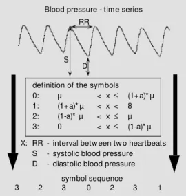
Furthermore, three further measures of non-linear dynamics were calculated from symbolic dynamics (20,21). Symbolic dy-namics is based on the transformation of the time series into symbols. By such transfor-mation a part of the detailed infortransfor-mation included in the time series is lost, but impor-tant information about dynamics within the time series remains. Depending on the cho-sen number of symbols and on the data length, words of different length are formed on the basis of the symbols. Figure 1 shows the transformation of the variability time series into a symbol time series. The original time series are transformed into symbols in accordance to the given definition depend-ing on the mean µ and on the parameter a.
The parameter a determines the permitted deviation from the mean and was set at 0.05. Words consisting of four symbols {0,1, 2,3} with the length of three were analyzed. The following parameters were extracted from the word distribution: fwshannon, Shannon-entropy (a well-known measure for complexity; 21); wpsum13, percentage of words containing only the symbols 1 and 3 and wsdvar, word variability.
Baro re ce pto r re fle x
The spontaneous baroreflex was meas-ured by the method of Parlow et al. (19). The time series of systolic BP and HR were examined in parallel for the three following signal scores respectively, where HR is de-creased while BP is inde-creased. The number and the increase of the spontaneous changes were then recorded.
Statistical m e tho ds
Since the calculated parameters were partly not normally distributed and since normal distribution is a precondition for many statistical procedures, the parameters were initially transformed logarithmically. They were then analyzed by the Kolmogorov-Smirnov test for normal distribution, and
Blood pressure - time series RR
S D
symbol sequence definition of the symbols 0: µ < x£ (1+a)* µ 1: (1+a)* µ < x < 8 2: (1-a)* µ < x£ µ 3: 0 < x£ (1-a)* µ
X: RR - interval betw een tw o heartbeats S - systolic blood pressure D - diastolic blood pressure
3 2 3 0 2 3 1
Figure 1 - The basic principle of sym bolic dynam ics, i.e., t he transformation into symbols. For each value of the tn-tn-1
this hypothesis was confirmed for all param-eters. Thereafter, univariate analyses of vari-ance were performed for the separation of control animals from the knockout animals. This was done for both the whole groups and the gender-matched subgroups. The crite-rion for significance was 5%.
To examine sex specificity, gradual dis-criminate analyses were applied additionally to differentiate between groups.
Re sults
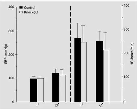
To study the interrelation between AngII and the Mas-gene product we investigated mean BP and HR in Mas-deficient and wild-type mice. No significant differences for either parameter were found in males or females (Figure 2). Irrespective of genotype, male mice exhibited higher BP values.
Using a high resolution beat-to-beat BP record it was possible to visualize the vari-abilities of HR and BP for single animals. Representative BP and HR recordings of control and Mas-deficient mice of both sexes are presented graphically in Figures 3 and 4.
The decrease in HRV was particularly vis-ible in female knockout mice, while the in-crease in BVP was particularly visible in males.
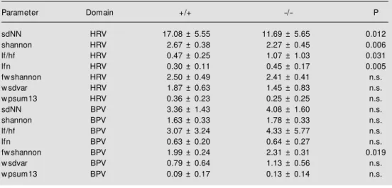
These recordings were used to quantita-tively analyze HR and BP variance, i.e., beat-to-beat dynamics. The extracted param-eters characterizing BPV and HRV are sum-marized in Table 1. HRV differed between knockout and control animals in the stan-dard procedures. A significant reduction in basic variance (sdNN, shannon) and an in-crease of lfn and consequently of lf/hf were observed in Mas-deficient mice.
BPV did not differ between groups apart from one non-linear parameter (fwshannon). However, the fwshannon parameter shows that an intermittent reduction of variance is connected with its increase. The shannon entropy calculated from the distributions of words fwshannon is a suitable measure of the complexity of the time series. Higher values of this entropy refer to a higher com-plexity in the corresponding tachogram and lower values to lower ones.
The influence of gender on parameters of HRV and BPV is presented in Table 2. Here as well, the standard parameters of the time and frequency domain were significantly dif-ferent in HRV. sdNN and shannon were reduced in female knockout animals, indi-cating a decreased basic variability. In male
Mas-deficient animals, on the other hand, lfn and lf/hf were significantly increased.
Surprisingly, BPV exhibited different re-sults. While in female animals there were no significant differences between knockout and control animals, in male animals there were highly significant differences in all param-eters with the exception of the frequency-area parameters. The basic variability (sdNN, shannon) was increased in knockout ani-mals, as also were the values for lfn and lf/hf. Furthermore, the non-linear parameters showed clear changes in variance which also characterize an increase in variability.
The sex-specific differences in HRV and
S
B
P
(
m
m
H
g
)
400
H
R
(
b
e
a
ts
/m
in
)
400
300
200
100
0
Knockout Control
300
200
100
0
BPV between control and Mas-deficient mice were found for all animals of the respective groups. A gradual discrimination analysis provided a separation of 100% for all male animals only in terms of the parameters lfn (HRV) and sdNN (BPV). For the female animals a separation of 80% was obtained in terms of the parameters sdNN (HRV), shannon (HRV) and lf/hf (HRV).
The spontaneous baroreflex of the ani-mals was also examined to see if the ob-served differences in variability are accom-panied by changes in this parameter (Table 3). Apparently, there were sex-specific dif-ferences in frequency and average increase of the baroreflex between +/+ or -/- animals, which, however, did not reach statistical significance.
D iscussio n
The central finding of this study was the marked difference in HRV and BPV in Mas -deficient mice compared to control animals, which was particularly visible when the data were analyzed according to gender. The ex-tent of the effect is the reason why male animals of the knockout and control groups could be classified completely by discrimi-nation analysis despite the relatively low number of animals per group and the rela-tively low sampling frequency (250 Hz) which, in combination, led to a relatively high scattering within groups. For the female animals the recognition/identification rate was 80%, demonstrating the less dramatic differences between female knockout and control animals. We cannot exclude an in-fluence of anesthesia on the measured car-diovascular phenotype since at least the heart rate itself is usually decreased under anes-thetic conditions, but treatment with chloral hydrate led to the same results (data not shown). However, the differences between mice of distinct genotypes and gender were significant and should reflect the genetic differences between these animals. Since
450
400
350
300
250
200
150
100
50
0
1 n 0
250
200
150
100
50 HRV
4000
BPV
Female M as +/+ mmHg
ms
A
0 250
200
150
100
50 HRV
4000
BPV
Female M as -/- mmHg
ms 450
400
350
300
250
200
150
100
50
0
1 n
Figure 3 - Continuous measurement of heart rate (HR) in beats/min and systolic blood pressure (BP) in mmHg in a female w ild-type (A) and a female M as-deficient animal (B) for 16 s. HRV and BPV, Heart rate and blood pressure variabilities, respectively.
Mas-deficient mice showed an increased anxiety-like behavior (16) measurements carried out on conscious mice may be influ-enced by these behavioral abnormalities.
HRV was significantly reduced in knock-out animals of both sexes as shown by the parameter sdNN which is a measure of the dynamic status of a time series. In addition, the autonomic balance was shifted in favor of the sympathetic tone. This is indicated by the higher level of the parameter lfn repre-senting the normalized low frequency power, i.e., the frequency components in the
1.0-Hz band divided by the frequency com-ponents in the 0.18-2.0-Hz band.
The BPV picture was much more com-plex. In an evaluation including all animals there was no difference between the knock-out and the control group apart from the marker fwshannon. The higher value of fwshannon seen in Mas-deficient mice rep-resents a higher degree of dynamics and is typical for a healthy behavior, whereas a lower value is related to pathological changes. If analysis of the data were left at this point,
the impression could arise that the influence of Mas-deficiency on BPV is much lower than on HRV. However, sex-specific evalu-ation revealed a more distinctive picture.
All parameters of the time domain and non-linear dynamics showed clear differ-ences, being significantly elevated in knock-out males. This increase was not accompa-nied by changes in lf or hf and was not measurable by standard spectral methods.
In female mice, the differences between knockout animals and the control group were not as clear as in male animals. However, the trends showed a decrease of basic variability (sdNN, shannon) and an increase of sympa-thetic tone (lfn, lf/hf) which was opposite to the effects in male mice.
The measurements of baroreflex sensi-tivity confirmed the sex-specific character of BPV, although the differences did not reach statistical significance. In male knock-out animals we registered an increase of baroreflex sensitivity as well as an increased number of baroreflex activities as a conse-quence of increased BPV, while in female animals we observed a decrease of both parameters with reduced HRV.
What is the reason for the gender-specif-ic differences in HRV and BPV between animals of different genotypes? There is ob-viously no difference in the expression level or distribution of the Mas protooncogene between male and female mice (data not shown). Interestingly, gender has been shown to be a main determinant of HRV also in humans (22-24). However, the physiologi-cal mechanisms linking sex to HRV are still elusive although a role of estrogens in this phenomenon has been suggested (25).
The impact of gender on cardiovascular variability points to the more general impor-tance of genetic influences on these param-eters. A genetic component in HRV has already been confirmed by twin studies in humans (26,27). However, the genes involved have not yet been detected in humans. To this purpose, transgenic animal models have
450
400
350
300
250
200
150
100
50
0
1 n
0 250
200
150
100
50 HRV
4000
BPV
M ale M as +/+ mmHg
ms
0 250
200
150
100
50 HRV
4000
BPV
M ale M as -/- mmHg
ms 450
400
350
300
250
200
150
100
50
0
1 n
Figure 4 - Continuous measurement of heart rate (HR) in beats/min and systolic blood pressure (BP) in mmHg in a male w ild-type (A) and a male M as-deficient animal (B) for 16 s. HRV and BPV, Heart rate and blood pressure variabilities, respectively.
A
Table 2 - Parameters of heart rate variability (HRV) and blood pressure variability (BPV).
Parameters of HRV and BPV in control (+/+) and M as-deficient (-/-) animals specified by gender. Values are reported as means ± SD; P, statistical significance; n.s., nonsignificant.
Parameter Domain M ale Female
+/+ -/- P +/+ -/- P
SdNN HRV 15.76 ± 6.47 10.70 ± 4.87 n.s. 18.39 ± 4.55 12.35 ± 6.31 0.016 Shannon HRV 2.59 ± 0.47 2.19 ± 0.52 n.s. 2.75 ± 0.25 2.32 ± 0.42 0.016 lf/hf HRV 0.48 ± 0.32 1.45 ± 1.53 0.023 0.46 ± 0.16 0.81 ± 0.45 n.s. lfn HRV 0.30 ± 0.13 0.49 ± 0.19 0.013 0.30 ± 0.08 0.41 ± 0.14 n.s. Fw shannon HRV 2.49 ± 0.55 2.43 ± 0.50 n.s. 2.50 ± 0.48 2.40 ± 0.37 n.s. w sdvar HRV 1.65 ± 0.79 1.44 ± 0.85 n.s. 2.10 ± 0.34 1.46 ± 0.88 n.s. w psum13 HRV 0.29 ± 0.25 0.25 ± 0.28 n.s. 0.43 ± 0.19 0.25 ± 0.25 n.s. sdNN BPV 2.94 ± 0.73 5.15 ± 1.75 0.011 3.78 ± 1.85 3.36 ± 1.05 n.s. shannon BPV 1.55 ± 0.23 1.97 ± 0.29 0.01 1.70 ± 0.40 1.64 ± 0.29 n.s. lf/hf BPV 2.67 ± 1.88 4.42 ± 6.07 n.s. 3.46 ± 4.35 5.85 ± 6.62 n.s. lfn BPV 0.66 ± 0.16 0.52 ± 0.34 n.s. 0.61 ± 0.25 0.72 ± 0.19 n.s. fw shannon BPV 1.87 ± 0.18 2.36 ± 0.39 0.014 2.11 ± 0.23 2.28 ± 0.26 n.s. w sdvar BPV 0.49 ± 0.30 1.38 ± 0.57 0.006 1.08 ± 0.76 0.96 ± 0.50 n.s. w psum13 BPV 0.02 ± 0.02 0.18 ± 0.18 0.003 0.15 ± 0.22 0.09 ± 0.09 n.s. Table 1 - Parameters of heart rate variability (HRV) and blood pressure variability (BPV).
Parameters of HRV and BPV, summarized for control (+/+) and knockout (-/-) animals of both genders. Values are reported as means ± SD; P, statistical significance; n.s., nonsignificant.
Parameter Domain +/+ -/- P
sdNN HRV 17.08 ± 5.55 11.69 ± 5.65 0.012
shannon HRV 2.67 ± 0.38 2.27 ± 0.45 0.006
lf/hf HRV 0.47 ± 0.25 1.07 ± 1.03 0.031
lfn HRV 0.30 ± 0.11 0.45 ± 0.17 0.005
fw shannon HRV 2.50 ± 0.49 2.41 ± 0.41 n.s.
w sdvar HRV 1.87 ± 0.63 1.45 ± 0.83 n.s.
w psum13 HRV 0.36 ± 0.23 0.25 ± 0.25 n.s.
sdNN BPV 3.36 ± 1.43 4.08 ± 1.60 n.s.
shannon BPV 1.63 ± 0.33 1.78 ± 0.33 n.s.
lf/hf BPV 3.07 ± 3.24 4.33 ± 5.77 n.s.
lfn BPV 0.63 ± 0.20 0.64 ± 0.27 n.s.
fw shannon BPV 1.99 ± 0.24 2.31 ± 0.31 0.019
w sdvar BPV 0.79 ± 0.64 1.13 ± 0.56 n.s.
w psum13 BPV 0.09 ± 0.17 0.13 ± 0.14 n.s.
Table 3 - Spontaneous baroreflex.
Spontaneous baroreflex in control (+/+) and M as-deficient (-/-) animals of both genders. Values are reported as means ± SD. There w ere no statistically significant differences.
M ales Females
+/+ -/- +/+
-/-Averaged number 9.4 ± 5.2 14.5 ± 14.0 7.3 ± 7.3 4.9 ± 5.0
been employed successfully. Recently, the ß1-adrenergic receptor (28), the
GTP-bind-ing protein Gsa (29) and the
muscarinic-gated potassium channel GIRK4 (30) have been demonstrated to cause alterations in HRV when their expression was enhanced or ablated in transgenic or knockout mice, respectively. The Mas-deficient mice repre-sent the fourth transgenic animal model ana-lyzed in this respect.
Mas could influence HRV in different ways. First, it is thought to be involved in the signaling of AngII (14,15) which by itself is an important regulator of BP and HR. In particular, the missing low-frequency fluc-tuations in the HRV and BPV tachograms indicate a reduction in the impact of angio-tensins on cardiovascular regulation in Mas -deficient mice. Secondly, Mas has been shown to influence neuronal activity since
its ablation leads to a more sustained LTP in the hippocampus (16). Therefore, also neu-rons involved in the control of HR may be affected by the absence of Mas. Thirdly, it has recently been shown that Mas signals via pathways involving mitogen-activated pro-tein kinases, like jnk and p38 kinase (31), which are also activated by adrenergic re-ceptors via G-proteins (32). The modulation of the signal transduction of these proteins shown to have a role in HRV (29) might be one mechanism by which Mas affects HRV. A reduced HRV together with an in-creased sympathetic tone, as found in Mas -deficient animals, is predictive for an in-creased cardiac risk in human beings (2,3). Further studies are needed to show whether this is also true in mice and whether Mas may be an interesting target for preventive drug treatment.
Re fe re nce s
1. Wolf M W, Varigos GA, Hunt D & Sloman JG (1978). Sinus arrhythmia in acute myo-cardial infarction. M edical Journal of Aus-tralia, 2: 52.
2. Kleiger RE, M iller JP, Bigger Jr JT & M oss AJ (1987). Decreased heart rate variability and its association w ith increased mortal-it y af t er acut e m yocardial inf arct ion. American Journal of Cardiology, 59: 256-262.
3. Algra A, Tijssen JGP, Roelandt JRTC, Pool J & Lubsen J (1993). Heart rate variability from 24-hour electrocardiography and the 2-year risk for sudden death. Circulation, 88: 180-184.
4. Bigger Jr JT, Fleiss JL, Steinman RC, Rolnitzky LM , Kleiger RE & Rottman JN (1992). Frequency domain measures of heart period variability and mortality after myocardial infarction. Circulation, 85: 164-171.
5. Singh N, M ironov D, Amstrong PW, Ross AM & Langer A (1996). Heart rate variabil-ity assessment early after acute myocar-dial infarction. Circulation, 93: 1388-1395. 6. Tsuji H, Larson M G, Venditti FJ, M anders ES, Evans JC, Feldman CL & Levy D (1996). Impact of reduced heart rate vari-ability on risk for cardiac events. Circula-tion, 94: 2850-2855.
7. Zuanet t i G, Neilson JM M , Lat ini R, Santoro E, M aggioni AP & Ew ing DJ (1996). Prognostic significance of heart rate variability in post-myocardial infarc-tion patients in the fibrinolytic era. Circula-tion, 94: 432-436.
8. Korpelainen JT, Sotaniemi KA, Huikuri HV & M yllya VV (1996). Abnormal heart rate variability as a manifestation of autonomic dysfunction in hemispheric brain infarc-tion. Stroke,27: 2059-2063.
9. Young D, W aitches G, Birchmeier C, Fasano O & Wigler M (1986). Isolation and characterization of a new cellular on-cogene encoding a protein w ith multiple potential transmembrane domains. Cell, 45: 711-719.
10. Bunnem ann B, Fuxe K, M et zger R, M ullins J, Jackson TR, Hanley M R & Ganten D (1990). Autoradiographic local-ization of mas proto-oncogene mRNA in adult rat brain using in situ hybridization. Neuroscience Letters, 114: 147-153. 11. M artin KA, Grant SGN & Hockfield S
(1992). The mas proto-oncogene is devel-opmentally regulated in the rat central ner-vous system. Developmental Brain Re-search, 68: 75-82.
12. M etzger R, Bader M , Ludw ig T, Berberich C, Bunnemann B & Ganten D (1994).
Ex-pression of the mouse and rat mas proto-oncogene in the brain and peripheral tis-sues. FEBS Letters, 357: 27-32. 13. Villar AJ & Pedersen RA (1994). Parental
imprinting of the M as protooncogene in mouse. Nature Genetics, 8: 373-379. 14. Jackson TR, Blair AC, M arshall J, Goedert
M & Hanley M R (1988). The mas onco-gene encodes an angiotensin receptor. Nature,335: 437-440.
15. Ambroz C, Clark AJL & Catt KJ (1991). The mas oncogene enhances angiotensin-induced [Ca2+]i responses in cells w ith
pre-existing angiotensin II receptors. Bio-chimica et Biophysica Acta, 1133: 107-111.
16. Walther T, Balschun D, Voigt JP, Fink H, Zuschratter W, Birchmeier C, Ganten D & Bader M (1998). Sustained long-term po-tentiation and anxiety in mice lacking the M as protooncogene. Journal of Biological Chemistry, 273: 11867-11873.
17. Denny JB, Polan-Curtain J, Wayner J & Amstrong DL (1991). Angiotensin II blocks hippocampal long term potentiation. Brain Research, 567: 321-324.
227-262.
19. Parlow J, Viale JP, Annat G, Hughson R & Quintin L (1995). Spontaneous cardiac baroreflex in humans. Hypertension, 25: 1058-1068.
20. Voss A, Dietz R, Fiehring H, Kleiner HJ, Kurths J, Saparin P, Vossing HJ & Witt A (1993). High resolution ECG, heart rate variability and nonlinear dynamics: tools for high risk stratification. In: Computers in Cardiology. IEEE Computer Society Press, Los Alamitos, 261-264.
21. Voss A, Kurths J, Kleiner HJ, Witt A, W essel N, Saparin P, Ost erziel KJ, Schurath R & Dietz R (1996). The applica-tion of methods of non-linear dynamics for the improved and predictive recogni-tion of patients threatened by sudden car-diac death. Cardiovascular Research, 31: 419-433.
22. Ryan SM , Goldberger AL, Pincus SM , M ietus J & Lipsitz LA (1994). Gender- and age-related differences in heart rate dy-namics: are w omen more complex than men? Journal of the American College of Cardiology, 24: 1700-1707.
23. Stein PK, Kleiger RE & Rottman JN (1997). Differing effects of age on heart rate vari-ability in men and w omen. American Jour-nal of Cardiology, 80: 302-305.
24. Umetani K, Singer DH, M cCraty R & Atkinson M (1998). Tw enty-four hour time domain heart rate variability and heart rate: relations to age and gender over nine decades. Journal of the American College of Cardiology, 31: 593-601.
25. Huikuri HV, Pikkujamsa SM , Airaksinen KE, Ikaheimo M J, Rantala AO, Kauma H, Lilja M & Kesaniemi YA (1996). Sex-re-lated differences in autonomic modula-tion of heart rate in middle-aged subjects. Circulation, 94: 122-125.
26. Busjahn A, Voss A, Knoblauch H, Knoblauch M , Jeschke E, W essel N, Bohlender J, M cCarron J, Faulhaber HD, Schuster H, Dietz R & Luft FC (1998). Angiotensin-converting enzyme and an-giotensinogen gene polymorphisms and heart rate variability in tw ins. American Journal of Cardiology, 81: 755-760. 27. Voss A, Busjahn A, Wessel N, Schurath R,
Faulhaber HD, Luft FC & Dietz R (1996). Familial and genetic influences on heart rate variability. Journal of Electrocardiol-ogy, 29 (Suppl): 154-160.
28. M ansier P, M édigue C, Charlot t e N, Vermeiren C, Coraboeuf E, Deroubai E, Ratner E, Chevalier B, Clairambault J, Carré F, Dahkli T, Bertin B, Briand P, Strosberg D & Sw ynghedauw B (1996).
Decreased heart rate variability in trans-genic mice overexpressing atrial ß1-adre-noreceptors. American Journal of Physiol-ogy, 271: 1465-1472.
29. Uechi M , Asai K, Osaka M , Smith A, Sato N, Wagner TE, Ishikaw a Y, Hayakaw a H, Vatner DE, Shannon RP, Homcy CJ & Vatner SF (1998). Depressed heart rate variability and arterial baroreflex in con-scious transgenic mice w ith overexpres-sion of cardiac Gsa. Circulation Research, 82: 416-423.
30. Wickman K, Nemec J, Gendler SJ & Clapham DE (1998). Abnormal heart rate regulation in GIRK4 knockout mice. Neu-ron, 20: 103-114.
31. Zohn IE, Sym ons M , Chrzanow ska-Wodnicka M , Westw ick JK & Der CJ (1998). M as oncogene signaling and trans-formation require the small GTP-binding protein Rac. M olecular and Cellular Biol-ogy, 18: 1225-1235.