Assessment of the cardiovascular
effects of electroconvulsive therapy in
individuals older than 50 years
1Departamento de Cardiologia, Instituto do Coração, 2Instituto de Psiquiatria, Faculdade de Medicina,
Universidade de São Paulo, São Paulo, SP, Brasil J.Y. Takada1, M.C. Solimene1,
P.L. da Luz1, C.J. Grupi1,
D.M.A. Giorgi1,
S.P. Rigonatti2, D.O. Rumi2,
L.H.W. Gowdak1 and
J.A.F. Ramires1
Abstract
To evaluate the impact of electroconvulsive therapy on arterial blood pressure, heart rate, heart rate variability, and the occurrence of ischemia or arrhythmias, 38 (18 men) depressive patients free from systemic diseases, 50 to 83 years old (mean: 64.7 ± 8.6) underwent electroconvulsive therapy. All patients were studied with simulta-neous 24-h ambulatory blood pressure and Holter monitoring, starting 18 h before and continuing for 3 h after electroconvulsive therapy. Blood pressure, heart rate, heart rate variability, arrhythmias, and ischemic episodes were recorded. Before each session of electrocon-vulsive therapy, blood pressure and heart rate were in the normal range; supraventricular ectopic beats occurred in all patients and ventricular ectopic beats in 27/38; 2 patients had non-sustained ven-tricular tachycardia. After shock, systolic, mean and diastolic blood pressure increased 29, 25, and 24% (P < 0.001), respectively, and returned to baseline values within 1 h. Maximum, mean and minimum heart rate increased 56, 52, and 49% (P < 0.001), respectively, followed by a significant decrease within 5 min; heart rate gradually increased again thereafter and remained elevated for 1 h. Analysis of heart rate variability showed increased sympathetic activity during shock with a decrease in both sympathetic and parasympathetic drive afterwards. No serious adverse effects occurred; electroconvulsive therapy did not trigger any malignant arrhythmias or ischemia. In middle-aged and elderly people free from systemic diseases, electro-convulsive therapy caused transitory increases in blood pressure and heart rate and a decrease in heart rate variability but these changes were not associated with serious adverse clinical events.
Correspondence
P.L. da Luz
Instituto do Coração, HC, FM, USP Av. Dr. Enéas C. Aguiar, 44 Bloco II, 2º andar, Sala 2 05403-000 São Paulo, SP Brasil
Fax: +55-11-3069-5447 E-mail: [email protected]
Publication supported by FAPESP.
Received July 16, 2004 Accepted May 23, 2005
Key words
•Electroconvulsive therapy •Blood pressure
•Heart rate •Arrhythmia
•Myocardial ischemia
Introduction
Electroconvulsive therapy consists of in-ducing a controlled convulsive seizure by electric stimulation of the brain. Cerletti and
disorders, drug-resistant patients and those more likely to have adverse effects such as pregnant and childbearing women, and the elderly (1,2).
The rate of adverse effects is usually low and mortality is estimated at 1:1000 to 1:10,000 cases (3,4); almost 2/3 of these deaths are of cardiovascular origin and there-fore cardiac risk must be assessed (5). The main cardiac complications are asystole, car-diac rupture, and myocardial infarction (3); other complications include subdural he-matoma and subarachnoid hemorrhage (4). The most common adverse effect is a tran-sient memory deficit after seizure, although many patients complain that their memory never returns completely (2).
In a review of the literature we did not find any detailed study on the cardiovascular effects of electroconvulsive therapy in indi-viduals older than 50 years. Thus, the aim of the present study was to assess the impact of electroconvulsive therapy on systemic arte-rial blood pressure, heart rate and heart rate variability and the possible occurrence of cardiac arrhythmias and/or ischemia in indi-viduals older than 50 years.
Material and Methods
The study was approved by the Institu-tional Ethics Committees. From March 1998 to July 2001, 120 patients older than 50 years were referred to our institution for electroconvulsive therapy for major depres-sion, resistant to drug therapy. Patients were excluded if they presented at least one of the following conditions: severe arterial hyper-tension (6), cardiac and non-cardiac dis-eases, persistent electrocardiographic alter-ations that could hinder the analysis of ven-tricular repolarization (bundle branch block, marked left ventricular hypertrophy, atrio-ventricular block, cardiac pacemaker) or continued use of medications. We selected 38 patients, 18 men and 20 women, aged 50 to 83 years (mean: 64.7 ± 8.6); 14 patients
(37%) were older than 65 years. Patients, or their close relatives, gave written informed consent to participate in the study.
Study protocol
Drugs were withheld 3 to 7 days before electroconvulsive therapy. All patients were submitted to detailed clinical evaluation by one of the investigators, and to simultaneous 24-h blood pressure and electrocardiographic (Holter) monitoring. Monitoring started 18 h before electroconvulsive therapy and con-tinued for at least 3 h after the procedure.
Thymatron device has a basic exit mode of 25 to 504 mC and operational exit mode of 50 to 1000 mC, and the load control is calibrated in percentages. The devices also monitored the electroencephalographic ac-tivity and recorded the tonic-clonic seizures. Electrical stimulation lasted less than 1 min. Seizures were considered to be effective when lasting more than 20 s (1). Blood pressure, electrocardiographic tracing and oxygen saturation index were monitored before and during each convulsive seizure because ventilation was not interrupted dur-ing convulsion. The mean duration of the convulsive seizure was 38 ± 5 s. After the seizures, all patients remained under medi-cal and nursing care until complete recov-ery. All patients received food prior to hos-pital discharge.
Ambulatory blood pressure monitoring was performed with Spacelabs 90207 de-vices (Spacelabs, Redmont, WA, USA), pro-grammed to make measurements every 10 min, except during sleeping hours (23:00 pm to 6:00 am), when the measurements were made every 20 min. Systolic, diastolic and mean blood pressure was recorded and monitoring lasted at least another 3 h after electroconvulsive therapy.
Data were analyzed using Spacelabs soft-ware. Baseline values were obtained as the mean values of 6 measurements before shock; after the electric stimulus, we considered the mean values of the following 6 measure-ments. During shock, the peak values of systolic, diastolic and mean blood pressure were noted and compared to the mean values before and after shock. Data were excluded from analysis if any artifacts were present.
Electrocardiographic Holter monitoring was performed with Marquette 800 portable amplitude-modulated two-channel devices (Marquette 9428; Marquette Medical Sys-tems Milwaukee, WI, USA). Two bipolar leads, CM5 (exploring electrode in the V5 position) and CM1 or modified D2 (explor-ing electrode in an inferior lead) were used
(7). To obtain the neutral value, an electrode was placed close to the last costal arches, next to the midclavicular line.
After the monitor was withdrawn, the tapes were analyzed with the MARS system (Marquette). Maximum, minimum and mean heart rate values, as well as heart rate varia-bility, were determined, and the occurrence of ST-segment deviations and arrhythmias was recorded. Baseline heart rate values were obtained as the mean values recorded 10 min before shock. Heart rate was then recorded minute-to-minute until 30 min after shock.
Heart rate variability was determined by frequency-domain analysis according to the guidelines of the American Heart Associa-tion (8). Holter monitoring data were ana-lyzed by the MARS system. For the analysis of heart rate variability we used the meas-urements of seven 4-min intervals before shock, a 4-min interval during shock and seven 4-min intervals after shock. The fol-lowing frequency bands were extracted from the power spectrum: total power or wide band (WB): 0.003-1.7 Hz; very low fre-quency (VLF): 0.003-0.04 Hz; low frefre-quency (LF): 0.04-0.15 Hz; high frequency (HF): 0.15-0.4 Hz. The LF/HF ratio was also de-termined. Data are reported as milliseconds (ms) and the values before and after shock were compared.
Arrhythmias were diagnosed and ana-lyzed manually; supraventricular and ven-tricular extrasystoles, and the atrioventricu-lar and intraventricuatrioventricu-lar conduction defects were recorded. The number of extrasystoles/ hour was compared before and after shock considering the total recording time.
An ischemic episode was defined as a transient ST-segment elevation, or horizon-tal or downsloping ST-segment depression from baseline of 1 mm or more, measured 60 to 80 ms after the J point, and lasting 1 min or more (7,8).
symp-tom and to press the event buttons under these situations. The event buttons of both recorders made it possible to identify the moment when the electrical stimulus was triggered.
All data recorded from ambulatory moni-toring were analyzed by expert technicians and reviewed by the investigators.
Statistical analysis
Data are reported as mean ± SD. Re-peated measures one-way ANOVA was used to compare heart rate and heart rate variabil-ity between time points (9,10). The level of significance was set at 0.05. The Wilcoxon rank sum test was used to compare the inci-dence of arrhythmias and blood pressure before, during and after shock (9,10). Is-chemic episodes were not subjected to sta-tistical analysis because of the small number of events.
Results
Twenty-four-hour monitoring
Blood pressure, heart rate and heart rate variability curves showed a circadian pat-tern, except during shock. The highest val-ues of mean systolic, diastolic and mean blood pressure occurred between 9:00 and 10:00 am (154.2 ± 19.9, 116.0 ± 13.4, and
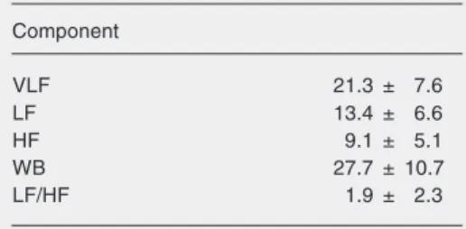
96.0 ± 10.7 mmHg, respectively) and the lowest values occurred between 3:00 and 4:00 am (117.0 ± 16.1, 87.2 ± 13.0, and 70.1 ± 12.0 mmHg, respectively); the mean de-creaseat night was 10.4% in systolic blood pressure and 9.9% in diastolic blood pres-sure, including 9 (23.6%) patients with mild arterial hypertension. Heart rate varied from 121.9 ± 19.6 bpm (between 10:00 and 11:00 am) to 57.8 ± 13.5 bpm (between 5:00 and 6:00 am). The frequency bands of heart rate variability are listed in Table 1.
All patients had at least one isolated su-praventricular extrasystole; 4 (10.5%) had more than 10/h and 2 (5.3%) presented non-sustained atrial tachycardia before and after shock. Ventricular extrasystoles occurred in 27 (72.9%) patients; 2 (5.3%) had an iso-lated episode of non-sustained ventricular tachycardia before shock. One asymptomat-ic patient (2.6%) had, on average, 1028 ven-tricular extrasystoles/h before shock and 1046 after shock. Another patient (2.6%) had in-termittent left bundle branch block during electrocardiographic recording, which was not associated with symptoms or shock. There were no other arrhythmias.
Holter recordings demonstrated silent myocardial ischemia in 3 patients. Two of them had ischemic episodes before and after shock; one had 10 episodes and the other 44 at different times during the recording pe-riod and also immediately after shock. The third patient had two brief episodes of ische-mia only after the procedure. All 3 patients remained asymptomatic during ischemia and did well after electroconvulsive therapy.
Electrical shock was identified in both recordings through a line of interference coinciding with the pressing of the event buttons. Patients were subjected to electro-convulsive therapy at different times but the time of shock was adjusted to 9:00 am.
Effects of electroconvulsive therapy
Blood pressure changes are depicted in Table 1. Spectral components of 24-h heart rate
variability.
Component
VLF 21.3 ± 7.6
LF 13.4 ± 6.6
HF 9.1 ± 5.1
WB 27.7 ± 10.7
LF/HF 1.9 ± 2.3
Figure 1. Systolic blood pressure increased 29%, from 128.3 ± 13.2 mmHg 1 h before shock, to 165.6 ± 31.5 mmHg during shock (P < 0.001), returning to 129.8 ± 14.1 mmHg after 1 h. Mean blood pressure varied from 98.7 ± 11.2 mmHg 1 h before shock to 123.0 ± 25.9 mmHg during shock, a 25% increase (P < 0.001), returning to 101.0 ± 10.7 mmHg after 1 h.
Diastolic blood pressure increased 24%, from 83.2 ± 10.6 to 103.5 ± 23.2 mmHg during shock (P < 0.001), returning to 84.6 ± 10.5 mmHg after 1 h. There was no differ-ence between blood pressure values before and after shock.
Heart rate changes are depicted in Figure 2. Maximum, minimum and mean heart rate values were always higher during shock com-pared to baseline (P < 0.001). Mean peak of maximum heart rate (133.6 ± 20.2 bpm) occurred in the first minute after shock and was significantly higher (58%) than the base-line value (84.3 ± 14.1 bpm). Maximum heart rate decreased gradually until the 5th minute after shock (95.5 bpm); after 15 min, it increased again but its values were lower compared to shock. Minimum heart rate in-creased 29% at the first min after shock (74.4 ± 13.7 to 95.9 ± 18.6 bpm) and then gradually decreased; after 8 min, it increased again and after 25 min it was similar to peak values. Mean heart rate increased 46% from 79.5 ± 13.8 to 116.5 ± 18.2 bpm in the first min after shock, then gradually decreased until the 5th min (91.7 bpm); after 14 min, it increased again.
Heart rate variability is depicted in Fig-ures 3 and 4. All components increased dur-ing shock and then decreased (Figure 3). LF/ HF ratio did not change significantly (P = 0.177); the lowest value (1.46) occurred im-mediately after shock and the highest (1.95), 12 to 16 min later. The highest mean values were recorded during shock (VLF = 50.9 ± 31.8 ms, LF = 22.7 ± 18.5 ms, HF = 11.8 ± 7.1 ms, and WB = 58.8 ± 35.4 ms). The lowest values were recorded 8 to 16 min
Figure 1. Blood pressure before, during and after shock. N = 33 patients. SBP = systolic blood pressure; MBP = mean blood pressure; DBP = diastolic blood pres-sure (mmHg). *P < 0.001 compared to before and after shock periods (Wilcoxon rank sum test).
Figure 2. Heart rate before, during and after shock. N = 32 patients. HRmax = maximum heart rate; HRm = mean heart rate; HRmin = minimum heart rate; B = baseline; S = shock. *P < 0.01 compared to all other periods (repeated measures one-way ANOVA).
Figure 3. Frequency bands of heart rate variability before, during and after shock. N = 28 patients. WB = wide band; VLF = very low frequency; LF = low frequency; HF = high frequency; 4-min interval before (-7 to -1), during (0) and after (1 to 7) shock. *P < 0.01 compared to all other periods (repeated measures one-way ANOVA).
B S 1 2 3 4 5 6 7 8 9 10 11 12 13 14 15 16 17 18 19 20 21 22 23 24 25 26 27 28 29 30 140
130
120
110
100
90
80
70
HR (bpm)
HRmax
Time (min)
HRmin HRm
-7 -6 -5 -4 -3 -2 -1 0 1 2 3 4 5 6 7
65
55
45
35
25
15
5
ms
LF
Intervals
after shock (VLF = 12.1 ± 5.6 ms, LF = 8.8 ± 5.6 ms, HF = 5.5 ± 2.9 ms, and WB = 19.1 ± 7.4 ms). Figure 4 shows the pattern of heart rate and heart rate variability during and after shock.
Arrhythmias and ischemia
The type of arrhythmias that occurred before electroconvulsive therapy did not change after shock. The supraventricular extrasystoles/h were 8.8 ± 23.8 before and 5.1 ± 12.5 after shock (P = 0.685); ventricu-lar extrasystoles/h increased from 42.0 ± 197.2 to 46.8 ± 200.3 (P = 0.016), without adverse effects. One patient had transient ST-segment changes only after shock; they lasted 2.0 and 2.25 min and were suggestive of ischemia. Two other patients had epi-sodes of silent ischemia during the recording time, not necessarily related to shock.
Discussion
Since the very beginning of electrocon-vulsive therapy, it was recognized that the procedure induced tachycardia and arterial hypertension (11,12), but the consequences of these effects were not analyzed in depth. In the present study, we started monitoring patients one day before electroconvulsive
therapy in order to identify cardiovascular changes not related to the procedure; the observed spontaneous events would then be compared to those directly caused by shock. At the time of shock, we observed sig-nificant increases in blood pressure and heart rate. These changes may be due to myotonic reflexes, direct stimulation of the sympa-thetic nervous system and norepinephrine release from the adrenal medullae (13,14). It is still debatable whether or not the seizures may have a direct effect on the observed cardiocirculatory changes; patients with spontaneous convulsions have smaller in-creases in blood pressure and heart rate whereas patients undergoing electrosive therapy but not experiencing convul-sions (frustrated seizure) have the highest reported increases in these parameters (15).
Long-lasting convulsions do not necessarily induce the most significant increases in blood pressure and heart rate(16).
It is possible that the autonomic over-drive may not be the only fact responsible for cardiocirculatory changes; ganglionic blockade (17) and elevated circulating cat-echolamines may play a role (18) and ß-blockers may counteract these effects (19).
Our results are similar to those obtained by Rumi et al. (20), who reportedsignificant increases in blood pressure and heart rate during shock in young adults, returning to baseline values after 25 min. Our patients continued to have higher heart rates 1 h after shock and their peak values of blood pres-sure were smaller. These differences may be related to particular cardiovascular responses in the elderly as compared to young people (21); there is an age-related reduction in tonic cardiac vagal inhibition of heart rate and cardiac output, as well as elevated sym-pathetic activity (22,23) and counteracting vascular α-adrenergic desensitization,
ex-plaining why the responses to stress are at-tenuated in the elderly (23).
We recorded significantly increased blood pressure and heart rate after shock; Figure 4. Maximum heart rate, high frequency and low frequency bands of heart rate
these sudden elevations may increase the risk of cardiovascular events such as myo-cardial infarction, brain hemorrhage and stroke, especially in older patients. Lack of adequate control of cardiocirculatory param-eters may lead to long-lasting cognitive de-fects (24), mainly because of cerebral edema and the passage of neurotoxic macromol-ecules through the blood-brain barrier dur-ing cerebral hypoperfusion (25). The routine use of drugs that attenuate the hypertensive peak and tachycardia induced by electro-convulsive therapy is controversial and only recommended for patients with associated cardiovascular diseases (26,27).
We did not observe a long-lasting sinus pause or transient bradyarrhythmias imme-diately after shock, but they may occur and death due to asystole has been reported (28). Electric shock is associated with intense va-gal stimulation even when convulsion does not occur, suggesting that the shock and not the convulsion is responsible for the para-sympathetic discharge (29) which can be prevented by atropine (1).
The analysis of heart rate variability dur-ing shock is important because significant changes in autonomic function have been described in depressive disorder (30). Some investigators have observed that, after elec-troconvulsive therapy, clinical improvement is associated with decreased levels of plasma catecholamines after the procedure (18). In addition, there are reports demonstrating that electroconvulsive therapy may influence the recovery of parasympathetic function (31) and improve depression.
In the spectral analysis of heart rate (32,33), the LF/HF ratio reflects sympatho-vagal imbalance (34). Decreased heart rate variability is considered to be a strong and independent predictor of cardiovascular mortality (33). Major depressive disorder may also be associated with reduced vagal modulation (30) and some investigators have reported the influence of treating depression on sympathovagal imbalance (31,35).
Major depression is predictive of cardiac events and death even when there is no evident cardiac disease (36), leading to the hypothesis that the reduction of vagal activ-ity in this affective disorder may trigger arrhythmic events (33). In our study, the LF/ HF ratio did not change significantly, lead-ing us to conclude that both components were well balanced. The high frequency com-ponent did not increase during shock but it decreased 2 min after it, demonstrating a decrease in parasympathetic activity at that time. The low frequency component, par-tially reflecting sympathetic activity, in-creased after shock and then dein-creased. These changes demonstrated that the sympathetic activity predominated during electric stimu-lation. Vagal activity did not change during shock when compared to the period before it; on the other hand, it decreased more slowly than sympathetic activity after shock and reached the lowest values 16 min there-after. Both high and low frequency compo-nents decreased after shock; heart rate re-mained elevated, but was lower than during electroconvulsive therapy. This fact could be related to increased circulating catechol-amines (18).
it analyzed heart rate variability specifically at the time of electrical stimulation.
Electroconvulsive therapy did not trig-ger malignant arrhythmias in our patients, in agreement with the results obtained by Rumi et al. (20) in young people without systemic diseases. In contrast, Zielinski et al. (37) reported the prevalence of arrhythmias in patients with previous cardiac diseases.
We observed episodes of silent myocar-dial ischemia in 3 patients at different times during the 24-h recordings and not necessar-ily related to shock. It is possible that these patients had definite coronary artery dis-ease, although they did not experience car-diac events. Gould et al. (38) described elec-trocardiographic changes suggesting myo-cardial infarction in a 75-year-old woman submitted to electroconvulsive therapy who did not exhibit myocardial necrosis or peri-carditis. Other investigators have reported
the same observation (39). In animal mod-els, electroconvulsive therapy induced in-creases in blood pressure and heart rate, ventricular ectopic beats and T-wave inver-sions during hypothalamic stimulation (40).
Clinical implications
In healthy individuals older than 50 years, electroconvulsive therapy can be considered a safe therapeutic method, although it causes transient but significant increases in blood pressure and heart rate and sympathetic stim-ulation. No significant arrhythmias or ische-mia or other adverse clinical effects occurred during and after the procedure. Neverthe-less, we think that elderly people with preex-isting organic diseases are at risk for cardio-vascular events because of the observed car-diocirculatory changes and should be care-fully evaluated.
References
1. Moreno DH, Moreno RA, Rigonatti SP et al. (2000). A eletroconvul-soterapia. In: Rigonatti SP & Rosa MA (Editors), Indicação e Prática da Eletroconvulsoterapia. Lemos Editorial, São Paulo, SP, Brazil. 2. Fink M (2001). Convulsive therapy: a review of the first 55 years.
Journal of Affective Disorders, 63: 1-15.
3. Stoudmire A (1995). Cardiovascular morbidity and ECT. American Journal of Psychiatry, 152: 1697-1698.
4. Abrams R (1997). The mortality rate with ECT. Convulsive Therapy, 13: 125-127.
5. Rumi DO, Takada JY & Solimene MC (2000). Efeitos sobre os sistemas orgânicos, avaliação clínica e contra-indicações. In: Rigo-natti SP & Rosa MA (Editors), Indicação e Prática da Eletroconvul-soterapia. Lemos Editorial, São Paulo, SP, Brazil.
6. Joint National Committee on Prevention, Detection, Evaluation and Treatment of High Blood Pressure (1997). The Sixth Report of the Joint National Committee on Prevention, Detection, Evaluation and Treatment of High Blood Pressure. Archives of Internal Medicine, 157: 2413-2444.
7. Solimene MC, Ramires JA, Gruppi CJ et al. (1993). Prognostic significance of silent myocardial ischemia after a first uncomplicated myocardial infarction. International Journal of Cardiology, 38: 41-47. 8. Crawford MH, Bernstein SJ, Deedwania PC et al. (1999). ACC/AHA Guidelines for Ambulatory Electrocardiography. A Report of the American College of Cardiology/American Heart Association Task Force on Practice Guidelines (Committee to Revise the Guidelines for Ambulatory Electrocardiography). Developed in collaboration with the North American Society for Pacing and Electrophysiology.
Journal of the American College of Cardiology, 34: 912-948.
9. Rosner B (1986). Fundamentals of Biostatistics. 2nd edn. PWS Publishers, Boston, MA, USA.
10. Timm NH (1975). Multivariate Analysis with Applications in Educa-tion and Psychology. Brooks/Cole, Monterey, CA, USA.
11. Bellet S, Kershbaum A & Furst W (1941). The electrocardiogram during electric shock treatment of mental disorders. American Jour-nal of the Medical Sciences, 201: 167.
12. Cleckley H, Hamilton WP, Woodbury RA et al. (1942). Blood pres-sure studies in patients undergoing convulsive therapy. Southern Medical Journal, 35: 375.
13. Mulgaokar GD, Dauchot PJ, Duffy JP et al. (1985). Noninvasive assessment of electroconvulsive-induced changes in cardiac func-tion. Journal of Clinical Psychiatry, 46: 479-482.
14. Swartz CM (2000). Physiological response to ECT stimulus doses.
Psychiatry Research, 97: 229-235.
15. Partridge BL, Weinger MB & Hauger I (1991). Is the cardiovascular response to electroconvulsive therapy due to the electricity or the subsequent convulsion? Anesthesia and Analgesia, 72: 706-709. 16. Fu W, Stool LA, White PF et al. (1997). Acute hemodynamic
re-sponses to electroconvulsive therapy are not related to the duration of seizure activity. Journal of Clinical Anesthesia, 9: 653-657. 17. Petrides G, Maneksha F, Zervas I et al. (1996). Trimethaphan
(Arfonad) control of hypertension and tachycardia during electro-convulsive therapy: a double-blind study. Journal of Clinical Anes-thesia, 8: 104-109.
19. Castelli I, Steiner LA, Kaufmann MA et al. (1995). Comparative effects of esmolol and labetalol to attenuate hyperdynamic states after electroconvulsive therapy. Anesthesia and Analgesia, 80: 557-561.
20. Rumi DO, Solimene MC, Takada JY et al. (2002). Electrocardio-graphic and blood pressure alterations during electroconvulsive therapy in young adults. Arquivos Brasileiros de Cardiologia, 79: 149-160.
21. Huang KC, Lucas LF, Tsueda K et al. (1989). Age-related changes in cardiovascular function associated with electroconvulsive thera-py. Convulsive Therapy, 5: 17-25.
22. Pfeifer MA, Weinberg CR, Cook D et al. (1983). Differential changes of autonomic nervous system function with age in man. American Journal of Medicine, 75: 249-258.
23. Jones PP, Shapiro LF, Keisling GA et al. (2001). Altered autonomic support of arterial blood pressure with age in healthy men. Circula-tion, 104: 2424-2429.
24. Zervas IM, Calev A, Jandorf L et al. (1993). Blood pressure, memory, and electroconvulsive therapy. Convulsive Therapy, 9: 14-22. 25. Bolwig TG, Hertz MM, Paulson OB et al. (1977). The permeability of
the blood-brain barrier during electrically induced seizures in man.
European Journal of Clinical Investigation, 7: 95-100.
26. McCall WV (1993). Antihypertensive medications and ECT. Convul-sive Therapy, 9: 317-325.
27. Maneksha FR (1991). Hypertension and tachycardia during electro-convulsive therapy: to treat or not to treat? Convulsive Therapy, 7: 28-35.
28. Malik MO (1972). Fatal heart block and cardiac arrest following ECT. A case report. British Journal of Psychiatry, 120: 69-70. 29. Wells DG, Zelcer J & Treadrae C (1988). ECT-induced asystole
from a subconvulsive shock. Anaesthesia and Intensive Care, 16: 368-373.
30. Grippo AJ, Moffitt JA & Johnson AK (2002). Cardiovascular alter-ations and autonomic imbalance in an experimental model of de-pression. American Journal of Physiology, 282: 1333-1341. 31. Nahshoni E, Aizenberg D, Sigler M et al. (2001). Heart rate
variabil-ity in elderly patients before and after electroconvulsive therapy.
American Journal of Geriatric Psychiatry, 9: 255-260.
32. Eckberg DL (1997). Sympathovagal balance. A critical appraisal.
Circulation, 96: 3224-3232.
33. Malik M (1998). Heart rate variability. Current Opinion in Cardiology, 13: 36-44.
34. Malliani A, Lombardi F & Pagani M (1994). Power spectrum analysis of heart rate variability: a tool to explore neural regulatory mechan-isms. British Heart Journal, 71: 1-2.
35. Schultz SK, Anderson EA & Van de Borne P (1997). Heart rate variability before and after treatment with electroconvulsive therapy.
Journal of Affective Disorders, 44: 13-20.
36. Pratt LA, Ford DE, Crum RM et al. (1996). Depression, psychotropic medication, and risk of myocardial infarction: prospective data from the Baltimore ECA follow-up. Circulation, 94: 3123-3129.
37. Zielinski RJ, Roose SP, Devanand DP et al. (1993). Cardiovascular complications of ECT in depressed patients with cardiac diseases.
American Journal of Psychiatry, 150: 904-909.
38. Gould L, Gopalaswamy C, Chandy F et al. (1983). Electroconvulsive therapy-induced ECG changes simulating a myocardial infarction.
Archives of Internal Medicine, 143: 1786-1787.
39. Cockey GH & Conti CR (1995). Electroconvulsive therapy-induced transient T-wave inversions on ECG. Clinical Cardiology, 18: 418-420.
40. Melville KI, Blum B, Schuster HE et al. (1963). Cardiac ischemic changes and arrhythmias induced by hypothalamic stimulation.