Abstract
Resistance to chemotherapy in cancer cells is mainly mediated by overexpression of P-glycoprotein (Pgp), a plasma membrane ATP-binding cassette (ABC) transporter which extrudes cytotoxic drugs at the expense of ATP hydrolysis. Pgp consists of two homologous halves each containing a transmembrane domain and a cytosolic nucleotide-binding domain (NBD) which contains two consensus Walker motifs, A and B, involved in ATP binding and hydrolysis. The protein also contains an S signature characteristic of ABC transport-ers. The molecular mechanism of Pgp-mediated drug transport is not known. Since the transporter has an extraordinarily broad substrate specificity, its cellular function has been described as a hydrophobic vacuum cleaner. The limited knowledge about the mechanism of Pgp, partly due to the lack of a high-resolution structure, is well reflected in the failure to efficiently inhibit its activity in cancer cells and thus to reverse multidrug resistance (MDR). In contrast to the difficulties encountered when studying the full-length Pgp, the recom-binant NBDs can be obtained in large amounts as soluble proteins. The biochemical and biophysical characterization of recombinant NBDs is shown here to provide a suitable alternative route to establish struc-ture-function relationships. NBDs were shown to bind ATP and analogues as well as potent modulators of MDR, such as hydrophobic steroids, at a region close to the ATP site. Interestingly, flavonoids also bind to NBDs with high affinity. Their binding site partly overlaps both the ATP-binding site and the steroid-interacting region. There-fore flavonoids constitute a new promising class of bifunctional modulators of Pgp.
Co rre spo nde nce
A. Di Pietro
Laboratoire de Biochimie Structurale et Fonctionnelle Institut de Biologie et
Chimie des Protéines, UPR 412-CNRS 7, Passage du Vercors
69367 Lyon Cedex 07
France
Fax: + 33-478-61-7873 E-mail: a.dipietro@ ibcp.fr
Research supported by CNRS (UPR 412 and PCV97-129), Université
Claude Bernard-Lyon I, Fondation de France (No. 96003891), Région Rhône-Alpes (Emergence 97027309), Association pour la Recherche contre le Cancer (No. 9147), Ligue Nationale Contre le Cancer, Fondation pour la Recherche Médicale (No. 20000400-01).
Received March 18, 1999 Accepted May 7, 1999
Ke y wo rds
·P-glycoprotein (Pgp)
·Multidrug resistance
·Anticancer chemotherapy
·ATP
·Steroids
·Flavonoids
P-glyco pro te in-m e diate d re sistance
to che m o the rapy in cance r ce lls:
using re co m binant cyto so lic
do m ains to e stablish structure
-functio n re latio nships
Laboratoire de Biochimie Structurale et Fonctionnelle,
Institut de Biologie et Chimie des Protéines, UPR 412-CNRS, Lyon, France A. Di Pietro, G. Dayan,
The structure o f Pgp: still a m o de l
cDNA sequences of human MDR1, mouse
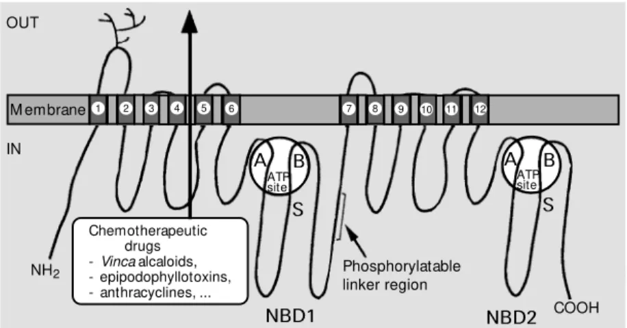
mdr1 and Chinese hamster pgp1 have led to the prediction of protein secondary-struc-ture elements and transmembrane segments which have allowed a topology model (Fig-ure 1) to be established. The protein is com-posed of 1276 amino acids in mice, has a 141-kDa molecular mass in the absence of glycosylation, and is composed of two ho-mologous halves. The two halves share 43% sequence identity and 78% similarity and are most likely to have evolved as a result of gene duplication (5). Hydrophobicity analy-sis indicates that each half contains a hydro-phobic domain with 6 transmembrane a-helices (6) involved in chemotherapeutic drug efflux, and a cytosolic domain, nucle-otide-binding domain 1 (NBD1) or NBD2, containing an ATP-binding site with charac-teristic Walker motifs A and B (7) and the S signature of ABC (ATP-binding cassette) transporters (8). A central sequence which connects the two homologous halves of the protein, called linker region, contains in vitro phosphorylatable serine residues, and the first extracellular loop is heavily N- gly-cosylated.
A low-resolution structure (2.5 nm) has been obtained for Pgp by electron micros-copy and single-particle image analysis (9). It shows a wide hydrophilic pore surrounded by a ring of transmembrane segments with a six-fold symmetry. The pore is closed on the cytosolic side by the two NBDs and the internal loops which connect the transmem-brane spans. The cup-shaped chamber within the membrane has been proposed to include an opening allowing a lateral entry of drug substrates to be effluxed.
The crystal structure of HisP, the ATP-binding subunit of bacterial histidine per-mease, has recently been solved (10), and the structures of HisP and the NBDs are likely to be similar. HisP has an L-shape consisting of two thick arms: one side of arm
1 2 3 4
M embrane
S
NBD2 NBD1
NH2
COOH OUT
IN
Phosphorylatable linker region Chemotherapeutic
drugs -Vinca alcaloids, - epipodophyllotoxins, - anthracyclines, ...
5 6
A B
ATP site S
11 12 9
7 8 10
A B
ATP site Anticance r che m o the rapy: the pro ble m o f m ultidrug re sistance
Cancer cell resistance to cytotoxic drugs is considered to be one of the major ob-stacles to successful chemotherapy. Some tumors are intrinsically resistant to the treat-ments whereas others progressively acquire multidrug resistance (MDR) to a variety of structurally and functionally unrelated drugs including anthracyclines, Vinca alkaloids, epipodophyllotoxins, taxanes and actinomy-cin D.
The best studied mechanism of MDR (1) is the overexpression of P-glycoprotein (Pgp) (2), a 170-kDa plasma membrane protein that functions as an ATP-driven drug efflux pump in cancer cells. A family of Pgp iso-forms is encoded by the mdr genes which are located on human chromosome 7, mouse chromosome 5 or Chinese hamster chromo-some 1. Only the MDR1 isoform can confer the MDR phenotype to humans, whereas the two isoforms mdr1 (or mdr1b) and mdr3 (or mdr1a) in mice or pgp1 and pgp2 in Chinese hamsters can cause the same phenotype. An additional isoform, MDR2 (or MDR3) in humans, mdr2 in mice or pgp3 in Chinese hamsters, cannot confer MDR and appears to be involved in the transport of phospho-lipids, especially phosphatidylcholine (3,4).
I, including ß1, ß2 and ß4-ß7 strands (cf. Figure 4), is involved in dimerization, whereas the other side contains the ATP-binding pocket. The side-chain of a tyrosine residue located between ß1 and ß2 appears to form a stacking interaction with the ad-enine ring. The residues of the P-loop, con-taining the Walker motif A, and of the fol-lowing N-terminal segment of the a1 helix are mainly involved in the formation of hy-drogen bonds with the ATP phosphates. Arm II contains helices a3-a9 which include the S signature, and is likely to interact with the membrane components of the transporter. The two arms of the L are connected by a six-stranded ß-sheet (ß3 and ß8-ß12).
ATPase activity and drug transpo rt
Although the exact mechanism of drug extrusion is not known, it is well established that Pgp functions as an ATP-dependent pump since: i) Pgp overexpression leads to both a lowered intracellular accumulation and an increased efflux of drugs and these effects are abolished by either intracellular ATP depletion or mutations within the Walker motifs A and B, ii) photoreactive drug analogues label Pgp, and iii) single-point mutations within either transmembrane spans or cytosolic loops differentially change the relative specificity towards transported drugs.
The ATPase activity of Pgp has been determined in enriched membranes of resist-ant cells and from a purified transporter reconstituted into liposomes. Both measure-ments resulted in values of a few micro-moles/min x mg protein for maximal rate and around 1 mM for Km(MgATP). The maximal rate was dependent on the addition of phospholipids: it was either increased by phosphatidylethanolamine or phosphatidyl-choline, or inhibited by phosphatidylserine or phosphatidylinositol. Pgp was found to be naturally associated with phosphatidyletha-nolamine and phosphatidylserine, but only
also been proposed (18), but only demon-strated for the mdr2/MDR2 transporter which uses phosphatidylcholine as a substrate (3,4). The stoichiometry between transported sub-strate and hydrolyzed ATP was generally found to be close to unity (19) for a broad range of substrates such as colchicine, 86Rb+ -valinomycin, vinblastine, daunorubicin or rhodamine 123.
The physio lo gical ro le o f Pgp
The pattern of distribution of the iso-forms in different tissues has provided infor-mation about their physiological role. High levels of gene and protein expression of human MDR1 were detected in medulla, adrenal glands, kidney, jejunum, colon, as well as in capillary endothelium from testes and blood-brain barrier. In contrast, the MDR2 isoform is almost exclusively located in the liver. In mice, mdr1 is mainly ex-pressed in adrenal glands, placenta and gravid uterus, whereas mdr3 is found in lung, diges-tive tube and blood-brain barrier, and mdr2 is restricted to liver and muscles.
Role in detoxification. Pgp is predomi-nantly localized in apical membranes of epi-thelia, on the luminal surface of jejunum and colon, kidney proximal tubules, bile and pan-creas canalicules, and secretory epithelial cells of the gravid endometrium. Such a polarized expression is consistent with a role in natural cellular detoxification. Transgenic mice with deletion of the mdr3 gene show that the absence of Pgp from the blood-brain barrier endothelium is correlated to a high increase in brain intoxication by ivermectin, which did not cause any effect in control animals (20). In addition, intravenous injec-tion of vinblastine leads to a 22-fold in-creased accumulation in the brain due to a lowered efflux rate. Therefore, it is reason-able to conclude that a physiological func-tion of Pgp is the extrusion of cell poisons in order to detoxify tissue. This function might be especially important in the capillary
en-dothelium of the blood-brain barrier.
Transport of hormones. The location of mdr1 Pgp in pancreatic ductules, pregnant mouse endometrium and adrenal cortex sug-gests its involvement in the transport of nor-mal substrates such as steroid hormones. Pgp is indeed able to transport cortisol, aldo-sterone, dexamethasone and corticosterone (21,22).
Phospholipid translocation. Transgenic mice with a deleted mdr2 gene exhibit se-vere hepatic disease due to progressive de-struction of ductule and bile canalicular cells. A complete absence of phospholipids is ob-served in the bile of homozygous mdr 2-/-mice. It has been proposed that the human MDR2 isoform might facilitate phosphati-dylcholine translocation from the inner to the outer leaflet of canalicular plasma mem-branes. The phosphatidylcholine accumu-lated on the outer leaflet can then be com-plexed by biliary salts excreted by canalicu-lar cells. A simicanalicu-lar function has been pro-posed for mouse mdr2 (3).
Po st-translatio nal mo dificatio ns
cross-resistance to drugs. However, the much lower occurrence of resistant cell lines sug-gests that glycosylation might contribute to accurate folding and targeting of Pgp to the plasma membrane. In contrast, glycosyla-tion does not protect against proteolytic cleav-age since the usual 18-h half-life of the trans-porter is not affected.
Phosphorylation. Among the first scien-tific findings about Pgp was the fact that it is present as a phosphorylated protein; there-fore the extent of phosphorylation was then used as a marker for protein maturation. Since human MDR1 Pgp, either purified from resistant KB-V1 cells or overexpressed as a recombinant protein, was phosphorylat-ed in vitro by protein kinase C, the latter was proposed to modulate multidrug resistance in animal cells (25). Protein kinase A and other, less-characterized, protein kinases were also able to phosphorylate Pgp, whereas phosphatases 1 and 2A are involved in de-phosphorylation. Pgp phosphorylation, which was either increased by the addition of pro-tein kinase activators or decreased by inhibi-tors, was assumed to be correlated to cell multidrug resistance by changing the rate of drug transport, modifying specificity towards transported drugs, or affecting the stability of the transporter. However, most of these effectors have a low specificity, and have been used at concentrations which are vastly superior to those needed to achieve a selec-tive inhibition of protein kinases (26). In addition, recent experiments with mutated phosphorylatable serines have shown that phosphorylation/dephosphorylation pro-cesses do not play an essential role in Pgp-mediated multidrug resistance (27), although it was suggested that the binding affinity of some drugs might depend on the state of phosphorylation. Despite the existence of about forty consensus sites within Pgp (25), the four serine residues that can be phospho-rylated by protein kinases A and C are all clustered in the linker region present in the central part of the transporter sequence (cf.
Figure 1). This indicates that the linker re-gion is quite accessible to protein kinases.
Multidrug transpo rte rs be lo ng to the family o f ABC transpo rte rs
The ABC (ATP-binding cassette) trans-porter superfamily is characterized by the simultaneous presence of the Walker motifs A and B, involved in ATP binding and hy-drolysis, and of the S signature involved in transport. ABC transporters play an impor-tant role both in eukaryotes and prokaryotes, especially in pathologies such as parasitic diseases, mycoses, familial cholestasis, eye diseases, hyperinsulinemic glycemia, adre-noleukodystrophy, cystic fibrosis and can-cer cell resistance to chemotherapeutic drugs, as well as bacterial resistance to antibiotics. More than one hundred ABC transporters, also called traffic ATPases (28), have been identified in different species such as bacte-ria, yeasts, protozoan parasites, insects, plants and higher animals (29). Transported sub-strates are very different, ranging from a large polypeptide (HlyB) to a simple ion (cystic fibrosis transmembrane conductance regulator, CFTR) and include sugars (MalEFGK, AraFGH), amino acids (HisJMPQ, GlnHPQ) and cytotoxic drugs (multidrug transporters).
Structural and functional homology of ABC transporters. All ABC transporters ex-hibit a common structural organization with a total of four domains, two of them cyto-plasmic and two membranous, which are synthesized either separately as two, three or four individual subunits, mainly in prokary-otes, or fused to form a single large polypep-tide, mainly in eukaryotes. The two hydro-phobic domains are generally formed by six transmembrane spans (6), but exceptions in-clude the Escherichia coli maltose permease where MalF contains eight spans and the
(MRP) transporter contains a third trans-membrane domain with five transtrans-membrane spans near the N-terminal end which is ex-tracellular (30). The two cytosolic domains of about two hundred amino acids, NBD1 and NBD2, contain the ATP-binding sites with three consensus sequences conserved among species. The Walker motifs A and B (7) are characteristic of many ATP-binding proteins. Motif A is also called glycine-rich loop or P-loop and has the consensus sequence GX4GKT/S (with X being any amino acid), which is involved in binding the ATP phosphates. Motif B, with the se-quence Z4D (with Z being any hydrophobic amino acid), forms a ß-strand and the aspar-tate residue generally interacts with the mag-nesium ion chelated to the nucleotide phos-phates. A third consensus motif, the S sig-nature of ABC transporters (8), is found just upstream of motif B. This segment is about fifteen amino acids in length and gen-erally starts with the sequence LSGG. There are other sequence similarities particularly when the N-terminal and C-terminal halves of the same transporter are compared. Like MRP, some ABC transporters have an addi-tional domain: CFTR contains a regulatory R domain which changes conformation when phosphorylated, whereas MalK includes a
C-terminal extension of which the function is unknown. The bacterial permeases con-tain a fifth, periplasmic, subunit which binds the substrate to be imported with high af-finity and specificity.
ABC transporters are able to transport a substrate against its concentration gradient by using the energy generated by ATP hy-drolysis. They are therefore expected to obey a common overall mechanism (31) despite the fact they are operating in either substrate import (bacterial permeases) or substrate export (multidrug transporters) or ion chan-nelling (CFTR).
ABC multidrug transporters. In addition to Pgp, mammalian cancer cells use MRP1, a 190-kDa glycoprotein, to extrude cytotoxic
drugs (30). MRP1 appears to have an even lower substrate specificity than Pgp and is also able to transport anionic compounds complexed to glutathione, glucuronate or sulfate. The role of several recently identi-fied isoforms is still to be elucidated (32). The smaller-sized ARA transporter might be involved in resistance to anthracyclines.
In yeasts, multidrug resistance was first described in Saccharomyces cerevisiae as pleiotropic drug resistance (PDR) (33). Two families of multidrug transporters were genetically and biochemically characterized in S. cerevisiae (34). One family, exempli-fied by PDR5 and SNQ2, includes proteins which resemble Pgp except that the trans-membrane domains follow, instead of pre-ceding, the NBDs in the polypeptide se-quence. The second family, including YCF1 and YOR1, appears to be more closely re-lated to MRP and CFTR. A PDR5 homo-logue has been described for the pathogen
Candida albicans.
In protozoan parasites, the resistance to chloroquine of Plasmodium falciparum re-sponsible for malaria is due to a rapid efflux of the drug and involves several genes (35). The pfmdr1 gene encodes the 162-kDa P-glycoprotein homologue 1 (Pgh1) trans-porter (36). This protein is only indirectly involved in the transport mechanism since it is thought to regulate the intralysosomal pH by means of chloride ion channelling (35). In contrast, a direct mechanism for the extru-sion of quinine, mefloquine and halofantine has been demonstrated for the Pgh1 trans-porter. Two types of genes were found in
Leishmania strains: ltpgp A in L. tarantolae
and lmpgp A in L. major. Both genes are located in a small extrachromosomal circu-lar DNA, called H circle, frequently ampli-fied upon selection with a hydrophilic drug (37). The ltpgp A protein of L. tarantolae
and tcpgp2 from Trypanosoma cruzi (38) show a significant sequence homology with MRP (39). The transporters encoded by
and ltmdr1 in L. tropica exhibit high se-quence identity with mammalian Pgp (40).
In bacteria, the resistance of Lactococcus lactis to amphipathic organic cations is me-diated by Lmr A, a half ABC transporter consisting of one transmembrane domain and one cytosolic domain. This protein func-tions as a homodimer and confers the same MDR phenotype as Pgp to transfected hu-man cells (41). A similar transporter is found in a number of bacteria and might be in-volved in resistance to antibiotics.
Failure to inhibit Pgp with modulators which are also substrates
Many efforts have been undertaken in the search for compounds which are able to inhibit the Pgp-mediated efflux of drugs in order to restore their intracellular cytotoxic-ity in tumors. Those compounds are called reversers, modulators or chemosensitizers. A number of compounds are able to reverse multidrug resistance in vitro, such as cal-cium channel blockers, calmodulin antago-nists, hydrophobic peptides, lysosomotrophic agents, protein kinase inhibitors, antibiotics, hormones, and flavonoids (1,14,42). Numer-ous other compounds with unrelated struc-tures are also able to modulate multidrug resistance such as ionophores, oligodeoxy (ribo)nucleotides, noncytotoxic analogues of anticancer agents like N-acetyl-daunorubicin, vinblastine derivatives, or inhibitors of hy-pertension like reserpine. A comparison of structurally different modulators resulted in mainly three similarities required for action: a nitrogen heteroatom and two planar, aro-matic, rings (43). The modulatory effects of reserpine analogues on Pgp was highly de-pendent on the relative orientation of the three components.
However, the mechanism of action of modulators is poorly known. Some of them are thought to interact with the drug-binding sites and thus behave as competitive inhibi-tors. Unfortunately, most modulators
includ-ing verapamil, cyclosporin A, FK506 and others (42) are transported by Pgp. There-fore, they are not real inhibitors but rather behave as pseudo-substrates that only lower the rate of anticancer-drug efflux. Hence, a permanent effect would require high con-centrations of such compounds which would cause unendurable side-effects, such as car-diotoxicity and hypotension for verapamil, or immunosuppression for cyclosporin A. Recent reviews have tried to classify all compounds known to be modulators (44). Steroids are an interesting series of com-pounds with differential effects: some, mainly hydrophilic compounds, behave as substrates and are transported by Pgp, whereas others, more hydrophobic derivatives, are not trans-ported and modulate vinblastine efflux (45).
No n-transpo rte d po te ntial mo dula-to rs: hydro pho bic ho rmo ne s and flavo no ids
Co-ad-ministration with adriamycin, vinblastine, etoposide or doxorubicin indeed produced a positive cell chemosensitization to the drug. Flavonoids constitute another class of modulators thought not to be transported by Pgp. They are natural compounds present in plants which are known to exhibit general antiproliferative effects against cancer cells with low in vivo intrinsic cytotoxicity (48). Flavonoids have been reported to differen-tially affect cytotoxic drug efflux from cancer cells: hydroxylated compounds such as galangin, kaempferol and quercetin were found to increase the efflux of 7,12-dimethylbenz(a)anthracene or adriamycin (49), whereas quercetin and hydrophobic derivatives inhibited the efflux of rhodamin 123 (50) and Hoechst 33342 (51). Flavonoid inhibitors were found to bind to the ATP sites of several other ATP-utilizing proteins. This is certainly due to the structural
similar-ity between flavonoids and the adenine moi-ety of ATP as demonstrated by crystallo-graphic studies (52,53). Those findings strongly suggest that the ATP sites of Pgp also serve as binding sites for flavonoids, and therefore it can be assumed that fla-vonoids are not transported by Pgp. In ex-periments using purified reconstituted Pgp, the flavonoid quercetin was shown both to inhibit ATPase activity (51) and to bind to two cooperative drug-binding sites; this re-sulted in either inhibition of Hoechst 33342 transport or stimulation of rhodamin 123 transport (16).
Re co mbinant cyto so lic do mains o f Pgp: de sign, o ve re xpre ssio n in bacte ria and purificatio n
The main difficulty in establishing Pgp structure-function relationships is the low
Figure 2 - Determination of molecular boundaries of recombinant NBD from the prediction of hydrophobicity, accessibility, antigenicity and secondary structures. The 350-708-amino acid sequence from mouse P-glycoprotein encoded by the mdr1 gene w as analyzed using the ANTHEPROT program (54). M dr, M ultidrug resistance.
a helix
ß sheet
350 400 450 500 550 600 650 700
Hydrophobicity 12.9
0
7.8
0
100 000
0
350 400 450 500 550 600 650 700
Accessibility
Antigenicity
Secondary structures
phosphorylatable linker region
643 PGNNAYGS LSEEFEVE M dr3
M dr2 L..E.CK. 395
FKNVHFN .SD...S M dr3 M dr2
...I..S
amount of pure protein available for studies. In natural sources, the protein is present at low concentrations and no efficient way to heterologously overexpress Pgp has yet been found. An additional problem is protein pu-rification which is, in general, tedious and time-consuming for membrane proteins. A suitable alternative way to address this prob-lem was to attempt the overexpression of the recombinant cytosolic NBDs which have been predicted to be soluble. We have de-cided to work with mdr1 Pgp from mouse adrenal glands where the corresponding mRNAs are expressed at above-average lev-els.
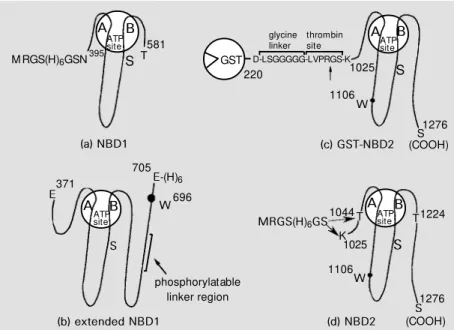
Four different recombinant domains were prepared: i) N-terminal NBD1, ii) extended NBD1 including a phosphorylatable linker sequence, iii) C-terminal NBD2, and iv) shorter NBD2 (cf. Figures 3 and 4). cDNAs corresponding to the different domains were obtained by reverse-transcriptase PCR from polyA+ mRNAs purified from total RNAs of mouse adrenal glands using specific oligo-nucleotide primers. Alternatively, fragments of the complete mdr1 cDNA (provided by Pr. P. Gros, Montreal, Canada (5)) were amplified by direct PCR and cloned into inducible bacterial expression plasmids. Pro-tein expression was achieved using Escheri-chia coli strains JM105, JM109 or BL21 (DE3).
The domain limits were predicted using the ANTHEPROT software (54), as illus-trated in Figure 2 for NBD1. The domains were designed taking into account the fol-lowing aspects: 1) all domains contained the Walker motifs A and B, and the S signature since they are characteristic for ABC trans-porters, 2) the hydrophobicity was predicted and minimized to increase protein solubility and to aid protein purification, 3) secondary structure elements were predicted and se-quence boundaries were chosen to be out-side the helical or strand regions to avoid structural disturbance of the recombinant protein, 4) designed oligonucleotides had to
be sufficiently different from the correspond-ing regions of the mdr2 and mdr3 genes, also present in adrenal glands, to avoid a cross-reaction.
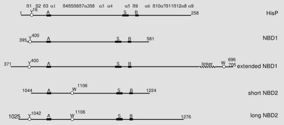
Three different lengths of the NBD1 were designed in this way, with the N-terminal end set at Asn-395 and the C-terminal one at either Ser-643, Glu-613 or Thr-581. Extended NBD1 stretched from Glu-371 to Glu-705, including the phosphorylatable linker region and Trp-696. A long NBD2 was designed from Lys-1025 to C-terminal Ser-1276, and a shorter one from Thr-1044 to Thr-1224, both proteins containing Trp-1106 (Figure 3, c and d). A schematic sequence alignment of the different recombinant domains with HisP, a cytosolic subunit of an ABC trans-porter whose 3-D structure has recently been reported (10), is shown in Figure 4.
Binding o f nucle o tide s and analo gue s
The NBDs1 were obtained as fusion pro-teins with an N-terminal hexahistidine tag. The cDNAs were cloned into pQE-30 and protein expression was induced by isopro-pyl-1-thio-ß-D-galactopyranoside (IPTG). Protein purification involved cell lysis by
M RGS(H)6GSN395 S
A B
ATP site
S1276 (COOH) 581
T D-LSGGGGG-LVPRGS-K
220 1025
1106
S AATPB site
S A BATP site
T T
S1276 (COOH) 1106
1224 1044
K
W W
1025 705
E-(H)6 696 W 371
E
S
phosphorylatable linker region
MRGS(H)6GS A BATP
site
GST
(a) NBD1 (c) GST-NBD2
(b) extended NBD1 (d) NBD2
Figure 3 - Recombinant cytosolic domains of P-glycoprotein. glycine linker
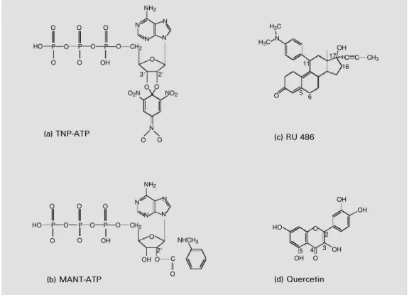
by derivatizing Tyr-400 according to results obtained with the full-length Pgp (56). Since NBD1 did not contain any tryptophan resi-due, the quenching of intrinsic fluorescence due to tyrosine residues was monitored to study the binding of 2'(3')-N -methylanthra-niloyl (MANT) derivative of ATP (Figure 5b), a fluorescent high-affinity nucleotide analogue (KD = 25 µM). There was evidence that the maximal quenching of about 15-20% was due at least in part to an interaction with Tyr-464. When the NBD1 and NBD2 sequences of the same protein are aligned, Tyr-464 corresponds to Trp-1106 which was shown to interact with the MANT group of MANT-ATP (see below). The competition observed between ATP and its analogues, TNP-ATP and MANT-ATP, indicates that all three nucleotides use the same binding site.
Inte ractio n with hydro pho bic ste ro ids
Extended NBD1 was fused to a C- termi-nal hexahistidine tag and was overexpressed after insertion of its DNA into a pT7-7 plas-mid. Despite many attempts to increase pro-tein solubility, such as lowering the temper-ature for bacterial growth and varying the IPTG concentration, recombinant extended-NBD1 was exclusively found in inclusion bodies. A soluble active form of the ex-French press treatment and affinity
chroma-tography on a nickel-nitrilotriacetic acid col-umn (55). The amount of soluble protein present in the supernatant indicated that, in the case of the three different NBD1s, the protein yield was indirectly correlated to the length of the protein domain. Therefore, the shorter domain, from Asn-395 to Thr-581, was prepared in high amounts for biochemi-cal and biophysibiochemi-cal studies.
The domain exhibited a low, but signifi-cant, ATPase activity with an extrapolated maximal rate of 25 nmol/min x mg protein and a Km(ATP) of 2.1 mM. A similar rate was obtained for the recombinant nucle-otide-binding fold of CFTR. The activity of Pgp NBD1 was competitively inhibited by micromolar concentrations of the 2',3'-O -(2,4,6-trinitrophenyl) (TNP) derivative of ATP, a high-affinity fluorescent nucleotide analogue (KD = 2.2 µM by enhancement of extrinsic fluorescence) whose structure is shown in Figure 5a. In addition, chemical modification by N-ethylmaleimide of the single cysteine residue, present in the Walker motif A, increased the Km(ATP) by 6-fold. This is probably due to steric hindrance caused by the modification, since the corre-sponding serine residue in HisP forms a hydrogen bond with the ß-phosphate of ATP (10). The NBD1 was labelled upon photoir-radiation in the presence of radioactive 8.7 µM 8-azido-ATP and 5 mM MgCl2, possibly
371
1025
HisP
extended NBD1 NBD1
short NBD2
long NBD2 705
696 W ß1
Y16
ß2 ß3 ß4ß5ß6ß7a2ß8
A S B
1 258
a1 a3 a4 a5ß9 a6 ß10a7ß11ß12a8 a9
Y400 A S B
395 581
Y400 A S B linker
A S B
1044 W1106 1224
Y1042 A W1106 S B 1276
tended NBD1 was obtained when the pro-tein was solubilized from inclusion bodies. This procedure involved solubilization with a rather low (1.5 M) guanidine concentration and a quick 20-fold dilution in phosphate buffer, pH 9, containing 0.5 M NaCl, 10-20% glycerol, and 0.01% 6-O-(N -heptylcar-bamoyl)-methyl-a-D-glucopyranoside (HECAMEG). There are two pieces of evi-dence for the guanidine-solubilized extended NBD1 being correctly folded: 1) NBD1 and extended NBD1 had similar binding affini-ties for nucleotides and analogues, and 2) NBD1 purified from inclusion bodies and the same protein purified from the superna-tant had similar properties (57). Trp-696 exhibited fluorescence properties character-istic for being in a quite hydrophobic envi-ronment, with maximal emission at a rela-tively low wavelength, 325 nm upon excita-tion at 295 nm; this further confirmed accu-rate folding, at least around Trp-696. Inter-action with MANT-ATP produced both a quenching of extended NBD1 intrinsic
fluo-rescence and a fluofluo-rescence resonance en-ergy transfer between Trp-696 and the MANT group of bound MANT-ATP. The 57% maxi-mal efficiency indicated that donor and ac-ceptor fluorophores were at a distance of about 17-23 Å (57).
The quenching of Trp-696 fluorescence was also used to monitor interaction with steroids. A marked effect was observed with hydrophobic steroids, known not to be trans-ported by Pgp, with the following efficiency: RU 486 (cf. Figure 5c), megestrol acetate > D6-progesterone > medroxyprogesterone ac-etate > progesterone > 5ß-pregnanedione. In contrast, a very low interaction was detected with hydrophilic hydroxylated derivatives, known to be transported by Pgp: triamcino-lone < cortisol, dihydrotestosterone, corti-costerone, 11a-hydroxyprogesterone < tes-tosterone, dexamethasone, 17a -hydroxy-progesterone, triamcinolone acetonide < 11-deoxycorticosterone. The same preference was observed for the binding of these com-pounds to full-length Pgp. The overall
re-NH2
Figure 5 - Chemical structure of compounds w hich bind to cyto-solic NBDs. TNP, 2' ,3' -O-(2,4, 6-trinitrophenyl); M ANT, 2' (3'
)-N-methylanthraniloyl. HO P
N N
N N
P P
O O O CH2
O
HO P
N N
N N
P P
O O O
NH2
CH2
O
CH3
3 2
O O O2N NO2
N
O O
O O OH
(a) TNP-ATP
NHCH3
C
O O OH OH
O O
2
(b) MANT-ATP
H3C
H3C N
OH C C 11
O 5 6
17 16
(c) RU 486
OH
2
OH
OH OH
HO O
O 3 4 5
(d) Quercetin
O O O
sults indicate that hydrophobic substituents increase steroid binding affinity for Pgp, prevent their transport and increase their ability to inhibit drug efflux, whereas hy-droxyl groups at positions 11, 16, 17 and 21 reduce binding affinity and inhibitory ef-fects and allow the steroids to be transported (45).
The almost complete quenching of ex-tended NBD1 intrinsic fluorescence by RU 486 suggested that Trp-696 is located within, or in close proximity to, the steroid-interact-ing region. The quenchsteroid-interact-ing was unaffected by preincubation with 10 mM ATP, indicat-ing that the two sites are distinct. In contrast, a complete antagonism was observed with MANT-ATP since its binding, as monitored by enhancement of extrinsic fluorescence, was prevented by preincubation with RU 486, and bound MANT-ATP was displaced upon addition of the antiprogestin. This sup-ports the existence of a cytosolic steroid-interacting region adjacent to the ATP-bind-ing site (Figure 6, A and B). The mutually exclusive binding of MANT-ATP against
RU 486 could be due to overlapping of the steroid-interacting region by the hydropho-bic MANT group of the nucleotide analogue bound to the ATP site (Figure 6C), although a distant conformational change cannot be completely excluded.
Bifunctio nal inte ractio ns with flavo no ids
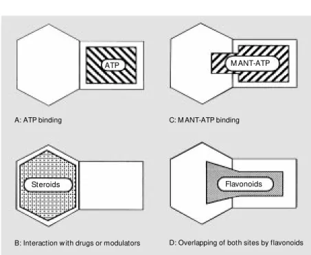
The cDNA of NBD2 was cloned into pGEX-KT, and the domain was obtained as a glutathione S-transferase fusion protein (cf. Figure 3b) which was purified on a glutathione-Sepharose 4B column. The pro-tein was found to bind ATP and its high-affinity derivative, TNP-ATP (58). However, the NBD2 appeared to be quite unstable when separated from the glutathione S -trans-ferase after thrombin cleavage, and thus could not be extensively studied. NBD2 was then generated as an N-terminally tagged hexa-histidine fusion protein and was found to be soluble and stable under the conditions men-tioned above, which allowed the preparation of large amounts of pure protein. The solu-bility appeared to be higher for a shorter version (from Thr-1044 to Thr-1224) of the domain (cf. Figure 3d). The intrinsic fluores-cence of the domain due to the single Trp-1106 was highly quenched with MANT-ATP, showing also an efficient fluorescence resonance energy transfer. A similar NBD2 from Chinese hamster ovary Pgp was ob-tained in fusion to maltose-binding protein. NBD2 intrinsic fluorescence was also highly quenched upon interaction with fla-vonoids (59). The binding affinities of dif-ferent classes of flavonoids could be ranked in the following order: flavones (quercetin (cf. Figure 5d), apigenin) > flavanones (naringenin) > isoflavone (genistein) > gly-cosyl-flavones (rutin). Within flavones, hy-droxyl groups at positions 3 and 5 in close proximity to the ketone at position 4 mark-edly increased the binding affinity. They might contribute to a more precise mimick-Figure 6 - Proposed schematic model of NBDs show ing the relative positions of different
nucleotide- and effector-binding sides. M ANT, 2' (3' )-N-methylanthraniloyl.
ATP M ANT-ATP
Flavonoids A: ATP binding
Steroids
C: M ANT-ATP binding
ing of the adenine moiety of ATP, which has already been observed for the flavonoid-liganded 3-D structures of cycline-depend-ent kinase 2 (52) and Hck tyrosine kinase (53). Flavonoid binding to NBD2 was in-deed partly prevented by preincubation with ATP or displaced upon ATP addition (59). The binding affinity was further increased by disrupting the C ring to give chalcones, and by substituting the B ring with a hydro-phobic halogen atom (60). In addition, fla-vonoid binding also partly antagonized the binding of the steroid RU 486. Since the steroid-interacting region is assumed to be close to the ATP site, bound flavonoids most likely cover both the ATP site and the vicinal steroid site. Therefore, flavonoids constitute a new class of MDR modulators which un-dergo bifunctional interactions with the cy-tosolic domains of Pgp (Figure 6D).
Inte re st o f re co m binant cyto so lic do mains fo r the study o f Pgp struc-ture -functio n re latio nships and fo r drug de sign
In addition to the ATP-binding site, NBDs also contain sequence segments which are likely to carry out other physiological
func-tions, such as signal transduction. The study of proteins mutated around the Walker motif B in Pgp, or Ste6 or malK, provided evi-dence that drug binding and ATP hydrolysis generate cellular signals. NBDs also contain part of drug-binding sites as demonstrated by photoaffinity labelling with either iodoaryl azidoprazosin or 3'-p -benzoyldihydrocin-namoyl-taxol. The similar behavior of the two NBDs towards steroids and especially towards flavonoids supports the existence of 2 modulator-binding sites (Figure 6). This is consistent with conclusions drawn from nu-merous kinetic and labelling experiments of membrane-inserted Pgp with modulators and drug substrates.
The overall results indicate that Pgp NBDs are active and can be prepared in high a-mounts as purified proteins which permit crystallization trials. The presence of specif-ic binding sites for ATP offers the possibility to search for selective bifunctional modula-tors able to cover both the ATP-binding site and the vicinal steroid-interacting region. Identification of reactive residues at both binding sites might allow a more rational design and synthesis of highly potent and selective inhibitors.
Re fe re nce s
1. Gottesman M M & Pastan I (1993). Bio-chemistry of multidrug resistance medi-ated by the multidrug transporter. Annual Review of Biochemistry, 62: 385-427. 2. Endicott JA & Ling V (1989). The
biochem-istry of P-glycoprotein-mediated multi-drug-resistance. Annual Review of Bio-chemistry, 58: 137-171.
3. Ruetz S & Gros P (1994). Phosphatidyl-choline translocase: a physiological role for the mdr2 gene. Cell, 77: 1071-1081. 4. van Helvoort A, Smith AJ, Sprong H,
Fritzche I, Schinkel AH, Borst P & van M eer G (1996). M DR1 P-glycoprotein is a lipid translocase of broad specificity, w hile M DR3 P-glycoprotein specifically translo-cates phosphatidylcholine. Cell, 87: 507-517.
5. Gros P, Croop J & Housman D (1986). M ammalian multidrug resistance gene: complete sequence indicates strong ho-mology to bacterial transport proteins.
Cell, 47: 371-380.
6. Kast C, Canfield V, Levenson R & Gros P (1996). Transmembrane organization of mouse P-glycoprotein determined by epi-tope insertion and immunofluorescence.
Journal of Biological Chem istry, 271: 9240-9248.
7. Walker JE, Saraste M , Runsw ick M J & Gay NJ (1982). Dist ant ly relat ed se-quences in the a- and ß-subunits of ATP synthase, myosin, kinases and other ATP-requiring enzymes and a common nucle-otide binding fold. EM BO Journal, 1: 945-951.
8. Higgins CF, Hiles ID, Salmond GPC, Gill DR, Dow nie JA, Evans IJ, Holland IB, Gray L, Buckel SD, Bell W & Hermodson M A (1986). A family of related ATP-binding subunits coupled to many distinct biologi-cal processes in bacteria. Nature, 323: 448-450.
9. Rosenberg M F, Callaghan R, Ford RC & Higgins CF (1997). Structure of the multi-drug resistance P-glycoprotein to 2.5 nm resolution determined by electron micros-copy and image analysis. Journal of Bio-logical Chemistry, 272: 10685-10694. 10. Hung L-W, Wang IX, Nikaido K, Liu P-Q,
Reconstitu-tion of drug-stimulated ATPase activity follow ing co-expression of each half of human P-glycoprotein as separate poly-peptides. Journal of Biological Chemistry, 269: 7750-7755.
12. Azzaria M , Schurr E & Gros P (1989). Dis-crete mutations introduced in the pre-dicted nucleotide-binding sites of the
mdr1 gene abolish its ability to confer multidrug resistance. M olecular and Cel-lular Biology, 9: 5289-5297.
13. Senior AE & Bhagat S (1998). P-glycopro-tein show s strong catalytic cooperativity betw een the tw o nucleotide sites. Bio-chemistry, 37: 831-836.
14. Leveille-Webster CR & Arias IM (1995). The biology of the P-glycoproteins. Jour-nal of M embrane Biology, 143: 89-102. 15. Tang-Wai DF, Brossi A, Arnold LD & Gros
P (1993). The nitrogen of the acetamido group of colchicine modulates P-glycopro-tein-mediated multidrug resistance. Bio-chemistry, 32: 6470-6476.
16. Shapiro AB & Ling V (1997). Positively cooperative sites for drug transport by P-glycoprotein w ith distinct drug specifici-ties. European Journal of Biochemistry, 250: 130-137.
17. Bolhuis H, van Veen HW, Poolman B, Driessen AJM & Konings WN (1997). M echanisms of multidrug transporters.
FEM S M icrobiology Review s, 21: 55-84. 18. Higgins CF & Gottesman M M (1992). Is
the m ultidrug transporter a flippase?
Trends in Biochemical Sciences, 17: 18-21.
19. Stein WD (1997). Kinetics of the multi-drug transporter (P-glycoprotein) and its reversal. Physiological Review s, 77: 545-590.
20. Schinkel AH, Smit JJM , van Tellingen O, Beijnen JH, Wagenaar E, van Deemter L, M ol CAAM , van der Valk M A, Robanus-M aandag EC, Riele HPJ, Bern AJRobanus-M & Borst P (1994). Disruption of the mouse
mdr1a P-glycoprotein gene leads to a de-ficiency in the blood-barrier and to in-creased sensitivity to drugs. Cell, 77: 491-502.
21. Ueda K, Okamura N, Hirai M , Tanigaw ara Y, Saeki T, Kioka N, Komano T & Hori R (1992). Human P-glycoprotein transports cortisol, aldosterone, and dexametha-sone, but not progesterone. Journal of Biological Chemistry, 267: 24248-24252. 22. Wolf DC & Horw itz SB (1992).
P-glycopro-t ein P-glycopro-t ransporP-glycopro-t s corP-glycopro-t icosP-glycopro-t erone and is photoaffinity-labelled by the steroid. In-ternational Journal of Cancer, 52: 141-146.
23. Bruggem ann EP, Germ ann UA,
Gottesman M M & Pastan I (1989). Tw o different regions of P-glycoprotein are photoaffinity labeled by azidopine. Jour-nal of BiologicalChemistry, 264: 15483-15488.
24. Evans GL, Ni B, Hrycyna CA, Chen D, Ambudkar SV, Pastan I, Germann UA & Gottesman M M (1995). Heterologous ex-pression systems for P-glycoprotein: E. coli, yeast and baculovirus. Journal of Bioenergetics and Biomembranes, 27: 43-52.
25. Germann UA, Chambers TC, Ambudkar SV, Pastan I & Gottesman M M (1995). Effects of phosphorylation of P-glycopro-tein on multidrug resistance. Journal of Bioenergetics and Biomembranes, 27: 53-60.
26. Smith CD & Zilfou JT (1995). Circumven-tion of P-glycoprotein-mediated multiple drug resistance by phosphorylation modu-lators is independent of protein kinases.
Journal of Biological Chem istry, 270: 28145-28152.
27. Germann UA, Chambers TC, Ambudkar SV, Licht T, Cardarelli CO, Pastan I & Gottesman M M (1996). Characterisation of phosphorylation-defective mutants of human P-glycoprotein expressed in mam-malian cells. Journal of Biological Chemis-try, 271: 1708-1716.
28. Doige CA & Ames GF-L (1993). ATP-de-pendent transport systems in bacteria and humans: relevance to cystic fibrosis and multidrug resistance. Annual Review of M icrobiology, 47: 291-319.
29. Higgins CF (1992). ABC transporters: From microorganisms to man. Annual Re-view of Cellular Biology, 8: 67-113. 30. Deeley RG & Cole SPC (1997). Function,
evolution and structure of multidrug resis-tance protein (M RP). Seminars in Cancer Biology, 8: 193-204.
31. Welsh M J, Robertson AD & Ostedgaard LS (1998). The ABC of a versatile engine.
Nature, 396: 623-624.
32. Borst P, Kool M & Evers R (1997). Do cM OAT (M RP2), other M RP homologues, and LRP play a role in M DR? Seminarsin Cancer Biology, 8: 205-213.
33. Balzi E & Goffeau A (1991). M ultiple or pleiotropic drug resistance in yeast. Bio-chimica et Biophysica Acta, 1073: 241-252.
34. Decottignies A & Goffeau A (1997). Com-plete inventory of the yeast ABC proteins.
Nature Genetics, 15: 137-145.
35. Rubio JP & Cow man AF (1996). The ATP binding cassette (ABC) gene family of
Plasmodium falciparum. Parasitology To-day, 12: 135-140.
36. Foote SJ, Thompson JK, Cow man AF & Kemp DJ (1989). Amplification of the mul-tidrug resistance gene in some chloro-quine-resistant isolates of P. falciparum.
Cell, 57: 921-930.
37. Ouelette M , Fase-Fow ler F & Borst P (1990). The amplified H circle of metho-trexate-resistant Leishmania tarentolae
contains a novel P-glycoprotein gene.
EM BOJournal, 9: 1027-1033.
38. Dallagiovanna B, Gamarro F & Castanys S (1996). M olecular characterization of a P-glycoprotein-related tcpgp2 gene in Try-panosomacruzi. M olecular and Biochemi-cal Parasitology, 75: 145-157.
39. Ullman B (1995). M ultidrug resistance and P-glycoproteins in parasitic protozoa. Jour-nal ofBioenergetics and Biomembranes, 27: 77-84.
40. Chiquero M J, Perez-Victoria JM , O’Valle F, Gonzalez-Ros JM , del M oral RG, Ferragut JA, Castanys S & Gamarro F (1998). Altered drug membrane perme-ability in a multidrug-resistant Leishmania tropica line. Biochemical Pharmacology, 55: 131-139.
41. van Veen HW, Callaghan R, Soceneantu L, Sardini A, Konings WN & Higgins CF (1998). A bacterial antibiotic-resistance gene that complements the human multi-drug-resistance P-glycoprotein gene. Na-ture, 391: 291-295.
42. Sikic BI (1997). Pharmacologic approaches to reversing multidrug resistance. Semi-nars in Hematology, 34: 40-47.
43. Zamora JM , Pearce HL & Beck WT (1988). Physiological-chemical properties shared by compounds that modulate multidrug resistance in human leukemic cells. M o-lecularPharmacology, 33: 454-562. 44. Seelig A (1998). A general pattern for
sub-strate recognition by P-glycoprotein. Eu-ropean Journal ofBiochemistry, 251: 252-261.
45. Barnes KM , Dickstein B, Culter GB, Fojo T & Bates SE (1996). Steroid transport, ac-cumulation, and antagonism of P-glyco-protein in multidrug-resistant cells. Bio-chemistry, 35: 4820-4827.
46. Gruol DJ, Zee M C, Trotter J & Bourgeois S (1994). Reversal of multidrug resistance by RU 486. Cancer Research, 54: 3088-3091.
47. Callaghan R & Higgins CF (1995). Interac-tion of tamoxifen w ith the multidrug re-sistance P-glycoprotein. British Journal of Cancer, 71: 294-299.
(Edi-tor), The Flavonoids: Advances in Re-search Since 1986. Chapman & Hall, Lon-don, 619-652.
49. Critchfield JW, Welsh CJ, Phang JM & Yeh GC (1994). M odulation of adriamycin in accumulation and efflux by flavonoids in HCT-15 colon cells. Act ivat ion of P-glycoprotein as a putative mechanism.
Biochemical Pharmacology, 48: 1437-1445.
50. Scam bia G, Ranellet t i FO, Benedet t i Panici P, De Vincenzo R, Bonanno G, Ferrandina G, Piantelli M , Bussa S, Rumi C, Cianfriglia M & M ancuso S (1994). Quercet in pot ent iat es t he ef f ect of adriamycin in a multidrug-resistant M CF-7 human breast-cancer cell line: P-glycopro-tein as a possible target. Cancer Chemo-therapy andPharmacology, 34: 459-464. 51. Shapiro AB & Ling V (1997). Effect of
quercetin on Hoechst 33342 transport by purified and reconstituted P-glycoprotein.
Biochemical Pharmacology, 53: 587-596. 52. De Azevedo WF, M ueller-Dieckmann H-J, Schulze-Gahmen U, Worland PJ, Sausville E & Kim S-H (1996). Structural basis for
specificity and potency of a flavonoid in-hibitor of human CDK2, a cell cycle ki-nase. Proceedings of the National Acade-my of Sciences, USA, 93: 2735-2740. 53. Sicheri F, M oarefi I & Kuryan J (1997).
Crystal structure of the Src family tyrosine kinase Hck. Nature, 385: 602-609. 54. Geourjon C & Deléage G (1995).
ANTHEPROT 2.0: a three-dimensional module fully coupled w ith protein se-quence analysis methods. Journal of M o-lecular Graphics, 13: 209-212.
55. Dayan G, Baubichon-Cortay H, Jault J-M , Cortay J-C, Deléage G & Di Pietro A (1996). Recombinant N-terminal nucle-otide-binding domain from mouse P-gly-coprotein: overexpression, purification and role of cysteine-430. Journal of Bio-logical Chemistry, 271: 11652-11658. 56. Sankaran B, Bhagat S & Senior AE (1997).
Photoaffinity labelling of P-glycoprotein catalytic sites. FEBS Letters, 417: 119-122.
57. Dayan G, Jault J-M , Baubichon-Cortay H, Baggetto LG, Renoir JM , Baulieu EE, Gros P & Di Pietro A (1997). Binding of steroid
modulators to recombinant cytosolic do-main from mouse P-glycoprotein in close proximity to the ATP site. Biochemistry, 36: 15208-15215.
58. Baubichon-Cortay H, Baggetto LG, Dayan G & Di Pietro A (1994). Overexpression and purification of the carboxyl-terminal nucleotide-binding domain from mouse P-glycoprotein. Strategic location of a tryp-tophan residue. Journal of Biological Chemistry, 269: 22983-22989.