Cytosolic Phospholipase A2 Modulates TLR2
Signaling in Synoviocytes
Randi M. Sommerfelt‡, Astrid J. Feuerherm‡, Trine Skuland, Berit Johansen*
Department of Biology, Lipid Signaling Group, Norwegian University of Science and Technology, Trondheim, Norway
‡These authors share first authorship on this work. *berit.johansen@ntnu.no
Abstract
Rheumatoid arthritis (RA) is an autoimmune disease characterized by chronic synovitis leading to destruction of cartilage and bone. PLA2 enzymes are key players in inflammation regulating the release of unsaturated fatty acids such as arachidonic acid (AA), a precursor of pro-inflammatory eicosanoids. Several lines of evidence point to toll-like receptors (TLRs) as drivers of synovitis and joint destruction in RA. However, few studies have ad-dressed the implication of PLA2 activity downstream TLR activation in the synovium. Here, we aimed to characterize PLA2 enzyme involvement in TLR2-induced signaling in synovial fibroblast-like cells. TLRs1-7 and a range of sPLA2, iPLA2 and cPLA2 enzymes were found to be transcriptionally expressed in cultured synoviocytes. Activation of TLR2/1 and TLR2/6 led to phosphorylation of cPLA2αat Ser505, and induced AA release and PGE
2production; effects that were attenuated by cPLA2αinhibitors. In contrast, sPLA2 inhibitors did not affect AA or PGE2release. cPLA2αinhibitors furthermore attenuated TLR-induced expres-sion of IL-6, IL-8 and COX2. COX1/2 inhibitors attenuated TLR2/6-induced IL-6 transcrip-tion and protein productranscrip-tion comparable to cPLA2αinhibition. Moreover, exogenously PGE2 added alone induced IL-6 production and completely rescued IL-6 transcription when added simultaneously with FSL-1 in the presence of a cPLA2αinhibitor. Our results demon-strate for the first time that cPLA2αis involved in TLR2/1- and TLR2/6-induced AA release, PGE2production and pro-inflammatory cytokine expression in synoviocytes, possibly through COX/PGE2-dependent pathways. These findings expand our understanding of cPLA2αas a modulator of inflammatory molecular mechanisms in chronic diseases such as RA.
Introduction
Rheumatoid arthritis (RA) is a complex systemic inflammatory disease characterized by chron-ic synovitis and irreversible destruction of cartilage and bone. The aetiology of RA is unclear, but genetic, epigenetic and environmental factors are involved in triggering and/or exacerbat-ing RA synovitis [1,2]. Fibroblasts are believed to play an important role in chronic
OPEN ACCESS
Citation:Sommerfelt RM, Feuerherm AJ, Skuland T, Johansen B (2015) Cytosolic Phospholipase A2 Modulates TLR2 Signaling in Synoviocytes. PLoS ONE 10(4): e0119088. doi:10.1371/journal. pone.0119088
Academic Editor:Bruno Lourenco Diaz, Universidade Federal do Rio de Janeiro, BRAZIL
Received:June 2, 2014
Accepted:January 9, 2015
Published:April 20, 2015
Copyright:© 2015 Sommerfelt et al. This is an open access article distributed under the terms of the
Creative Commons Attribution License, which permits unrestricted use, distribution, and reproduction in any medium, provided the original author and source are credited.
Data Availability Statement:All relevant data are within the paper.
Funding:This work was supported by the Norwegian Research Council, BIA program; Health Region Mid-Norway and Avexxin AS. The funders had no role in study design, data collection and analysis, decision to publish, or preparation of the manuscript.
inflammation [3], and RA fibroblast-like synoviocytes (FLS) actively promote inflammation and joint destruction [4].
Lipid metabolites derived from the unsaturatedΩ6 fatty acid arachidonic acid (AA) play pivotal roles in inflammation [5]. The eicosanoid prostaglandin E2 (PGE2) is metabolized from
AA by the cyclooxygenase (COX) enzymatic pathway, and is a key regulator of immunopathol-ogy and chronic inflammation [6]. PGE2is abundantly detected in synovial fluid of arthritic
joints [7], and the effective symptomatic relief in RA patients by non-steroid anti-inflammato-ry drugs (NSAIDs) targeting the COX enzymes is in large part due to decreased PGE2synthesis
[8]. Phospholipase A2 (PLA2) enzymes act to hydrolyze membrane phospholipids at thesn-2 position to liberate unsaturated fatty acids from cellular membranes, and are thus important regulators of lipid signaling [9]. To date, the PLA2 enzyme family includes 16 groups compris-ing more than 30 members. Based on their biochemical properties, PLA2 enzymes are subdi-vided into distinct types; Ca2+-dependent secretory PLA2s (sPLA2s), Ca2+-dependent cytosolic PLA2s (cPLA2s), Ca2+-independent cytosolic PLA2s (iPLA2s), platelet-activating factor acetyl hydrolases (PAF-AHs), lysosomal PLA2 and adipose-PLA2 [9]. Cytosolic group IVa PLA2 (cPLA2α) is activated by intracellular calcium and by phosphorylation in response to various extracellular stimuli. cPLA2αis selective for AA-containing acyl chainsin vitro[10], and is considered a central enzyme in AA-derived eicosanoid production [9]. sPLA2 and iPLA2 also contribute to AA release, although they do not display the same acyl chain specificity as cPLA2α[11,12]. Due to its arachidonyl selectivity, cPLA2αis believed to play a key role in in-flammatory disease, a view supported by the findings that cPLA2α-deficient mouse models are resistant to various inflammatory diseases including asthma, pulmonary fibrosis and CIA-in-duced arthritis [13–16]. Moreover, inhibitors targeting cPLA2αdecelerate disease progression in CIA mice [17,18]. However, through which mechanisms cPLA2α-deficiency or inhibition prevent disease progression is not fully understood.
Toll-like receptors (TLRs) are pattern recognition receptors (PRRs), constituting a major part of the innate immune system sensing pathogen associated molecular patterns (PAMPs) on invading pathogens [19]. Moreover, TLRs can induce non-infectious inflammation by sens-ing endogenous molecules released in response to tissue damage or necrosis (damage associat-ed molecular patterns, DAMPs), and elevatassociat-ed TLR activation is associatassociat-ed with several inflammatory, autoimmune and non-infectious diseases including RA [20]. The TLR2 family of receptors (TLR1, TLR2, TLR6) is located on the cell surface. TLR2 dimerizes with TLR1 or TLR6 to recognize a range of PAMPs and DAMPs [20], of which several, including bacterial li-poproteins [2] and heat-shock proteins [21,22], are detected in RA joints. In FLS from RA pa-tients, TLRs including TLR2 and 6 levels are significantly elevated compared to patients with non-inflammatory arthritis [23], and TLR2 is found in excess at sites of pannus invasion and cartilage and bone erosion [24]. Accordingly, TLR2 activation is believed to play a role in chronic inflammation and joint destruction in RA.
TLR2 ligands are reported to activate PLA2 in human leukocytes and murine macrophages [25,26]. However, interactions between PLA2 enzymes and TLR2 signaling in synoviocytes are hitherto not well described. Here, we propose that cPLA2αis a major regulator of TLR2-in-duced AA release and PGE2production in human synoviocytes. In contrast, sPLA2
involve-ment was not found. Furthermore, we demonstrate that cPLA2αinhibition attenuates TLR2-induced expression of inflammatory cytokines, suggesting a regulatory role of cPLA2α
in synovial TLR responses.
Materials and Methods
Reagents
PBS was from Oxoid. DNAse- and RNAse-free water was from VWR. Recombinant human TNF and IL-6 ELISA Duoset were from R&D systems. Quantitect primer assays for TLR1-7 and 18S were from Qiagen. QuantiTect Reverse Transcription kit, RNeasy minikit, Leupeptin, pepstatin and LightCycler 480 SYBR Green I Master mix were from Roche Molecular Bio-chemicals. RNAlaterwas from Life technologies. FSL-1 and Pam3CSK4were from Invivogen. [3H]-arachidonic acid ([3H]-AA), and liquid scintillation cocktail Ultima Gold were from NEN Perkin Elmer. AVX002 and Inhibitor 28 were provided by Avexxin AS (Trondheim, Nor-way). Arachidonyl trifluoromethyl ketone (AACOCF3, ATK) was from Enzo Life Sciences. Varespladib (LY315920) was from Selleckchem. CAY10502, CAY10590 and PGE2ELISA kit
were from Cayman Chemicals. Phospho-cPLA2 (Ser505) antibody was from Cell Signal Tech-nology.α-tubulin antibody was from Santa Cruz Biotechnology. Polyclonal goat anti-mouse immunoglobulins horse radish peroxidase-conjugated secondary antibody was from Dako. Hybond-C nitrocellulose membranes were from GE healthcare. NuPAGE gel system (10% Bis-Tris gel) was from Invitrogen. Hybond-C nitrocellulose membranes were from GE healthcare. SuperSignal West Femto Maximum Sensivity substrate was from Thermo Scientific. All other reagents were from Sigma-Aldrich.
Cell culture
The human synovial sarcoma derived cell line SW982 (ATCC, London, UK) was maintained at 37°C with 10% CO2in a humidified atmosphere in Dulbecco's Modified Eagle Medium
(DMEM) supplemented with 10% FBS, 0.1 mg/mL gentamicin and 0.3 mg/mL L-glutamine. Experiments were performed at 3 days post-confluence. Prior to experimental treatment, cells were serum-deprived in serum-free DMEM overnight, and all experiments were performed in serum-free DMEM. Inhibitors used were applied 2 hours before stimulation with FSL-1, Pam3CSK4or TNF.
[
3H]-arachidonic acid release assay
SW982 cells were labeled for 18 hours with [3H]AA in serum-free DMEM and washed in PBS prior to experimental treatment. Release of [3H]AA was analyzed in triplicates as previously described [27]. In short, cells were incubated for the desired period of time in the absence or presence of chemical inhibitors prior to stimulation with FSL-1 or Pam3CSK4. Supernatants
were removed and cleared of detached cells or cell debris by centrifugation. Cells attached in the wells were dissolved in 1M NaOH and all samples were assayed for fatty acid release by scintillation counting. Results are shown as released [3H]AA in supernatants relative to total [3H]AA incorporated into the cells. All experiments were performed in triplicates.
PGE
2and IL6 enzyme-linked immunosorbent assays (ELISA)
Serum-deprived SW982 cells were treated with FSL-1 or Pam3CSK4for 24 hours in the absence
or presence of chemical inhibitors. Supernatants were harvested, and PGE2and IL-6 levels
Real-time reverse-transcription polymerase chain reaction (qPCR)
One sample of post-natal full-term placenta was collected from St Olavs hospital (Trondheim, Norway). Small pieces of placenta were immediately placed in RNAlaterand stored at -80°C. No further permissions are required from Regional Committees for Medical and Health Research Ethics (REC, Norway) for use of anonymized biological material in the technical and methodo-logical work presented in current publication. Total RNA from placental and SW982 cells was isolated using RNeasy minikit according to kit protocol. RNA concentrations and integrity was monitored by NanoDrop spectrophotometric measurement (NanoDrop Technologies Inc.) and total RNA (1μg) was reverse transcribed using the QuantiTect Reverse Transcription kit
follow-ing the recommended protocol. qPCR analyses were carried out usfollow-ing the Lightcycler 480 system. Primers for PLA2 subgroups, IL-6, IL-8, COX2 and GAPDH were designed with Primer 3 soft-ware (Whitehead Institute for Biomedical Research, Cambridge, MA, USA). Primer sequences or Qiagen reference sequence for all primers applied are listed in Tables1and2, respectively. Disso-ciation curve analysis for each SYBR Green primer pair and reaction was performed to verify spe-cific amplification. Primer amplification efficiencies and Cq-values were calculated by the LinRegPCR software [28] and fold changes in mRNA expression were analyzed by the Qbase-PLUS software (Biogazelle) using GAPDH or 18S as reference gene.
Immunoblotting
Following FSL-1 or Pam3CSK4treatment for indicated times with or without chemical
inhibi-tors, cells were washed in PBS and lysed in buffer containing 50mM Tris pH 7.5, 150 mM NaCl, 10% glycerol, 0.5% Triton-X-100, 2mM EDTA, 40 mMβ-glycerophosphate, 100 mM NaF, 200μM Na3VO4, 10μg leupeptin, 1μM pepstatin and 1mM phenylmethylsulfonyl
fluo-ride. Lysates were cleared by centrifugation, separated by SDS-PAGE and transferred to Hybond-C nitrocellulose membranes by electrophoresis using the NuPAGE gel system (10%
Table 1. Overview of qPCR primers.
Gene Forward primer Reverse primer Product size (basepairs)
PLA2G2A aaggaagccgcactcagtta ttgcacaggtgattctgctc 186
PLA2G2D gatggtcaagcaagtgactgg gagcagtggatgttcccctg 213
PLA2G4A catgcccagacctacgattt cccaatatggctaccacagg 163
PLA2G4C tgccggagtctcatttgtcc gggtgaactcgaaccaggtc 141
PLA2G4D cgtcagatcgcccagaaaac ccaggaaggatgtctgtgtgt 177
PLA2G4F gagttggaggctcagaccag cagagaggtcaaagccaagg 178
PLA2G5 gccaaagagaaccccagag gccgtagaagccgtagtttg 154
PLA2G6A ttatgctgtccagggtgaca gagaacttcatggccgagtg 216
PLA2G6B tggagccatgcattttatga gacatgtggggtttcttgc 100
PLA2G6C ctggaacctgtgttggacct cggtgatatctgtggtcacg 146
PLA2G6D cgtggatgccttggtatgttc aagggtacgttgtcactcact 111
PLA2G6F taacgcccggttatgacttc tgctggctaggaggacctta 186
PLA2G7A attgacctggcatctcatggg ccaagacttgtcccctatttctg 117
PLA2G10 cctggcagtgcgtcaatca tgtactcagtttgggctaagca 114
PLA2G12A ggatgtggctctccactgtt tgccacaggtctcatagcac 100
IL-6 tgtgtgaaagcagcaaagag gcaagtctcctcattgaatcc 104
IL-8 gacatactccaaacctttccac cttctccacaaccctctgc 160
COX2 ggggatcagggatgaacttt tggctacaaaagctgggaag 172
GAPDH catcaagaaggtggtgaagcag tgtagccaaattcgttgtcatacc 191
Bis-Tris gel). Membranes were blocked in Tris-buffered saline containing 0.1% Tween 20 and 5% non-fat dry milk for 1 hour at room temperature before incubation with the primary anti-bodies overnight at 4°C. Following washing three times with Tris-buffered saline containing 20% Tween 20, target proteins were detected by horseradish peroxidase-conjugated secondary antibody (1 hour at room temperature) and SuperSignal West Femto Maximum Sensivity sub-strate. Blots were analyzed by the BioRad Image Lab software, and target protein band intensi-ties were normalized toα-tubulin.
Statistical analysis
For AA release and ELISA analysis, statistical analyses were performed by one-way analysis of variance (ANOVA) in conjunction with Bonferroni's post hoc test for multiple comparison (IBM SPSS Statistics 20 software) or Student t-test when appropriate. For qPCR data, statistical analyses were performed by the QbasePLUS software (Biogazelle). Differences were considered significant at p0.05.
Results
Synoviocytes express cPLA2, sPLA2 and iPLA2 enzymes, of which
cPLA2
α
is regulated by TLR2 ligands
Different human PLA2 isoforms are detected in various tissues and cell types. In RA synovium, cPLA2αand several sPLA2s are detected, and mRNA expression correlate to the presence of protein [29,30]. PLA2 transcriptional expression can further be increased by pro-inflammato-ry cytokines in cultured synovial cells [29,30]. Cellular enzymatic activity of PLA2 enzymes can be induced by various inflammatory stimuli including TLR ligands [25,26], but the down-stream effects of PLA2 enzyme activity in synoviocyte TLR signaling is not well known. To fur-ther characterize the repertoire of PLA2 enzymes expressed in synoviocytes, we investigated the transcriptional expression of 16 different PLA2 genes by qPCR. PLA2 subgroups GIIA, GIID, GIVA, GIVD, GIVF, GV, GVIA, GVIB, GVIC, GVIF, GVIIA, GX and GXIIA were first transcriptionally detected in placental tissue samples (Fig 1A) to ascertain primer function and specificity as placenta is reported to express a large repertoire of PLA2s including sPLA2s (GIIA, GIID, GV), cPLA2s (GIVA, GIVD), iPLA2 (GVI) and PLA2GVII [31–34]. All PLA2 subgroups were also detected in synoviocytes, except for GIID, GIVD and GIVF (Fig 1B), with the relative reciprocal expression profiles presented inFig 1C. Transcripts for cPLA2 enzymes GIVA and GIVC were detected at relatively high levels. Of the sPLA2s, GXIIA was the most highly expressed while GIIA, GV and GX were detected at very low levels. Six subgroups of iPLA2s, and also one PAF-AH, GVIIA, were detected.
Table 2. Overview of qPCR Primer Assays (Qiagen).
Gene Qiagen RefSeq accession Product size (basepairs)
TLR1 NM_003263.3 189
TLR2 NM_003264.3 154
TLR3 NM_003265.2 97
TLR4 NM_138554.3 68
TLR5 NM_003268.5 136
TLR6 NM_006068.4 151
TLR7 NM_016562.3 195
18S rRNA X03205.1 100
We next investigated if PLA2 enzyme mRNA levels were regulated in response to the TLR2/ 1 and TLR2/6 ligands Pam3CSK4and FSL-1. We found that GIVA was significantly
upregu-lated by the TLR2 ligands after 6 hours of stimulation, 2-fold by Pam3CSK4and 4-fold by
FSL-1 (Fig 1D). No significant change in mRNA expression was detected for any other PLA2 sub-groups. Thus, SW982 synoviocytes express a repertoire of PLA2 enzymes, of which cPLA2α
stand out as it is transcriptionally induced by activators of TLR2/1 and TLR2/6. Moreover, these results suggest activation of functional TLR1/2 and TLR2/6 dimers, leading to activation of gene transcription.
Synoviocytes express a repertoire of TLRs
FLS in RA synovium express a range of TLRs, whose transcriptional expression correlate to immunohistochemical staining [23]. In FLS from RA joints, TLR2 and TLR4 are regulated by pro-inflammatory stimuli such as tumor necrosis factor (TNF), a central pro-inflammatory Fig 1. SW982 synoviocytes express a variety of PLA2 enzymes.Transcriptional expression of PLA2 enzymes in placenta and unstimulated synoviocytes was determined by qPCR as described in the Methods section. PCR product specificity from placenta (A) and synoviocytes (B) were analyzed by gel electrophoresis according to fragment size. In unstimulated synoviocytes, expression profile of the detected PLA2 subgroups was calculated in reciprocal comparison to GAPDH (C). Data shown are mean±SD of 3 independent experiments, results are considered significant at p0.05. Means sharing a common alphabetical symbol did not differ significantly. Synoviocytes were treated with FSL-1 (100 ng/mL) or Pam3CSK4(200 ng/mL) for indicated time and
transcriptional expression of PLA2G4A was determined by qPCR as described in the Methods section (D). Results are concidered significantly upregulated (p*0.05) when compared to untreated control values.
cytokine in RA pathogenesis [24]. We characterized the expression of TLRs1-7 in SW982 syno-viocytes by qPCR. Transcripts for all seven TLR receptors were detected (Fig 2A), of which TLR2 was most abundantly expressed (Fig 2B). We next investigated whether TLR expression was regulated by TNF. TLR2 and TLR3 mRNA expression were induced in a time-dependent manner peaking at 6 hours (6- and 4-fold, respectively,Fig 2C). In contrast, TLR4 and TLR6 were time-dependently down-regulated by 50% in response to TNF after 12 hours (TLR6), 24 and 48 hours of stimulation (TLR4, TLR6). Thus, SW982 synoviocytes express TLRs 1–7, of which TLRs2, 3, 4 and 6 are transcriptionally regulated by TNF.
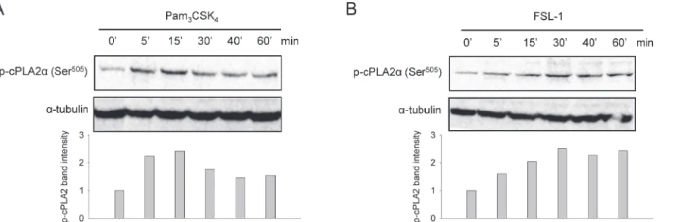
TLR2 ligands induce cPLA2
α
phosphorylation at Ser
505In response to pro-inflammatory stimuli, cPLA2αcan be activated by phosphorylation at Ser505[9]. To evaluate TLR2-induced cPLA2αactivation in synoviocytes, we analyzed cPLA2α
phosphorylation at Ser505by immunoblotting. Both Pam3CSK4and FSL-1 rapidly evoked
time-dependent cPLA2αphosphorylation, evident after 5 minutes of stimulation (Fig 3). Pam3CSK4-induced cPLA2αphosphorylation peaked after 15 min, to decrease after 30 min
(Fig 3A). In comparison, the FSL-1 response was slower; FSL-1 induced a maximum phosphor-ylation after 30 minutes that persisted throughout 60 min of stimulation (Fig 3B). These results suggest that Pam3CSK4and FSL-1 both activate cPLA2α.
Fig 2. SW982 synoviocytes express TLRs 1–7 gene transcripts.Transcriptional expression of TLRs 1–7 in unstimulated synoviocytes was determined by qPCR as described in the Methods section. qPCR products in untreated cells were analyzed by gel electrophoresis(A). Relative reciprocal mRNA
expression levels normalized to 18S (mean±SD from 3 independent experiments) (B). Differences are considered significant at p0.05. Means sharing a common alphabetical symbol do not differ significantly. Cells were treated with TNF (10 ng/mL) for indicated time(C). Results shown are mean±SD of three
independent experiments with 18S as reference gene. Results are concidered significantly upregulated (p*0.05) or downregulated (p#0.05) compared
to untreated controls at each timepoint.
Pam
3CSK
4- and FSL-1-induced AA release and PGE
2production
depend on cPLA2
α
activity, not sPLA2 activity
AA release and eicosanoid production induced by various TLR2 ligands is mediated by cPLA2αactivity in human leukocytes [25]. In murine macrophages and mast cells, TLR-in-duced cPLA2αactivity is modulated by sPLA2 GV [26,35]. However, little is known con-cerning TLR-induced PLA2 activity and lipid metabolism in synoviocytes. Having found that FSL-1 and Pam3CSK4stimulation lead to cPLA2αphosphorylation, we next investigated their
effect on AA release and PGE2production. In a time-dependent manner, AA release was
signif-icantly increased following 4 hours stimulation (3-fold by Pam3CSK4and 4-fold by FSL-1),
and persisted through 24 hours (Fig 4A). In dose-response experiments, FSL-1 was a more po-tent inducer of AA release than Pam3CSK4in most concentrations tested (p<0.001) (Fig 4B).
Subsequent PGE2production was increased 10-12-fold by both ligands with no significant
dif-ference between the two (Fig 4C). Thus, the increased PGE2levels in response to TLR2 ligands
correspond to the increased availability of AA.
To investigate the roles of sPLA2s and cPLA2αin TLR2-induced synoviocyte AA release and PGE2production, we next analyzed the effects of synthetic inhibitors of cPLA2α(Inhibitor
28 [36], CAY10502 [37], AVX002 [38,39], ATK [40],) or sPLA2 (CAY10590 [41], Varespladib [42]). In response to both TLR2 ligands, all cPLA2αinhibitors efficiently reduced AA release to untreated control level (Fig 4D). In contrast, sPLA2 inhibitors did not affect the release of AA (Fig 4D), as confirmed by dose-response (0.2–50μM) and time course (4 and 24 hours) experiments (results not shown). Corresponding results were found in PGE2production;
in-hibitors of cPLA2αpotently attenuated PGE2production in response to both Pam3CSK4and
FSL-1, whereas sPLA2 inhibitors did not (Fig 4C). Due to the lack of effect in synoviocytes, CAY10590 and Varespladib were evaluated in the keratinocyte cell line HaCaT known to ex-press functional sPLA2 enzymes that contribute to AA release [43]. CAY10590 and Varespla-dib completely blocked IL-1β-induced AA release (144.2 ± 3.8% and 104.0 ±6.8%, respectively, results not shown) indicating that the sPLA2 inhibitors are indeed effective. In summary, these results suggest that cPLA2αhas a more prominent role than sPLA2 enzymes in releasing AA for PGE2production downstream TLR2 activation.
Fig 3. TLR2 ligands induce cPLA2αphosphorylation at Ser505.Synoviocytes were treated with Pam3CSK4(200 ng/mL)(A)or FSL-1 (100 ng/mL)(B)for indicated periods of time. Cell lysates were prepared and analyzed by immunoblotting for activation of phospho-cPLA2α(Ser505) as described in the Methods
section. Immunoblotting forα-tubulin was performed to assess protein loading. Results shown are one representative of three independent experiments.
TLR2-induced gene transcription and protein production involve cPLA2
α
activity
TLR2 activation induce inflammation by upregulating pro-inflammatory genes such as IL-6, IL-8 and COX2 [19], but the role of cPLA2αactivity in this pathway is hitherto not well de-scribed. Protein levels of the pro-inflammatory interleukin (IL)-6 and IL-8 are both induced by TLR2 ligands in RA synovial cells [44,45]. By qPCR, we found a time-dependent increase in synoviocyte IL-6 gene transcription in response to Pam3CSK4and FSL-1, with maximum
in-duction at 3 hours of 40-fold and 50-fold, respectively (Fig 5A). A stronger induction was de-tected for IL-8 transcription, which was induced by ~70-fold by both Pam3CSK4and FSL-1
Fig 4. TLR2/1- and TLR2/6-induced AA release and PGE2production depend on cPLA2α, not sPLA2 activity.Synoviocytes were treated with Pam3CSK4or FSL-1 to determine AA release for indicated times (200 ng/mL and 100 ng/mL, respectively)(A), or in indicated concentrations(B)for 4 hours.
Release of AA was measured as described in the Methods section. Data presented are mean±SD of one representative of at least 3 independent
experiments, and statistical significance is indicated by p#0.05 when compared to untreated control samples. Cells were treated with inhibitors for cPLA2α
(5μM Inhibitor 28, 1μM CAY10502, 5μM AVX002, 5μM ATK); sPLA2 (10μM CAY10590, 10μM Varespladib) or COX1/2 (10μM Indomethasin) for 2 hours followed by FSL-1 (100 ng/mL) or Pam3CSK4(200 ng/mL) for 24 hours (C) or 4 hours (D). Release of PGE2(C) or AA (D) was measured by ELISA and AA
release assay, respectively, as described in the Methods section. Results shown are mean±SD of three independent experiments. Stimulated controls are
compared to untreated controls whereas inhibitor-treated samples are compared to stimulated controls. Statistical significance is indicated by p#0.001
when compared to untreated control samples; p*0.001 when compared to the respective ligand-stimulated samples(C); p#0.05 when compared to
untreated control samples; p*0.05 when compared to the respective TLR ligand-treated samples(D).
after 3 hours (Fig 5B). cPLA2αinhibitors attenuated IL-6 and IL-8 transcription by 40% and 30%, respectively, in response to both Pam3CSK4and FSL-1 (Fig 5C and 5D). For IL-6, these
results were confirmed at protein level; the detected increase in IL-6 protein levels (16-fold by Pam3CSK4and 20-fold by FSL-1) was attenuated by cPLA2αinhibitors by 80–90% in response to Pam3CSK4and by 30–50% in response to FSL-1 (Fig 5E). Reduced IL-6 transcription is thus reflected in reduced IL-6 protein production, although the reduction in Pam3CSK4-induced
IL-6 was far more prominent at protein level than at the transcriptional level.
In addition to IL-6 and IL-8, we found that both TLR2-ligands also induced COX2 gene transcription, peaking at 3 hours (Pam3CSK4) and 6 hours (FSL-1) (Fig 5F). When treated with
cPLA2αinhibitors (AVX002 and CAY10502), the TLR2 ligand-induced COX2 mRNA expres-sion was reduced by approximately 50% (Fig 5G). These results suggest a regulatory role of cPLA2αin TLR1/2- and TLR2/6-induced IL-6, IL-8 and COX2 gene expression, and may fur-ther indicate involvement of differential post-transcriptional regulating processes of IL-6 pro-tein [46].
Fig 5. TLR2 ligand-induced gene transcription and protein production involve cPLA2α.Synoviocytes were treated with Pam3CSK4(200 ng/mL) or FSL-1 (100ng/mL) for indicated time points (A,B,F), 3 hours (C,D,G) or 24 hours (E). Prior to stimulation, cells were pretreated with inhibitors of cPLA2α
(5μM AVX002, 1μM CAY10502, 2 hours). Expression of IL-6 (A,C), IL-8 (B,D) or COX2 (F,G) mRNA was analyzed by qPCR with GAPDH as reference gene, and secreted IL-6 protein (E) was analyzed by ELISA as described in the Methods section. Results shown are mean±SD of at least three independent experiments. Results are considered significant at p0.05. Transcription of IL-6, IL-8 and COX2 was found to be significant induced by both ligands at all time points (p#0.05) (A,B,F). Significance is indicated by p#0.05 when compared to untreated control, and by p
*0.05 when compared to the respective ligand-treated samples (C,D,E,G).
cPLA2
α
regulates FSL-1-induced IL-6 production through the COX/
PGE
2pathway
Many of the beneficial pharmacological effects of NSAIDs in RA patients are attributed the in-hibition of PGE2synthesis [8]. In the next series of experiments, we investigated if the COX/
PGE2pathway is involved in cPLA2α-dependent regulation of gene expression, focusing on
FSL-1-induced IL-6 levels.
Involvement of the COX enzymatic pathway was investigated using the dual COX1/2 inhib-itor Indomethasin. Indomethasin effectively diminished FSL-1-induced PGE2production (Fig
4C), supporting its clinical relevance. Indomethasin reduced FSL-1-induced IL-6 gene tran-scription by 30% (Fig 6A). This response was again reflected at protein level where Indometha-sin reduced FSL-1-induced IL-6 protein by 20% (Fig 6B). The effect of Indomethasin was comparable to that of AVX002 both at mRNA level and at protein level (Fig 5E) indicating that prostanoids produced downstream FSL-1-activated cPLA2αmay regulate FSL-1-induced IL-6 production. We next asked whether reduced PGE2levels due to cPLA2αinhibition (Fig 4C)
could account for effect of cPLA2αinhibition on IL-6 levels. When added alone, PGE2slightly,
but significantly induced both gene transcription and protein production of IL-6 (Fig 6A and 6B). When added in combination with AVX002, PGE2completely rescued the FSL-1-induced
IL-6 transcription (Fig 6A). In summary, these results for the first time suggest that PGE2is an
important mediator that regulates FSL-1-induced IL-6 production in synoviocytes.
Discussion
TLR2 signaling is proposed to promote joint destruction and synovitis in RA. In this work, we present for the first time the mechanism in which cPLA2αregulates several important media-tors of inflammation in response to TLR2 activation in synoviocytes, namely PGE2, COX2, IL6
and IL8.
There is little data available concerning TLR-induced PLA2 activity, AA mobilization and eicosanoid production in RA synoviocytes. In the current work we demonstrate that the TLR2/ 1 and TLR2/6 ligands Pam3CSK4and FSL-1 are potent inducers of synoviocyte cPLA2α
phos-phorylation, AA release and subsequent PGE2production, in line with responses reported in
Fig 6. PGE2regulates FSL-1-induced IL-6 production.Synoviocytes were treated with Indomethasin (10μM, 2 hours) or AVX002 (5μM, 2 hours) before stimulation with FSL-1 (100ng/mL) and/or PGE2for 3 hours (A) or 24 hours (B). IL-6 mRNA expression was analyzed by qPCR with GAPDH as reference
gene (A) and IL-6 protein was analyzed by ELISA (B) as described in the Methods section. Results presented are mean±SD of three independent experiments. Significance is indicated by p#0.05 when compared to unstimulated control, by p*0.05 when compared to FSL-1-stimulated control
human leukocytes [25]. Furthermore, cPLA2αinhibition effectively attenuated the TLR-in-duced AA release and PGE2production, suggesting a central role for cPLA2αin synoviocyte
AA metabolism. cPLA2αis expressed in RA synovium and cultured FLS, and its transcription is induced by various pro-inflammatory stimuli, including IL-1β, TNF and lipopolysaccharide [30,39,47]. Here, we report that TLR2/1 and TLR2/6 ligands induce synoviocyte cPLA2αgene transcription, indicating that TLR2 ligands activate cPLA2α-dependent AA mobilization di-rectly by increased enzyme activity, and indidi-rectly by transcriptional regulation andde novo synthesis as previously described [30].
In various cell types, cPLA2αand sPLA2 act in concert to release AA [43,48]. In murine macrophages and mast cells, TLR2-induced AA release is dependent on cPLA2α, amplified by GV sPLA2 [26,35]. However, in IL-1β-stimulated FLS, sPLA2s is not reported to contribute to PGE2production [49]. The latter finding corresponds to our results demonstrating a dominant
role for cPLA2α, and minor roles for sPLA2 enzymes in TLR2-induced AA release and PGE2
production in synoviocytes. It should however be noted that the sPLA2 inhibitors used in this work are selective for sPLA2 isotypes GIIA, GV and GX, but not GXII [9,42,50]. The role of sPLA2 GXIIA is to date unknown, but may withhold important household functions as it is de-tected at high levels in various tissues [51]. We found that GXIIA was the most highly express-ed sPLA2 indicating a hitherto unknown role in synoviocyte cell communication and function, a finding that deserves further investigation. The lack of response to sPLA2 inhibitors in AA re-lease and PGE2production may be explained by a low sPLA2 expression in cultured
synovio-cytes. RA synovial tissue expresses several sPLA2 isotypes, whereas cultured primary synovial cells only weakly express sPLA2s GIIA, GV and GX transcripts [29]. In agreement with these findings, we report a weak transcriptional sPLA2 expression and a lack of response to sPLA2 inhibitors, suggesting a subordinate role of endogenous sPLA2 enzymes in cultured synovio-cytes. Subgroups and splice variants of iPLA2 GVI is expressed in various cells and tissues [52], but their expression and function in the joint is not known. Expression profiles or functional roles of iPLA2 in RA are not previously described; the hereby reported high expression of sev-eral iPLA2 subgroups is an interesting finding which indicates a role for these enzymes in the synovium and should be investigated further.
The role of AA metabolites in chronic inflammation and RA disease progression is well es-tablished, and reducing PGE2levels by non-steroid anti-inflammatory drugs (NSAIDs)
target-ing COX enzymes is a well-known strategy for symptom relief and pain management in RA patients [53]. We have previously shown that cPLA2αinhibition normalize AA release, PGE2
levels and gene transcription in response to TNF stimulus in synoviocytes [39], a mechanism hereby extended to include FSL-1 and Pam3CSK4stimuli. Also, the herein described
attenuat-ing effect of cPLA2αinhibition on TLR-induced COX2 gene transcription is similar to our finding in the TNF-response [39], and suggest that cPLA2αinhibitors may target stimuli-in-duced PGE2production at two levels; directly by reducing AA substrate availability and
indi-rectly by reducing COX2 expression.
Expression levels of several TLRs, including TLR2 and TLR6, but not TLR1, in RA are in-creased compared to osteoarthritic (OA) synovium [23]. In synoviocytes, we detected tran-scripts for TLRs 1–7, of which TLR2 was the most abundantly expressed. We further show that TLR expression is regulated by TNF. The TNF-induced up- and down-regulation of TLR2 and TLR4 corresponds to findings in primary synovial fibroblasts from RA and OA joints [24] and support the use of SW982 synoviocytes as a validin vitromodel system to mechanistically in-vestigate synovitis. In the present study, FSL-1 was a more potent inducer of cPLA2α phos-phorylation and AA release than Pam3CSK4. This observation is in accordance with findings in
synovium [23]. However, post-translational regulatory mechanisms are known to impact TLR protein expression [54]. Caution must thus be taken in comparing transcriptional expression and levels of functional receptors when protein expression data is not available.
TLR2 is shown to regulate the expression of IL-6 in RA FLS [54], and the involvement of PLA2 in this pathway is to our knowledge a novel finding. IL-6 is a pluripotent cytokine in-volved in pannus formation, osteoclast and FLS activation in RA and plays a key role in chronic inflammation [55]. Increased levels of IL-6 are detected both in the joint and systemically in RA patients [56,57]. IL-6 deficiency provides protection against development of murine colla-gen-induced arthritis (CIA) [58,59], and anti-mouse IL-6 monoclonal antibody suppress CIA development [60]. In humans, anti-IL-6 therapy improves symptoms of RA [61,62]. Our re-sults show that TLR2-induced synoviocyte IL-6 production is partly controlled by cPLA2α, suggesting an additional beneficial therapeutic effect of cPLA2αinhibitors. The finding that cPLA2αinhibition was more efficient on IL-6 protein than mRNA in response to Pam3CSK4
than FSL-1 is an interesting finding. IL-6 mRNA stability and degradation are known to be reg-ulated in macrophages by mechanisms including TLR-induced RNase activity [46]. Differential regulation of such post-transcriptional mechanisms may thus influence IL-6 protein levels.
Our results further suggest involvement of PGE2in regulating FSL-1-induced IL-6
expres-sion. PGE2and its receptors are previously shown to regulate IL-1β- and TNF-induced IL-6
generation in human and murine FSL [63–66]. In our experiments, the effect of COX1/2 inhi-bition was comparable to cPLA2αinhibition in reducing IL-6 production. Furthermore, PGE2
resulted in a complete rescue of FSL-1- induced IL-6 transcription following cPLA2α inhibi-tion, suggesting that decreased PGE2levels at least in part account for the attenuating effect of
inhibiting cPLA2αactivity. This hypothesis is supported by the finding that exogenously added PGE2induced IL-6 expression both at transcriptional and protein levels. Even though
PGE2-induced IL-6 levels were very low compared to FSL-1-induced IL-6, our results
corre-spond to previously reported results in primary synovial fibroblasts [63] and support a role for PGE2in FSL-1-induced IL-6 production.
The chemokine IL-8 is an important contributor to angiogenesis in the rheumatic joint [67], and acts as a potent neutrophil attractant [68]. IL-8 is associated with the pathogenesis of arthritis, and RA FLS produce IL-8 in both early [69] and established phases of arthritis [70]. cPLA2αhas been reported to induce the expression of IL-8 in human lung fibroblasts [71], and is proposed to be a regulator of neutrophil recruitment and inflammation in murine collagen-induced arthritis [72]. This corresponds to our previous findings describing a regulatory role of cPLA2αin TNF-induced IL-8 expression in synovial fibroblasts [39]. Here, we show that cPLA2αalso regulates TLR-induced IL-8 expression, suggesting that cPLA2αmay act to mod-ulate angiogenesis and neutrophil attraction in synovitis.
In conclusion, our data demonstrate that cPLA2αregulates TLR2-induced lipid biosynthe-sis and pro-inflammatory gene expression in synoviocytes, in part through the COX/PGE2
pathway. Seen in context with our previous finding that cPLA2αalso regulates TNF-induced signaling [39], these results expand our understanding of cellular signaling mechanisms modu-lated by cPLA2αactivity and indicate a central regulatory role for cPLA2αin synovitis.
Acknowledgments
Author Contributions
Conceived and designed the experiments: RMS AJF BJ. Performed the experiments: RMS AJF TS. Analyzed the data: RMS AJF TS BJ. Contributed reagents/materials/analysis tools: BJ. Wrote the paper: RMS AJF BJ.
References
1. McInnes IB, Schett G. The pathogenesis of rheumatoid arthritis. N Engl J Med. 2011; 365: 2205–2219. doi:10.1056/NEJMra1004965PMID:22150039
2. Van Der Heijden IM, Wilbrink B, Tchetverikov I, Schrijver IA, Schouls LM, Hazenberg MP, et al. Pres-ence of bacterial DNA and bacterial peptidoglycans in joints of patients with rheumatoid arthritis and other arthritides. Arthritis Rheum. 2000; 43: 593–598. PMID:10728753
3. Buckley CD. Why does chronic inflammation persist: An unexpected role for fibroblasts. Immunol Lett. 2011; 138: 12–14. doi:10.1016/j.imlet.2011.02.010PMID:21333681
4. Bartok B, Firestein GS. Fibroblast-like synoviocytes: key effector cells in rheumatoid arthritis. Immunol Rev. 2010; 233: 233–255. doi:10.1111/j.0105-2896.2009.00859.xPMID:20193003
5. Harizi H, Corcuff JB, Gualde N. Arachidonic-acid-derived eicosanoids: roles in biology and immunopa-thology. Trends Mol Med. 2008; 14: 461–469. doi:10.1016/j.molmed.2008.08.005PMID:18774339 6. Kalinski P. Regulation of Immune Responses by Prostaglandin E2. J Immunol. 2012; 188: 21–28. doi:
10.4049/jimmunol.1101029PMID:22187483
7. Husby G, Bankhurst AD, Williams RC. Immunohistochemical localization of prostaglandin E in rheuma-toid synovial tissues. Arthritis Rheum. 1977; 20: 785–791. PMID:324481
8. Murakami M. Lipid Mediators in Life Science. Exp Anim. 2011; 60: 7–20. PMID:21325748
9. Dennis EA, Cao J, Hsu Y, Magrioti V, Kokotos G. Phospholipase A2 Enzymes: Physical Structure, Bio-logical Function, Disease Implication, Chemical Inhibition, and Therapeutic Intervention. Chem Rev. 2011; 111: 6130–6185. doi:10.1021/cr200085wPMID:21910409
10. Sundler R, Winstedt D, Wijkander J. Acyl-chain selectivity of the 85 kDa phospholipase A2 and of the release process in intact macrophages. Biochem J. 1994; 15: 455–458.
11. Singer AG, Ghomashchi F, Le Calvez C, Bollinger J, Bezzine S, Rouault M, et al. Interfacial Kinetic and Binding Properties of the Complete Set of Human and Mouse Groups I, II, V, X, and XII Secreted Phos-pholipases A2. J Biol Chem. 2002; 277: 48535–48549. PMID:12359733
12. Lio YC, Dennis EA. Interfacial activation, lysophospholipase and transacylase activity of group VI Ca2 +-independent phospholipase A2. Biochim Biophys Acta. 1998; 1392: 320–332. PMID:9630702 13. Uozumi N, Kume K, Nagase T, Nakatani N, Ishii S, Tashiro F, et al. Role of cytosolic phospholipase A2
in allergic response and parturition. Nature. 1997; 390: 618–622. PMID:9403692
14. Nagase T, Uozumi N, Ishii S, Kita Y, Yamamoto H, Ohga E, et al. A pivotal role of cytosolic phospholi-pase A2 in bleomycin-induced pulmonary fibrosis. Nat Med. 2002; 8: 480–484. PMID:11984592 15. Nagase T, Uozumi N, Ishii S, Kume K, Izumi T, Ouchi Y, et al. Acute lung injury by sepsis and acid
aspi-ration: a key role for cytosolic phospholipase A2. Nat Immunol. 2000; 1: 42–46. PMID:10881173 16. Hegen M, Sun L, Uozumi N, Kume K, Goad ME, Nickerson-Nutter CL, et al. Cytosolic phospholipase
A2alpha-deficient mice are resistant to collagen-induced arthritis. J Exp Med. 2003; 197: 1297–1302. PMID:12743172
17. Malaviya R, Ansell J, Hall L, Fahmy M, Argentieri RL, Olini JGC, et al. Targeting cytosolic phospholi-pase A2 by arachidonyl trifluoromethyl ketone prevents chronic inflammation in mice. Eur J Pharmacol. 2006; 539: 195–204. PMID:16712837
18. Tai N, Kuwabara K, Kobayashi M, Yamada K, Ono T, Seno K, et al. Cytosolic phospholipase A2 alpha inhibitor, pyrroxyphene, displays anti-arthritic and anti-bone destructive action in a murine arthritis model. Inflamm Res. 2010; 59: 53–62. doi:10.1007/s00011-009-0069-8PMID:19655230
19. Kawai T, Akira S. The role of pattern-recognition receptors in innate immunity: update on Toll-like re-ceptors. Nat Immunol. 2010; 11: 373–384. doi:10.1038/ni.1863PMID:20404851
20. Goh FG, Midwood KS. Intrinsic danger: activation of Toll-like receptors in rheumatoid arthritis. Rheuma-tology. 2012; 51: 7–23. doi:10.1093/rheumatology/ker257PMID:21984766
21. Huang Q-Q, Sobkoviak R, Jockheck-Clark AR, Shi B, Mandelin AM, Tak PP, et al. Heat Shock Protein 96 Is Elevated in Rheumatoid Arthritis and Activates Macrophages Primarily via TLR2 Signaling. J Immunol. 2009; 182: 4965–4973. doi:10.4049/jimmunol.0801563PMID:19342676
tissue. Differential regulation of hsp70 expression and hsf1 activation in synovial fibroblasts by proin-flammatory cytokines, shear stress, and antiinproin-flammatory drugs. J Clin Invest. 1998; 102: 302–311. PMID:9664071
23. Tamaki Y, Takakubo Y, Hirayama T, Konttinen YT, Goodman SB, Yamakawa M, et al. Expression of Toll-like Receptors and Their Signaling Pathways in Rheumatoid Synovitis. J Rheumatol. 2011; 38: 810–820. doi:10.3899/jrheum.100732PMID:21324962
24. Seibl R, Birchler T, Loeliger S, Hossle J, Gay R, Saurenmann T, et al. Expression and regulation of Toll-like receptor 2 in rheumatoid arthritis synovium. Am J Pathol. 2003; 162: 1221–1227. PMID: 12651614
25. Lindner SC, Köhl U, Maier TJ, Steinhilber D, Sorg BL. TLR2 ligands augment cPLA2αactivity and lead to enhanced leukotriene release in human monocytes. J Leukoc Biol. 2009; 86: 389–399. doi:10.1189/ jlb.1008591PMID:19401382
26. Ruipérez V, Astudillo AM, Balboa MA, Balsinde J. Coordinate Regulation of TLR-Mediated Arachidonic Acid Mobilization in Macrophages by Group IVA and Group V Phospholipase A2s. J Immunol. 2009; 182: 3877–3883. doi:10.4049/jimmunol.0804003PMID:19265167
27. Anthonsen MW, Andersen S, Solhaug A, Johansen B. Atypicalλ/ιPKC Conveys 5-Lipoxygenase/Leu-kotriene B4-mediated Cross-talk between Phospholipase A2s Regulating NF-κB Activation in Re-sponse to Tumor Necrosis Factor-αand Interleukin-1β. J Biol Chem. 2001; 276: 35344–35351. PMID: 11445585
28. Ruijter JM, Ramakers C, Hoogaars WMH, Karlen Y, Bakker O, van den Hoff MJB, et al. Amplification ef-ficiency: linking baseline and bias in the analysis of quantitative PCR data. Nucleic Acids Res. 2009; 37: e45. doi:10.1093/nar/gkp045PMID:19237396
29. Masuda S, Murakami M, Komiyama K, Ishihara M, Ishikawa Y, Ishii T, et al. Various secretory phospho-lipase A2 enzymes are expressed in rheumatoid arthritis and augment prostaglandin production in cul-tured synovial cells. FEBS J. 2005; 272: 655–672. PMID:15670148
30. Chi PL, Luo SF, Hsieh HL, Lee IT, Hsiao LD, Chen YL, et al. Cytosolic phospholipase A2 induction and prostaglandin E2 release by interleukin-1βvia the myeloid differentiation factor 88–dependent pathway and cooperation of p300, Akt, and NF-κB activity in human rheumatoid arthritis synovial fibroblasts. Ar-thritis Rheum. 2011; 63: 2905–2917. doi:10.1002/art.30504PMID:21702012
31. Johansen B, Rakkestad K, Balboa MA, Dennis EA. Expression of cytosolic and secreted forms of phos-pholipase A2 and cyclooxygenases in human placenta, fetal membranes, and chorionic cell lines. Pros-taglandins Other Lipid Mediat. 2000; 60: 119–125. PMID:10751642
32. Ohto T, Uozumi N, Hirabayashi T, Shimizu T. Identification of Novel Cytosolic Phospholipase A2s, Mu-rine cPLA2δ,ε, andζ, Which Form a Gene Cluster with cPLA2β. J Biol Chem. 2005; 280: 24576–
24583. PMID:15866882
33. Varastehpour A, Radaelli T, Minium J, Ortega H, Herrera E, Catalano P, et al. Activation of Phospholi-pase A2 Is Associated with Generation of Placental Lipid Signals and Fetal Obesity. J Clin Endocrinol Metab. 2006; 91: 248–255. PMID:16249288
34. Mosher AA, Rainey KJ, Riley B, Levinson HS, Vinturache AE, Wood SL, et al. Regulation of sPLA2-IID in Human Decidua: Insights Into the Complexity of the Prostaglandin Pathway in Labor. Reprod Sci. 2014.
35. Kikawada E, Bonventre JV, Arm JP. Group V secretory PLA2 regulates TLR2-dependent eicosanoid generation in mouse mast cells through amplification of ERK and cPLA2αactivation. Blood. 2007; 110: 561–567. PMID:17369491
36. Kokotos G, Feuerherm A, Barbayianni E, Shah I, Sæther M, Magrioti V, et al. Inhibition of Group IVA Cytosolic Phospholipase A2 by Thiazolyl Ketones In Vitro, Ex Vivo, and In Vivo. J Med Chem. 2014; 57: 7523–7535. doi:10.1021/jm500192sPMID:25152071
37. Ludwig J, Bovens S, Brauch C, Elfringhoff AS, Lehr M. Design and Synthesis of 1-Indol-1-yl-propan-2-ones as Inhibitors of Human Cytosolic Phospholipase A2α. J Med Chem. 2006; 49: 2611–2620. PMID: 16610804
38. Huwiler A, Feuerherm AJ, Sakem B, Pastukhov O, Filipenko I, Nguyen T, et al. Theω3-polyunsaturated fatty acid derivatives AVX001 and AVX002 directly inhibit cytosolic phospholipase A2 and suppress prostaglandin E2 formation in mesangial cells. Br J Pharmacol. 2012; 168: 1691–1701.
39. Sommerfelt RM, Feuerherm AJ, Jones K, Johansen B. Cytosolic phospholipase A2 regulates TNF-in-duced production of joint destructive effectors in synoviocytes PLOS One. 2013; 8: e83555. doi:10. 1371/journal.pone.0083555PMID:24349530
41. Antonopoulou G, Barbayianni E, Magrioti V, Cotton N, Stephens D, Constantinou-Kokotou V, et al. Structure–activity relationships of natural and non-natural amino acid-based amide and 2-oxoamide in-hibitors of human phospholipase A2 enzymes. Bioorg Med Chem. 2008; 16: 10257–10269. doi:10. 1016/j.bmc.2008.10.046PMID:18993078
42. Snyder DW, Bach NJ, Dillard RD, Draheim SE, Carlson DG, Fox N, et al. Pharmacology of LY315920/ S-5920, [[3-(Aminooxoacetyl)-2-ethyl-1-(phenylmethyl)-1H-indol-4-yl]oxy]acetate, a Potent and Selec-tive Secretory Phospholipase A2Inhibitor: A New Class of Anti-Inflammatory Drugs, SPI. J Pharmacol Exp Ther. 1999; 288: 1117–1124. PMID:10027849
43. Anthonsen MW, Solhaug A, Johansen B. Functional coupling between secretory and cytosolic phos-pholipase A2 modulates tumor necrosis factor-alpha- and interleukin-1beta-induced NF-kappa B acti-vation. J Biol Chem. 2001; 276: 30527–30536. PMID:11390371
44. Sacre SM, Andreakos E, Kiriakidis S, Amjadi P, Lundberg A, Giddins G, et al. The Toll-Like Receptor Adaptor Proteins MyD88 and Mal/TIRAP Contribute to the Inflammatory and Destructive Processes in a Human Model of Rheumatoid Arthritis. Am J Pathol. 2007; 170: 518–525. PMID:17255320 45. Kyburz D, Rethage J, Seibl R, Lauener R, Gay RE, Carson DA, et al. Bacterial peptidoglycans but not
CpG oligodeoxynucleotides activate synovial fibroblasts by toll-like receptor signaling. Arthritis Rheum. 2003; 48: 642–650. PMID:12632416
46. Matsushita K, Takeuchi O, Standley DM, Kumagai Y, Kawagoe T, Miyake T, et al. Zc3h12a is an RNase essential for controlling immune responses by regulating mRNA decay. Nature. 2009; 458: 1185–1190. doi:10.1038/nature07924PMID:19322177
47. Dieter P, Kolada A, Kamionka S, Schadow A, Kaszkin M. Lipopolysaccharide-induced release of ara-chidonic acid and prostaglandins in liver macrophages: Regulation by Group IV cytosolic phospholi-pase A2, but not by Group V and Group IIA secretory phospholiphospholi-pase A2. Cell Signal. 2002; 14: 199–
204. PMID:11812647
48. Balsinde J, Balboa MA, Dennis EA. Functional coupling between secretory phospholipase A2 and cy-clooxygenase-2 and its regulation by cytosolic group IV phospholipase A2. Proc Natl Acad Sci U S A. 1998; 95: 7951–7956. PMID:9653121
49. Hulkower KI, Hope WC, Chen T, Anderson CM, Coffey JW, Morgan DW. Interleukin-1βstimulates cyto-solic phospholipase A2 in rheumatoid synovial fibroblasts. Biochem Biophys Res Commun. 1992; 184: 712–718. PMID:1575744
50. Fraser H, Hislop C, Christie RM, Rick HL, Reidy CA, Chouinard ML, et al. Varespladib (A-002), a Secre-tory Phospholipase A2 Inhibitor, Reduces Atherosclerosis and Aneurysm Formation in ApoE−/−Mice. J Cardiovasc Pharmacol. 2009; 53: 60–65 doi:10.1097/FJC.0b013e318195bfbcPMID:19129734 51. Murakami M, Taketomi Y, Miki Y, Sato H, Hirabayashi T, Yamamoto K. Recent progress in
phospholi-pase A2 research: From cells to animals to humans. Prog Lipid Res. 2011; 50: 152–192. doi:10.1016/j. plipres.2010.12.001PMID:21185866
52. Winstead MV, Balsinde J, Dennis EA. Calcium-independent phospholipase A2: structure and function. Biochim Biophys Acta. 2000; 1488: 28–39. PMID:11080674
53. Crofford LJ. Specific cyclooxygenase-2 inhibitors: what have we learned since they came into wide-spread clinical use? Curr Opin Rheumatol. 2002; 14: 225–230. PMID:11981317
54. Philippe L, Alsaleh G, Suffert G, Meyer A, Georgel P, Sibilia J, et al. TLR2 Expression Is Regulated by MicroRNA miR-19 in Rheumatoid Fibroblast-like Synoviocytes. J Immunol. 2012; 188: 454–461. doi: 10.4049/jimmunol.1102348PMID:22105995
55. Choy E, Dayer JM. Therapeutic targets in rheumatoid arthritis: the interleukin-6 receptor. Rheumatolo-gy. 2010; 49: 15–24. doi:10.1093/rheumatology/kep329PMID:19854855
56. Usón J, Balsa A, Pascual-Salcedo D, Cabezas JA, Gonzalez-Tarrio JM, Martín-Mola E, et al. Soluble interleukin 6 (IL-6) receptor and IL-6 levels in serum and synovial fluid of patients with different arthropa-thies. J Rheumatol. 1997; 24: 2069–2075. PMID:9375862
57. Okamoto H, Yamamura M, Morita Y, Harada S, Makino H, Ota Z. The synovial expression and serum levels of interleukin-6, interleukin-11, leukemia inhibitory factor, and oncostatin M in rheumatoid arthri-tis. Arthritis Rheum. 1997; 40: 1096–1105. PMID:9182920
58. Alonzi T, Fattori E, Lazzaro D, Costa P, Probert L, Kollias G, et al. Interleukin 6 Is Required for the De-velopment of Collagen-induced Arthritis. J Exp Med. 1998; 187: 461–468. PMID:9463396
59. Sasai M, Saeki Y, Ohshima S, Nishioka K, Mima T, Tanaka T, et al. Delayed onset and reduced severi-ty of collagen-induced arthritis in interleukin-6–deficient mice. Arthritis Rheum. 1999; 42: 1635–1643. PMID:10446862
61. Levi M, Grange S, Frey N. Exposure-response relationship of tocilizumab, an anti-IL-6 receptor mono-clonal antibody, in a large population of patients with rheumatoid arthritis. J Clin Pharmacol. 2013; 53: 151–159. doi:10.1177/0091270012437585PMID:23436260
62. Maini RN, Taylor PC, Szechinski J, Pavelka K, Bröll J, Balint G, et al. Double-blind randomized con-trolled clinical trial of the interleukin-6 receptor antagonist, tocilizumab, in European patients with rheu-matoid arthritis who had an incomplete response to methotrexate. Arthritis Rheum. 2006; 54: 2817–
2829. PMID:16947782
63. Inoue H, Takamori M, Shimoyama Y, Ishibashi H, Yamamoto S, Koshihara Y. Regulation by PGE2 of the production of interleukin-6, macrophage colony stimulating factor, and vascular endothelial growth factor in human synovial fibroblasts. Br J Pharmacol. 2002; 136: 287–295. PMID:12010778
64. Honda T, Segi-Nishida E, Miyachi Y, Narumiya S. Prostacyclin-IP signaling and prostaglandin E2-EP2/ EP4 signaling both mediate joint inflammation in mouse collagen-induced arthritis. J Exp Med. 2006; 203: 325–335. PMID:16446378
65. Kawashima M, Ogura N, Akutsu M, Ito K, Kondoh T. The anti-inflammatory effect of cyclooxygenase in-hibitors in fibroblast-like synoviocytes from the human temporomandibular joint results from the sup-pression of PGE2 production. J Oral Pathol Med. 2013; 42: 499–506. doi:10.1111/jop.12045PMID: 23331485
66. Kunisch E, Jansen A, Kojima F, Löffler I, Kapoor M, Kawai S, et al. Prostaglandin E2 Differentially Mod-ulates Proinflammatory/Prodestructive Effects of TNF-αon Synovial Fibroblasts via Specific E Prosta-noid Receptors/cAMP. J Immunol. 2009; 183: 1328–1336. doi:10.4049/jimmunol.0900801PMID: 19542367
67. Koch AE, Volin MV, Woods JM, Kunkel SL, Connors MA, Harlow LA, et al. Regulation of angiogenesis by the C-X-C chemokines interleukin-8 and epithelial neutrophil activating peptide 78 in the rheumatoid joint. Arthritis Rheum. 2001; 44: 31–40. PMID:11212173
68. Hayashida K, Nank T, Girschick H, Yavuz S, Ochi T, Lipsky PE. Synovial stromal cells from rheumatoid arthritis patients attract monocytes by producing MCP-1 and IL-8. Arthritis Res. 2001; 3: 118–126. PMID:11178119
69. Takahashi Y, Kasahara T, Sawai T, Rikimaru A, Mukaida N, Matsushima K, et al. The participation of IL-8 in the synovial lesions at an early stage of rheumatoid arthritis. Tohoku J Exp Med. 1999; 188: 75–
87. PMID:10494903
70. Deleuran B, Lemche P, Kristensen M, Chu CQ, Field M, Jensen J, et al. Localisation of interleukin 8 in the synovial membrane, cartilage–pannus junction and chondrocytes in rheumatoid arthritis. Scand J Rheumatol. 1994; 23: 2–7. PMID:8108662
71. Pawliczak R, Logun C, Madara P, Lawrence M, Woszczek G, Ptasinska A, et al. Cytosolic phospholi-pase A2 group IVαbut not secreted phospholipase A2 group IIA, V, or X induces interleukin-8 and cy-clooxygenase-2 gene and protein expression through peroxisome proliferator-activated receptorsγ1 and 2 in human lung cells. J Biol Chem. 2004; 279: 48550–48561. PMID:15331599