ORIGINAL
ARTICLE
Miltefosine induces metacaspase and PARP genes
expression in
Leishmania infantum
Authors
Shahram Khademvatan1,2 Mohammad Javad Gharavi3 Jasem Saki1,4
1Department of Medical
Parasitology, Ahvaz Jundishapur University of Medical Sciences, Ahvaz, Iran
2Infectious and Tropical
Disease Research Center, Ahvaz Jundishapur University of Medical Sciences, Ahvaz, Iran
3Department of Medical
Parasitology and Mycology, Faculty of Allied Medicine, Tehran University of Medical Sciences, Tehran, Iran
4Cellular and Molecular
Research Center, Ahvaz Jundishapur University of Medical Sciences, Ahvaz, Iran
Submitted on: 02/02/2011 Approved on: 05/11/2011
Correspondence to: Jasem Saki
Department of Medical Parasitology
Jundishapur University of Medical Sciences Ahvaz, Iran PO Box: 613715794 Phone: (+98 611) 3367543-50 Fax: (+98 611) 333203 jasem.saki@gmail.com
We declare no conflict of interest.
©2011 Elsevier Editora Ltda. All rights reserved.
ABSTRACT
Objectives: Apoptosis is the process of programmed cell death (PCD) that occurs in both animal and plant cells. Protozoan parasites possess metacaspase and these caspase-related proteases could be in-volved in the PCD pathways in these organisms. Therefore we analyzed the activities of metacaspase and PARP genes in Leishmania infantum (MCAN/IR/96/LON49) treated with miltefosine. Materials and Methods: Anti-leishmania activity of miltefosine was studied by treatment of cultured promas-tigotes with various concentration of miltefosine. MTT assay and Annexin-V FLUOS staining by using FACS flow cytometry methods were used. Cytotoxic potential of HePC on the amastigots of L. infantum was evaluated in J774 cell line. In addition, metacaspase and PARP genes expression of treated L. infantum were studied. Results: Miltefosine led to dose-dependent death of L. infantum with features compatible with apoptosis. Over expression of metacaspase and PARP was seen 6 hr after treatment. Conclusions: Our study showed that miltefosine exerts cytotoxic effect on L. infan-tum via an apoptotic-related mechanism.
Keywords: Leishmania infantum; apoptosis; gene expression.
of caspase gene. Therefore these caspase-relat-ed proteases could be playing the same role in the PCD pathways in these organisms. Human protozoan parasites of the genera Plasmodium, Leishmania and Trypanosoma also possess genes encoding metacaspase that may be pre-sent redundantly in their genomes.1,9
Metacaspase has folds similar to caspase-3 and caspase-1 in its secondary structure.9 How-ever, it is functionally different from caspases. Thus, while caspases have an aspartic acid-di-rected substrate specificity at P1 position, some plant metacaspases have been found to possess a strict arginine/lysine substrate specificity.10 Also an arginine-directed specificity has also been shown for the Leishmania major metacas-pase expressed in yeast.11
The participation of metacaspase in PCD in yeast and plants has been studied and re-vealed that metacaspase play a crucial role in the induction of PCD in response to various stress agents, ageing and impairment of some biological functions.10,12-15 The possible role of metacaspase in the cell death pathway in pro-tozoa lead researchers to study this attractive issue. Study on Trypanosoma cruzi, Trypano-INTRODUCTION
Apoptosis is the process of programmed cell death (PCD) that occurs in both animal and plant cells.1 Integrity maintenance, cell popu-lation and differentiation are controlled by apoptosis. In addition to multicellular organ-isms, PCD was also studied in unicellular sys-tems. Several studies have shown that different drugs induce the Leishmania spp. to die, an apoptosis-like death.2-5 Anti-parasitic drugs kill the parasites by three distinct mechanisms: autophagy, necrosis and apoptosis.6,7 In proto-zoology, improvement of new drugs able to kill parasitic protozoa without interference in host cells is the main goal of the researchers.
In animal cells, activation of caspases (cysteine aspartate proteases) is a central foundation execution switch for apoptosis. In this route caspase induce cascades of reac-tions eventually leading to cell death. Caspases cleave their substrates after aspartate residues and the different caspases have different pep-tide recognition sites.8
soma brucei and Leishmania sp., showed that apoptosis-like death occur in these trypanosomatids.16-18
During apoptosis in metazoans, caspases cleave PARP [Poly (ADP-ribose) polymerase], a DNA repair enzyme. Treatment of Leishmania with hydrogen peroxide result-ed in a similar process involving the cleavage of a PARP-like protein.19
Miltefosine (HePc), originally developed as an anticancer drug, has been introduced with success for oral treatment of visceral leishmaniasis caused by Leishmania donovani in India. This drug has a low toxicity profile.20 Different inves-tigations on miltefosine showed induction of apoptosis-like cell death in Leishmania sp.21 In this study, we analyzed the activities of metacaspase and PARP genes in Leishmania in-fantum (MCAN/IR/96/LON49). This is the first report of the PARP and metacaspase activity in L. infantum treated by miltefosine.
MATERIALS AND METHODS
Materials
Annexin-V FLUOS staining kit, primers and Taq DNA polymerase were purchased from Roche-applied-science, Germany. RNX™ isolation reagent was purchased from Cinnagen Co., Tehran, IRAN, and cDNA synthesis kit was obtained from Fermentas, Vilnius, Lithuania. Miltefosine (1-O-hexadecylphosphocholine) with structural formula C21H46NO4P was prepared by Zentaris GmbH (Zentaris, GmbH, and Frankfurt,Germany). All other chemicals were obtained from Sigma (Sigma, Chemical Co., St. Louis, MO, USA).
L. infantum promastigotes culture
Briefly, 5×10⁵ cells/mL L. infantum promastigotes (MCAN/ IR/96/LON49), were cultured in RPMI1640 medium (pH 7.2, containing 25 mM HEPES) (Sigma, Chemical Co., St. Louis, MO, USA) supplemented with 10% heat inactivated fetal bo-vine serum and antibiotics at 24°C for 96 hr and subcultured at cell densities of 2×10⁷ to 2.5×10⁷ cells/mL. After subcul-turing, promastigotes were seeded in 96-well culture plates at a density of 2×10⁶ cells/mL and treated with HePC in final concentrations ranging from 1-100 μM. The plates were in-cubated at 25°C for 48h before MTT assay.
In vitro infection of cell line macrophages by
L. infantum
Cytotoxic potential of HePC on the amastigots of L. in-fantum was evaluated. J774 cell line was cultured in RPMI medium (containing 10% FCS, 2 mM L-glutamine and 100 μg/mL penicillin streptomycin) at 37°C with 5% CO2. Monolayer J774 cells were inoculated with L. infantum (MCAN/IR/96/LON49) in a ratio of five parasites per mac-rophage. After four hours incubation at 32°C, flasks were
washed two times to remove free promastigotes. Miltefosine was added in different concentrations (1, 2.5, 5, 10, 20, 30 μM) and then flasks were incubated for 48 hr in 32°C with 5% CO2. Microscopic slides were prepared from each cell suspension and stained by Giemsa (100 macrophages per treatment) to find the percentage of infected cells and the number of parasites per infected macrophage. The EC50 was defined for each strain as the effective dose of miltefosine that reduced the survival of leishmania parasites by 50%. Each test was done in triplicate.
In vitro cell cytotoxicity by colorimetric assay (MTT)
Colorimetric assay MTT [3-(4,5-methylthiazol-2-yl)-2,5 di-phenyltetrazolium bromide] performed to determination of relative numbers of live and death cells based on the optical absorbance of the treated and untreated samples.
The basis of this test is measurement of MTT dye (tetra-zolium) reduction into formazan by mitochondrial enzymes in viable cells.
Anti-leishmania activity of miltefosine was measured using the following formula:22 Viable cells (%) = (AT-AB) / (AC-AB) × 100. Where, AC is the absorbance of the untreat-ed samples, AT is the absorbance of the treatuntreat-ed samples, and AB is the absorbance of the blank. All values are means of triplicate wells. Results were expressed as the concentration that inhibited parasite growth by 50% (IC50).
Primer design, isolation of total RNA and cDNA synthesis
The sequences of the primers were designed for L. infantum: Metacaspase (610bp): forward primer 5-TGC CGG AAG GCG GCT CAT TC-3, reverse primer 5-CGC AGT GCG TTG CGC ATA CC-3; PARP (350bp): forward 5-TGC CGG AAG GCG GCT CAT TC-3, reverse primer 5-CGC AGT GCG TTG CGC ATA CC-3; and GAPDH (glyceraldehyde-3-phosphate dehy-drogenase.) primers were: forward primer 5-GTC TTC ACC ACC ATG GAG-3 and reverse 5-CCA AAG TTG TCA TGG ATG ACC-3. Total RNA was isolated from 1 x 10⁶ promastig-otes in post logarithmic phase using RNX™ isolation reagent ac-cording to the manufacturer’s instruction.
Complementary DNA was prepared from total RNA us-ing a reverse transcription system. Briefly, 1 µg of extracted RNA was added to 10 U RNAse inhibitor, 500 mM each of dNTP, 20 unit of M-MuLV reverse transcriptase, 160 pM of oligo (dT) primer, and 5 mM MgCl2 in a total volume of 20 µL. The reaction tube was incubated at 37°C for 1 hr, and followed by 10 min at 95°C for inactivate the enzyme.
Polymerase chain reaction (PCR) and quantification mRNA expression
pair, separately. Each reaction contained, in a total of 20 µL, 2 µL cDNA, 2 µL 10 x PCR buffer (100 mM Tris-HCl, pH 9.0, 500 mM KCl, 15 mM MgCl2,), 0.4 µL dNTP (10 mM), 0.5 µL of each primer (50 pm/µL), 0.5 µL Taq DNA polymerase (1 U/mL). Cycling parameters for GAPDH mRNA amplification was 94°C/30s, 65°C/45s and 72°C/30s for 30 cycles and for the amplification of metacaspase were 32 cycles and for the amplifi-cation of PARP were 30 cycles in a DNA Eppendorf Mastercycler gradient thermal cycler (Eppendorf-Netherland, Hinz, Ham-burg, Germany). After amplification, the PCR products (8 µL with 2 µL of a tracking dye) were run on a 1.5% agarose gel con-taining 1 mg/mL ethidium bromide. The products were scanned (Uvidoc, Gel Documentation System, Cambridge, UK) and the amount of PCR products present in each lane was determined using the Molecular Analyst software (Bio-Rad, Philadelphia, PA, USA) version 1.4. The intensity of bands was measured by densitometry and normalized based on the GAPDH expression.
FACS analysis for determination of phosphatidylserine (PS) externalization
For the detection of apoptotic and necrotic cells death, the Annexin-V FLUOS Staining Kit (Roche, Germany) was used according to the manufacturer’s protocol. Briefly, promastig-otes were washed in cold phosphate-buffered saline (PBS) (x 2) and centrifuged at 1,400 g for 10 min. Then, they were incubated for 15 minutes in dark and at room temperature in 100 μL of Annexin-V FLUOS in the presence of PI. FACS analysis was performed with a Becton Dickinson FACSCali-bur using the FL2-A (detecting fluorescence emission be-tween 585 and 642 nm), the forward scatter (FSC, cell size) and the side scatter detectors (SSC, cell granulometry or in-ternal complexity). Data were analyzed using the CellQuest software and the percentage of positive cells was determined for each sample.
Effect of miltefosine on the cell cycle
Parasites (1 x 10⁶ cells) were treated with an IC
50 dose of for 24, 36 and 48 h at 24°C; at each time point, cells were fixed in chilled 70% ethanol and kept at -20°C until analysis. After washing the cells in PBS, the resultant pellet was resuspend-ed in 500 mL DNase-free RNase (200 μg) and incubatresuspend-ed for 1 hr at 37°C. Cells were then stained with PI (40 μg) and incubated in the dark for 20 min at 20-25°C. Data acquisi-tion was carried out using a FACSCalibur and analyzed us-ing CELLQUEST PRO software.
RESULTS
Determination of the IC50 of miltefosine-mediated
death in L. infantum promastigotes
MTT assay was used for evaluation of the viability of L. infantum promastigotes.
Changing of MTT to formazan by mitochondrial en-zymes indicates the cell viability. Therefore low
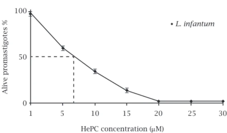
pro-duction of formazan predicts decreasing cell viability. Using HePC for treating Leishmania promastigotes demon-strated inhibition of parasite growth and the IC50 was 7 µmol for (MCAN/IR/96/LONDON49) promastigotes. Miltefosine showed a dose-dependent cytotoxic effect with almost 100% death at a concentration of about 22 μM (Figure 1).
In vitro effect of miltefosine in L. infantum amastigotes
ED50 (half maximal effective dose) for L. infantum amastigotes was determined after 48 hours exposure to different concentra-tion of miltefosine.
The percentage of infected J774 cell lines was evaluated by microscopic examination of at least 100 cells. The data repre-sent the means ± standard deviations (SDs) of three independ-ent experimindepend-ents. ED50 of miltefosine was 12.5 μM for L. infan-tum. In amastigote-infected macrophages [more than 93% of cells (parasite and macrophage)] were killed after 48 hours of incubation with 25 μM of miltefosine (Figure 2).
Figure 1: The viability of L. infantum promastigotes
(MCAN/IR/96/LONDON49) at various concentrations of HePC were assessed by MTT. Each point represents the mean of 3 independent determinations.
Figure 2: Effect of different concentrations of miltefosine
on the proliferation of L. infantum amastigotes. Microscopic
examination of at least 100 infected J774 was evaluated. Each
value represents the mean ± standard deviation (SDs) of three independent experiments.
L. infantum
HePC concentration (mM)
HePC concentration (mM)
Alive promastigotes %
Alive amastigotes %
1 5 10 15 20 25 30
1 5 10 15 20 25 30
100
50
0
Expression of metacaspase and PARP during treatment with miltefosine
Metacaspases are caspase-related cysteine-proteases that are present in organisms without caspases such as plants, yeast, and protozoan parasites. Since caspases are important effec-tor molecules in mammalian apoptosis, the possible role of metacaspases in PCD was evaluated in the L. infantum pro-mastigotes (MCAN/IR/96/LONDON 49). After treatment of promastigotes with IC50 (7 μmol) of miltefosine, metacas-pase gene expression was analyzed with RT-PCR in various time periods. Expression of metacaspase detected in 6, 18, 24, 36 and 48 hours post treatment with miltefosine. Over expression of metacaspase was seen 6 to 24 hours after treat-ment. Untreated cells expressed metacaspase gene steadily in several time points. In addition, L. infantum promastigotes (MCAN/IR/96/LONDON 49) were treated with 7 μmol milte-fosine for various time periods and the expression of PARP was studied by RT-PCR. The expression of PARP could be detected in all cells at 6, 18 and 24 hours post-treatment. However, PARP expression could not be detected in cells treated with miltefo-sine for 36 hr and the effect continued even 48 hr after treat-ment. This was observed when the L. infantum promastigotes were treated with miltefosine (Figure 3).
Figure 3:(A and B) Relative gene expression of L. infantum. (A)
Metacaspase gene expression in treated and control group. (B)
PARP gene expression in treated and control group in different time periods. The bars indicate the mean value of metacaspase and PARP gene expression for each group. The density of each band in PCR products was digitized using molecular analyst software for densitometry. This represents a level of expression for each gene in different time periods. The PARP and metacaspase gene expression responses to HePC (7 µM) were also expressed as the expression index defined as the ratio. Results are shown in
relation to GAPDH, defined as 100%. The values (mean ± SEM) are
derived from three independent experiments.
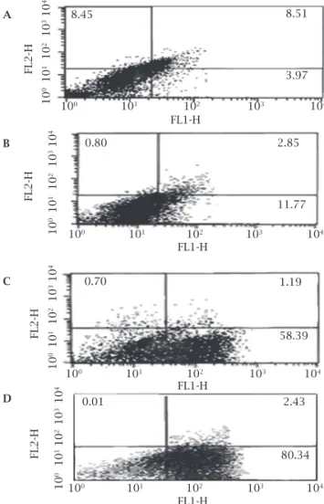
Figure 4: Flow cytometry analysis of promastigotes following
treatment with 7 µM HePC and after labeling with annexin-V and PI. Miltefosine did not lead to necrosis even after prolonged incubation, as the entire cells remained negative for PI. Lower right region (LR) belongs to apoptotic cells (annexin positive) and upper left region (UL) belongs to necrotic cells (PI positive). A, B, C and D flow cytometry analysis in 12, 18, 36 and 48 hours respectively. Values are percentages.
Detection of phosphatidylserine externalization and mode of cell death
During apoptoic cell death process, phosphatidylserine is translocated from the inner side to the outer layer of the plasma membrane in metazoan and unicellular cells. An-nexin V is routinely used to label externalization of phos-phatidylserine. Staining by Annexin V and PI simultane-ously can differentiate apoptotic, necrotic and living cells. After 36 and 48 hours of treatment, the percent of annexin-positive cells were 58% and 80%, respectively, whereas the corresponding figures in the control group were just 4% for both time points (Figure 4). The negative results of PI stain-ing in several time points showed that miltefosine did not induce necrosis even after prolonged incubation.
Miltefosine induces sub-G0/G1 phase cell-division arrest
To evaluate the ratio of pseudohypodiploid cells, flow cy-tometric analysis after cell permeabilization and labeling with PI was used. In a given cell, the amount of bound
6h 18h 24h 36h 48h
6 h 18 h 24 h 36 h 48 h Metacaspase PARP Treated Control Expression Indix Expression Indix Treated Control A B C D A B 10 6 2 -2 10 8 6 4 2 0 8.45 FL2-H FL2-H FL2-H FL2-H 8.51 3.97 0.80 FL1-H FL1-H 2.85 11.77 1.19 58.39 2.43 80.34 0.01 0.70
100 101 102 103 104
100 101 102 103 104
100 101 102 103 104
100 101 102 103 104
10
0
10
1 1
0
2 10 3 10
4
10
0 10
1
10
2
10
3 1
0
4
10
0 10
1
10
2
10
3 1
0
4
10
0 10
1
10
2
10
3 1
0
4
FL1-H
6 hr after treatment with 7 μM miltefosine, but cell death only occur 36 to 48 hr after treatment. It is assumed that cell death process of Leishmania promastigotes depends on metacaspase beginning the initial signaling pathway.
González et al.11 showed that L. major metacaspase has a role in cell death. These researchers showed that metacas-pase gene of Leishmania major (LmjMCA) was involved in yeast cell death, similar to Saccharomyces cerevisiae metacas-pase (YCA1), and that this function depends on its catalytic activity. These results suggest that in spite of probable dif-ferences in their catalytic activity, metacaspases are mem-bers of a family of peptidases that have a role in cell death.25 As metacaspases have a role in cell death they may become potential targets for anti-Leishmania medications. In mam-malians, caspases do not only function in PCD, but they also have functions in cell proliferation and differentiation.11
Ambit et al.26 found that LmjMCA is expressed in active-ly replicating amastigotes and procyclic promastigotes, but at a lower level in metacyclic promastigotes. Over expression of LmjMCA in promastigotes leads to severe growth retar-dation. Also they implicated L. major active metacaspase has effective role in the separation of the nucleus and the kineto-plast, function that could be independent of PCD.26
In this study, PARP and metacaspase gene expression increased simultaneously. This evolution could repair cell DNA damage. In the present study, increase of PARP gene expression reached its maximum 18 hr after exposure to miltefosine, but the slow decrease observed in the following several time points seemed to be due to DNA injury.
Breakdown of PARP, a DNA repair enzyme that cataly-ses the poly (ADP-ribosyl)ation of various nuclear proteins is a hallmark of metazoan apoptosis.27 Western blot analy-ses reported by Das et al.19 showed cleavage of a PARP-like protein during apoptosis in Leishmania treated by H2O2. This process blocked caspase inhibitors. Molecular size of dye correlates with the DNA content and thus DNA
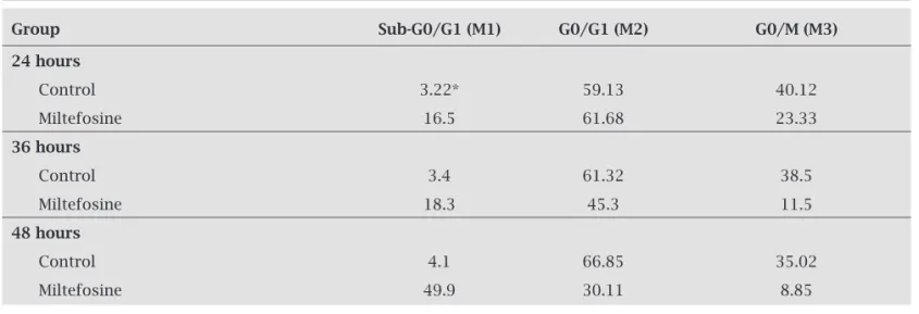
frag-mentation in apoptotic cells is translated by fluorescence intensity lower than that of G0/G1 cells, i.e. a sub-G0/G1 peak. Incubation of L. infantum promastigotes with 22 μM miltefosine for 24 hr indicated an increased proportion of cells in the sub-G0 G1 phase to 16.5% compared with 3.22% of controls (Table 1). In the HePc treatment, in 48 hr, the percentage of promastigotes in the sub-G0/G1 phase was increased to 49% compared to 4% in the control cells. In contrast, an increase in the cell number in sub-G0/G1 phase led to a decrease in the number of cells in the G2/M phase compared with untreated cells.
DISCUSSION
Leishmaniasis is one of the most significant causes of mor-bidity and mortality in several countries. The most severe form of leishmaniasis is visceral leishmaniasis (VL) or kala-azar, which is fatal in 90% of untreated patients. VL is more frequently observed in developing countries with an esti-mated incidence of 500,000 per year. L. infantum is wide-spread in the Mediterranean areas and causes lethal VL in children under 9 years old.23
The current medications for treatment of VL are penta-valent antimony, pentamidine and amphotericin B. How-ever, their use is limited due to their high toxicity and treat-ment failure in endemic areas.23 Miltefosine (HePC) has been proved to be an effective oral treatment for VL with fewer side effects and a cure rate of about 98%.20 Induc-tion of PCD is one of the advantages of miltefosine against other currently used drugs, including antimony.22,24 The role played by metacaspase in the cell death pathway in protozoa is less clear than for plants or yeast. Our study showed that miltefosine induces overexpression of metacaspase in L. in-fantum promastigotes. Expression of metacaspase increased
Table 1. Percentages of L. infantum promastigotes DNA content after treatment with 22 μM miltefosine for 24,
36 and 48 hours, respectively
Group Sub-G0/G1 (M1) G0/G1 (M2) G0/M (M3)
24 hours
Control 3.22* 59.13 40.12
Miltefosine 16.5 61.68 23.33
36 hours
Control 3.4 61.32 38.5
Miltefosine 18.3 45.3 11.5
48 hours
Control 4.1 66.85 35.02
Miltefosine 49.9 30.11 8.85
the PARP-like protein that was detected in Leishmania by antibody is smaller than the sizes of proteins in mamma-lian systems (78-kDa intact protein and 63-kDa cleaved fragments in Leishmania and 113-kDa intact protein and 85-kDa cleaved fragments in mammalian). L. major cysteine proteinase has been reported to process human nuclear PARP into a 40-kDa fragment.11
Verma et al.22 found that in the presence of miltefosine there is no cleavage of L. donovani PARP protein.22 Likewise, there is no cleavage of PARP in the presence of novobiocin.28
Flow cytometry analysis after labeling with PI showed that the amount of bound dye correlates with the DNA content and thus DNA fragmentation in apoptotic cells translates into fluorescence intensity lower than that of G0/G1 cells, i.e. a sub-G0/G1 peak. In promastigotes treated with miltefosine (7 μM in 24, 36 and 48 hr), the proportion of cells in the sub-G0/G1 phase increased compared to controls continuously (Table 1). Increase in sub-G0/G1 phase was accompanied by a decrease in the number of cells in the G2/M phase compared with untreated cells in several time points. Increased cells in sub-G0/G1 phase confirmed DNA degradation and apopto-sis in L. infantum exposed to miltefosine. In another study Khademvatan et al.29 showed DNA degradation of treated L. infantum with DNA ladder assay after 48 hr, that is at the end stage of apoptosis.29
In the present study, we have determined an IC50 of 7 μM for L. infantum promastigotes (MCAN/IR/96/LONDON49), which is lower than what had been reported for other Leish-mania species. The reported IC50 for two strains of L. dono-vani are different from our study. Verma et al.22 found IC
50 of 13 μM for L. donovani (MHOM/80/IN/Dd8), whereas Paris et al.24 found to be 25 μM IC50 for promastigote of L. donovani (MHOM/ET/67/HU3/L82).24 In contrast, this study showed that the Iranian strain of L. infantum promastigote was very sensitive to miltefosine. In another study, we showed that L. infantum was more sensitive compared to standard strain of L. majori.29
In conclusion our findings indicate that miltefisine can in-duce genes expression related to PCD.
The induction or inhibition route of apoptosis in Leishma-nia spp. is an interesting subject to study as it can identify po-tential targets for improving anti-Leishmania medications.
ACKNOWLEDGEMENTS
We would like to show our appreciation to the cooperation of all staff of Cellular and Molecular Research Center, Teh-ran University of Medical Sciences.
REFERENCES
1. Reape TJ, McCabe PF. Apoptotic-like regulation of pro-grammed cell death in plants. Apoptosis 2010; 15(3):249-56.
2. Kaur J, Singh N, Singh BK et al. Leishmania donovani: oral therapy with glycosyl 1,4-dihydropyridine analogue showing apoptosis like phenotypes targeting pteridine reductase 1 in intracellular amastigotes. Exp Parasitol 2010; 125(3):310-4. 3. Luque-Ortega JR, Rivas L Characterization of the
leishmani-cidal activity of antimicrobial peptides. Methods Mol Biol 2010; 618:393-420.
4. Mukherjee P, Majee SB, Ghosh S et al. Apoptosis-like death in Leishmania donovani promastigotes induced by diospyrin and its ethanolamine derivative. Int J Antimicrob Agents 2009; 34(6):596-601.
5. Kulkarni MM, McMaster WR, Kamysz W et al. Antimicrobial peptide-induced apoptotic death of leishmania results from calcium-de pend ent, caspase-independent mitochondrial tox-icity. J Biol Chem 2009 5; 284(23):15496-504.
6. Zangger H, Mottram JC, Fasel N. Cell death in Leishmania in-duced by stress and differentiation: programmed cell death or necrosis? Cell Death Differ 2002; 9(10):1126-39.
7. Bera A, Singh S, Nagaraj R et al. Induction of autophagic cell death in Leishmania donovani by antimicrobial peptides. Mol Biochem Parasitol 2003; 127(1):23-35.
8. Elmore S. Apoptosis: a review of programmed cell death. Toxi-col Pathol 2007; 35(4):495-516.
9. Uren AG, O’Rourke K, Aravind LA et al. Identification of pa-racaspases and metacaspases: two ancient families of caspase-like proteins, one of which plays a key role in MALT lympho-ma. Mol Cell 2000; 6:961-7.
10. Watanabe N, Lam E. Two arabidopsis metacaspases AtMCP1b and AtMCP2b are arginine/lysine-specific cysteine proteases and activate apoptosis-like cell death in yeast. J Biol Chem 2005; 280(15):14691-9.
11. González IJ, Desponds C, Schaff C et al. Leishmania major metacaspase can replace yeast metacaspase in programmed cell death and has arginine-specific cysteine peptidase activity. Int J Parasitol 2007; 37(2):161-72.
12. Liang Q, Li W, Zhou B. Caspase-independent apoptosis in yeast. Biochim Biophys Acta 2008; 1783(7):1311-9.
13. Büttner S, Eisenberg T, Herker E et al. Why yeast cells can un-dergo apoptosis: death in times of peace, love, and war. J Cell Biol 2006; 175(4):521-5.
14. Suarez MF, Filonova LH, Smertenko A et al. Metacaspase-de-pendent programmed cell death is essential for plant embryo-genesis. Curr Biol 2004; 14(9):339-40.
15. Hoeberichts FA, ten Have A, Woltering EJ. A tomato meta-caspase gene is upregulated during programmed cell death in Botrytis cinerea-infected leaves. Planta 2003; 217(3):517-22. 16. Kosec G, Álvarez VE, Aguero F et al. Metacaspases of
Trypa-nosoma cruzi: possible candidates for programmed cell death mediators. Mol Biochem Parasitol 2006; 145:18-28.
17. Szallies A, Kubata BK, Duszenko M. A metacaspase of Trypa-nosoma brucei causes loss of respiration competence and clon-al death in the yeast Saccharomyces cerevisiae. FEBS Lett. 2002; 517:144-50.
18. Mukherjee P, Majee SB, Ghosh S et al. Apoptosis-like death in Leishmania donovani promastigotes induced by diospyrin and its ethanolamine derivative. Int J Antimicrob Agents 2009; 34(6):596-601.
19. Das M, Mukherjee SB, Shaha C. Hydrogen peroxide induces apoptosis-like death in Leishmania donovani promastigotes. J Cell Sci 2001; 114(Pt 13):2461-9.
21. Shaha C. Apoptosis in ispecies & its relevance to disease patho-genesis Indian J Med Res 2006; 123(3):233-44.
22. Verma NK, Dey CS. Possible mechanism of miltefosine-medi-ated death of Leishmania donovani. Antimicrob Agents Chem-other 2004; 48(8):3010-5.
23. Desjeux P. Leishmaniasis: current situation and new perspec-tives. Comp Immunol Microbiol Infect Dis 2004;27(5):305-18. 24. Paris C, Loiseau PM, Bories C et al. Miltefosine induces apo-ptosis-like death in Leishmania donovani promastigotes. Anti-microb Agents Chemother 2004; 48(3):852-9.
25. Lamkanfi M, Festjens N, Declercq W et al. Caspases in cell survival, proliferation and differentiation. Cell Death Differ 2006; 14:44-55.
26. Ambit A, Fasel N, Coombs GH et al. An essential role for the Leishmania major metacaspase in cell cycle progression. Cell Death Differ 2008; 15(1):113-22.
27. Arvind L, Dixit VM, Konin EV. Apoptotic molecular machin-ery: vastly increased complexity in vertebrates revealed by ge-nome comparisons. Science 2001; 291:1279-84.
28. Singh G, Jayanarayan KG, Dey CS. Novobiocin induces apo-ptosis-like cell death in topoisomerase II over-expressing ar-senite resistant Leishmania donovani. Mol Biochem Parasitol 2005; 141(1):57-69.