Rev Bras Cardiol Invasiva. 2013;21(2):193-8
© 2013 Sociedade Brasileira de Hemodinâmica e Cardiologia Intervencionista. Published by Elsevier Editora Ltda. All rights reserved.
Coronary Artery Thrombus Recanalisation:
Documentation by Optical Coherence Tomography
Caused by Angiographic Haziness
Daniel Chamié
1, Alexandre Abizaid
2A
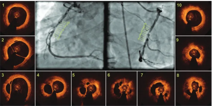
57-year-old male patient with a history of
myo-cardial infarction two months before was treated
conservatively. Coronary angiography evidenced a
single lesion in the middle segment of the right
coro-nary artery, with intraluminal haziness and preserved
distal flow (Thrombolyis in Myocardial Infarction [TIMI]
3) (Figure 1).
An evaluation with optical coherence tomography
was performed in order to assess the morphology and
1 Interventional cardiologist in the Department of Invasive Cardio
-logy of the Instituto Dante Pazzanese de Cardiologia. São Paulo, SP, Brazil.
2 Lecturer. Director of Invasive Cardiology Service of the Instituto Dante
Pazzanese de Cardiologia. São Paulo, SP, Brazil.
Correspondence to: Daniel Chamié. Av. Dr. Dante Pazzanese, 500 – Ibirapuera – São Paulo, SP, Brazil – CEP 04012-180
E-mail: [email protected]
Received: 04/16/2013 • Accepted on: 05/30/2013
Cardiovascular Intervention Image
composition of stenosis, in addition to guiding the
intervention procedure.
Figures 1 and 2 show a complex structure, with
the vascular lumen divided into multiple cavities
separated by high-intensity tissue and low attenuation
of the optical signal, suggesting the presence of an
organized, white, recanalized thrombus, which was
crossed using a 0.014-inch hydrophilic guide with
mouldable tip.
Chamie et al.
Coronary Artery Thrombus Recanalisation
Rev Bras Cardiol Invasiva. 2013;21(2):199-200
200