Recebido 28/01/2008 / Aceito 28/04/2008 Revista de Ciências
Farmacêuticas Básica e Aplicada
Journal of Basic and Applied Pharmaceutical Sciences
Rev. Ciênc. Farm. Básica Apl., v. 28, n.3, p. 335 - 340, 2007
ISSN 1808-4532
Antiproliferative effect of Pterogyne nitens on
melanoma cells
Regasini, L.O.1*, Lopes, A.A.1, Silva, D.H.S.1, Furlan, M.1, Young, M.C.M.2, Maria, D.A.3, Barreiro, E.J.4, Bolzani, V.S.1
1Departamento de Química Orgânica, NuBBE, Instituto de Química, Universidade Estadual Paulista, UNESP, Araraquara, SP, Brasil. 2Seção de Fisiologia e Bioquímica de Plantas, Instituto de Botânica, São Paulo, SP, Brasil.
3Serviço de Bioquímica, Divisão de Ciências Fisiológicas e Químicas, Instituto Butantan, São Paulo, SP, Brasil. 4Faculdade de Farmácia, LASSBio, Universidade Federal do Rio de Janeiro, UFRJ, Rio de Janeiro, RJ, Brasil.
*Corresponding author: Luis Octávio Regasini - Departamento de Química
Orgânica, NuBBE - Instituto de Química, Universidade Estadual Paulista, UNESP Rua Prof. Francisco Degni, s/nº CEP: 14801970 Araraquara -SP, Brasil - Telefone: +55 (16) 3301-6600 - Fax: +55 (16) 3322-7932 - e-mail address: [email protected]
ABSTRACT
As part of our program of bioprospecting for novel antitumor drug prototypes, twenty extracts and fractions obtained from Pterogyne nitens Tul. (Fabaceae, Caesalpinioideae) were screened for antiproliferative activity against B16F10 murine melanoma cells, by the MTT colorimetric assay. The strongest activity was found in EtOAc fractions from the flowers (IC50 = 0.35 µg/mL), fruits (IC50 = 0.34 µg/mL), leaves (IC50 = 0.33 µg/mL) and stems (IC50 = 0.29 µg/mL). Analysis by TLC and HPLC-DAD showed the presence of guanidine alkaloids, flavones and flavonols in the bioactive samples. Additionally, a phytochemical study of the EtOAc fraction of the stems afforded quercetin (1) and isoquercitrin (2), two flavonols with antiproliferative activity previously described in the literature. On the basis of these results, it can be concluded that P. nitens inhibits the growth of melanoma cells in vitro. Further investigations will be needed to assess the usefulness of the samples under study for the treatment of neoplasms and to characterize other bioactive compounds.
Keywords: antiproliferative; Pterogyne nitens; Caesalpinioideae; melanoma; flavonoids; Fabaceae.
INTRODUCTION
Cancer is an important public health problem that, according to the WHO, claims the lives of more than seven million people worldwide every year (Almeida et al., 2005). In particular, melanoma is the most aggressive form of skin cancer and advanced stages are inevitably resistant to conventional therapy. In particular, the inability of chemotherapy and other external stimuli to induce apoptosis
in melanoma cells leads to a selective advantage for tumor progression and metastasis formation, as well as resistance to therapy (La Porta, 2007).
Although a number of antineoplastic drugs are already available, their use is limited by a number of factors, such as low potency, poor solubility in water, emergence of resistant cancer and drug toxicity. There is thus a distinct need for new, safer and more effective anti-cancer agents (Srivastava et al., 2005). It is notable that natural products with diverse biological activities have contributed to the development of nearly 75% of the antineoplastic drugs in the modern pharmacopoeia. Several plant-derived compounds, for example, Vinca's alkaloids, podophyllotoxin, camptothecins and taxanes, provided important "leads" for chemotherapeutic treatment of many forms of cancer, including melanoma (Newman & Cragg, 2007).
toxicity to the DNA repair-deficient yeast (Saccharomyces cerevisiae) strain RS321 over the repair-proficient wild-type strain RAD+, suggesting a potential anti-cancer activity (Bolzani et al., 1995).
Thus, the aim of the current study is to screen the extracts and fractions of flowers, fruits, leaves, stems and non-alkaloid compounds from P. nitens for an antiproliferative effect on B16F10 murine melanoma cells.
MATERIALS AND METHODS
Plant material
Leaves, stems, flowers and fruits of Pterogyne nitens Tul. (family Fabaceae, subfamily Caesalpinioideae) were collected in the Botanic Institute of São Paulo (São Paulo State, Brazil) in May 2003. A voucher specimen (SP 204319) was deposited at the State Herbarium "Maria E. P. Kaufmann" of the Botanic Institute (São Paulo State, Brazil).
Extraction
Shade-dried, powdered plant material was first defatted with hexane and exhaustively extracted by steeping in ethanol. After filtering, the solvent was evaporated cold under reduced pressure to yield a thick syrup, which was dispersed in methanol-water (4:1) and then successively partitioned with ethyl acetate and n-butanol. Samples of the hexane and ethanol (EtOH) extracts and the ethyl acetate (EtOAc), n-butanol (BuOH) and lyophilized hydromethanolic fractions were tested for antiproliferative activity. The extracts and fractions were analyzed by TLC and HPLC-DAD, to screen them for classes of phytochemical constituents.
Thin layer chromatography (TLC) analysis
Thin-layer plates of F254 silica gel (Merck®) were used with the solvent system ethyl acetate: water : formic acid : acetic acid (100:27:11:11) or chloroform: methanol (3:1) as the mobile phase. The following sprays were used to develop the spots: Sakaguchi reagent (for guanidine compounds) (Weber, 1930), Dragendorff reagent, 10% ethanolic KOH, anisaldehyde-sulphuric acid reagent (AS) and 1% diphenylboric acid- -ethylamino ester in methanol (NP reagent). UV light at 254 and 366 nm was also used to make the spots visible (Wagner & Bladt, 1996).
High-performance liquid chromatography with diode-array detection (HPLC-DAD)
In order to characterize the constituents responsible for the bioactivity of extracts and fractions, the latter were analyzed by HPLC on a Supelcosil LC-18 column (250mm x 4.6mm i.d.; 5 m particle size) protected by a corresponding guard column (20mm x 4.6mm i.d.; 5 m), coupled to a
Varian Pro Star® system consisting of a ternary pump (model 240), a photodiode array detector (model 330) and an auto-sampler (model 410), controlled by a Star Chromatography® workstation (version 5.3). In order to ensure optimal peak resolution in the chromatograms, and hence efficient separation of the analytes in a reasonably short time (25 min), the loaded sample was eluted in the gradient mode with a mobile phase consisting of a mixture of acetonitrile and 0.02 M phosphate buffer (pH 6.53) in a proportion evolving from 10: 90 to 55: 45. The flow rate was 1.20 mL/min. The range of the photodiode array detector was 225-400 nm and the sensitivity of the method was maximized by using the wavelengths where the analyte peaks in the chromatograms were most pronounced, namely: 265 nm for guanidine alkaloids and 254 nm for phenolic compounds.
Isolation and identification of flavonols 1 and 2
The EtOAc fraction of stems (2.0 g) was s u b j ec te d to p r e p a r a ti v e G el P er me at io n C h r o mat o g r a p h y ( G P C ) o n a S ep h a d e x LH - 2 0 (Pharmacia®) column (185 x 6.0 cm i.d.) and eluted with
methanol. Fractions (30.0 mL) were collected and checked by TLC on silica gel F254 plates eluted with a mixture of chloroform: methanol: water (80: 18: 2). Subfractions 20-27 (330 mg) were purified by repeated column chromatography (CC) with silica gel (Merck®) eluted with chloroform: methanol (3: 1), furnishing quercetin (1; 33 mg) and isoquercitrin (2; 41 mg). The mo l ec u l ar s tr u c tu r e s o f t h e s e c o mp o u n d s w er e characterized by comparison with literature data, mainly
1H and 13C NMR values (Agrawall, 1989; Harborne &
Williams, 1994). The NMR spectra were obtained in DMSO-d6 solution, using a Varian INOVA 500® spectrometer (11.7 T), operating at 500 MHz for 1H and
125 MHz for 13C. Chemical shifts are given as values
(ppm) relative to tetramethylsilane (TMS) as internal standard.
Quercetin (1): yellow solid. 1H NMR H
(multiplicity; J in Hz; position): 12.5 (s; 5-OH), 7.66 (d; 2.0; H-2'), 7.52 (dd; 2.0 and 8.5; H-6'), 6.87 (d; 8.5; H-5'), 6.39 (d; 2.0; H-8), 6.17 (d; 2.0; H-6). 13C NMR
C (position):
175.8 (C-4), 163.8 (C-7), 160.7 (C-5), 156.1 (C-9), 146.8 (C-2), 147.6 (C-4'), 145.0 (C-3'), 135.7 (C-3), 119.9 (C-6'), 121.9 1'), 115.6 2' and C-5'), 103.0 10), 98.1 (C-6), 93.3 (C-8).
Isoquercitrin (2): yellow solid. 1H NMR H
(multiplicity; J in Hz; position): 13.2 (br s, 5-OH), 7.56 (dd; 2.0 and 8.5; H-6'), 7.55 (d; 2.5; H-2'), 6.83 (d; 8.5; H-5'), 6.17 (d; 1.5; H-6), 6.37 (d; 1.5; H-8), 5.43 (d; 7.5; H-1''), 3.24 (m; H-2''), 3.01 (m; H-3'' and H-4''), 3.34 (m; H-5''), 3.57 (br d; 11.0; H-6''). 13C NMR
C 177.4 4), 165.0
Cell line
The B16F10 murine melanoma cells originally derived from the C57BL/6 mouse strain were purchased from the American Type Culture Collection (Manassas, VA). The B16F10 cells were maintained in RPMI-1640 (Sigma Chemical Co., St Louis, MO, USA) supplemented with 10% heat-inactivated fetal calf serum (FCS), 100 U/mL of penicillin, 100 g/mL streptomycin and 2mM L-glutamine, in a humidified incubator under 5% CO2 at 37°C, and passaged at subconfluence.
MTT colorimetric assay
The B16F10 cells were harvested in exponential phase and seeded in 96-well flat-bottomed tissue culture microplates at a concentration of 5×10³ cells in 100 µL per
well. The cells were allowed to grow and stabilize for 24 h. Subsequently, the cells were treated with serial concentrations of extracts and fractions prepared in complete medium. Each treatment was performed in eight well replicates. Treated cells were washed with PBS and allowed to grow in complete medium for 24 h. After incubation, the cell viability was determined by MTT colorimetric assay. Briefly, 20 µL of MTT (3-[4,5-dimethylthiazol-2-yl]-2,5-diphenyltetrazolium bromide) reagent (Sigma Chemical Co., St Louis, MO, USA) was added to each well to make a final concentration of 1000 g/mL in the medium and incubated for 4 h at 37°C. Microplates were then centrifuged at 2,000 rpm for 10 min. Medium was aspirated from the wells and 100 µL of DMSO was added to each. Optical density was measured in an ELISA plate reader (Molecular Devices, Spectra Max 190 with Soft max Pro software) at 540 nm with a reference wavelength of 690 nm. Cell viability was plotted as a percentage of untreated control. Vinblastine was used as a reference antineoplastic drug (Mosmann, 1983; Wilson, 2000).
The results are expressed as means and represent eight independent experiments. Inhibitory concentration 50 (IC50) of each extract and fraction was determined from its concentration effect curve as the concentration that decreased the cell viability to 50 %.
RESULTS
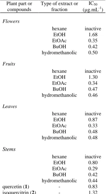
Two extracts (hexane and ethanol) and three fractions (ethyl acetate, n-butanol, hydromethanolic) of the flowers, fruits, leaves and stems were screened for inhibition of melanoma cells and the results are summarized in Table 1.
The low-polarity extracts obtained by extraction with hexane proved to be inactive, since the concentration at which this extract showed activity was over 1000 µg/mL. In general, EtOH extracts obtained from leaves and stems (IC50 ca. 0.80 µg/mL) showed stronger activity than did those from flowers and fruits (IC50 > 1.20 µg/mL). The ethyl acetate (EtOAc), n-butanol (BuOH) and hydromethanolic fractions obtained by liquid-liquid partition were found to be more active than the EtOH extracts. The BuOH and hydromethanolic
fractions (IC50 ca. 0.40 µg/mL) were slightly less effective than EtOAc fractions (IC50 ca. 0.30 µg/mL), suggesting that the potential antiproliferative compounds were in the medium-polarity fractions. The EtOAc fraction of the stems exhibited the best activity against melanoma cells (IC50 = 0.29 µg/mL) and most EtOAc fractions showed an activity considered good (IC50 < 0.35 µg/mL).
The results of phytochemical analysis by TLC of the extracts and fractions of P. nitens are shown in Table 2. According to the TLC analysis, alkaloids were detected in the EtOAc, BuOH and hydromethanolic fractions, which showed an orange spot in the presence of Dragendorff reagent, as well as a pink spot when sprayed with Sakaguchi reagent (positive for guanidine alkaloids). Alcoholic KOH (10%) can be used to reveal anthrones (yellow), anthraquinones (red) or coumarins (blue), depending on the color of the spot observed
Plant part or compounds
Type of extract or fraction
IC50
( g.mL-1)
Flowers
hexane inactive
EtOH 1.68
EtOAc 0.35
BuOH 0.42
hydromethanolic 0.50
Fruits
hexane inactive
EtOH 1.30
EtOAc 0.34
BuOH 0.47
hydromethanolic 0.46
Leaves
hexane inactive
EtOH 0.87
EtOAc 0.33
BuOH 0.48
hydromethanolic 0.48
Stems
hexane inactive
EtOH 0.80
EtOAc 0.29
BuOH 0.42
hydromethanolic 0.44
quercetin (1) - 0.83
isoquercitrin (2) - 1.32
Table 1 - Antiproliferative effect of Pterogyne nitens fractions and extracts on melanoma cells.
alkaloids and typical of the leaves of P. nitens (Bolzani et al., 1995). In addition, two classes of flavonoids were detected in these fractions (flavone and flavonol), as the UV spectrum exhibited two bands of maximum absorption in the ranges 240-285 nm (band II, primarily due to ring A) and 300-400 nm (band I, due to ring B). The UV spectrum assigned to flavones and flavonols displays similar bands II and I, between 304-350 nm and 328 -358 nm, respectively (Agrawall, 1989).
DISCUSSION
Higher plants are known to provide a wide range of natural compounds of potential interest (Fabricant & Farnsworth, 2001). Notwithstanding the breadth of chemodiversity, a number of unusual guanidine alkaloids have been found to display several different types of biological activity (Berlinck, 2002), including hypotensive (Botta et al., 2003) and hypoglycemic (Bailey & Day, 2004) effects, induction of nyctinastic movements in plants (Ueda et al., 2000) and cytotoxicity (Mavar-Manga et al., 2006).
In contrast, flavonoids are already widely recognized as a major class of secondary metabolites with a broad spectrum of pharmacological properties, due to their ability to act in redox processes (Havsteen, 2002).
The preliminary TLC and HPLC-DAD analysis O
O
OH
HO
OH
OH
OH O
O
OH
HO
OH
OH
O O HO
OH OH OH
(1) quercetin (2) isoquercitrin
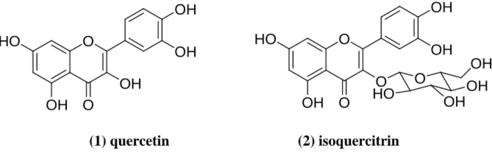
Figure 1. Molecular structures of flavonols isolated from the ethyl acetate fraction of Pterogyne nitens stems.
suggested that flavonoids and/or guanidine alkaloids may be responsible for the antiproliferative effect of the extracts and fractions of P. nitens on melanoma cells. The phytochemical fractionation of stems afforded two flavonoids (Figure 1), identified as quercetin (1 = 3,5,7,3',4'-pentahydroxy-flavone) and isoquercitrin (2 = quercetin 3-O-glucoside), which showed IC50 values of 0.83 µg/mL and 1.32 µg/mL, respectively. These flavonols, whose antiproliferative properties have been extensively demonstrated in the literature, occur widely in nature. Edwards and collaborators reported that a catechol-containing flavonoid (5,7,3',4'-tetrahydroxy-3-O-glycosylflavone) possessed antineoplastic activity towards Walker carcinoma 256 (Edwards et al, 1979). Quercetin has growth inhibitory effects on several malignant tumor cell lines in vitro, such as Ehrlich ascites, L1210 and P-388 leukaemia, NK/Ly ascites tumor, HeLa, gastric cancer (HGC-27, NUGC-2, MKN-7 and MKN-28), colon cancer, human breast cancer, human squamous, gliosarcoma, ovarian cancer and multi-drug-resistant human breast cancer (Harborne & Williams, 1994). It may be concluded from the results of this study that P. nitens has potent antiproliferative activity and could be an important source of potential antitumor agents, useful for developing new antineoplastic drugs, including guanidine alkaloids (Bolzani et al., 1995) and flavonols. In view of these findings, further chemical and pharmacological investigations to isolate and identify other secondary metabolites and to screen their mechanisms of action are recommended.
ACKNOWLEDGEMENTS
This work was funded by grants of the São Paulo State Research Foundation (FAPESP), within the Biota-FAPESP Program - The Biodiversity Virtual Institute Program (www.biota.org.br), grant no. 03/02176-7, awarded to V. S. B. (Principal Investigator), and other authors acknowledge the research and Ph.D. fellowships awarded by the Brazilian federal research and higher education funding bodies, CNPq and CAPES. The authors wish to thank Primeiro Simpósio Paulista de Farmacognosia (School of Pharmaceutical Sciences, UNESP at Araraquara, São Paulo State, Brazil) for their invitation to publish in the JBAPS.
Antiproliferative effect of P. nitens
under UV 365 nm (Wagner & Bladt, 1996), and none of the extracts or fractions showed detectable amounts of anthrones, anthraquinones or coumarins. On the other hand, spots that did not fluoresce under UV 254 or 365 nm could be seen on the TLC plates and that may indicate the presence of terpenes, steroids and their derivatives (saponins, cardiac glycosides, etc.) in the extracts and fractions. Only the hexane extracts appeared to have terpenes or terpenes derivatives. NP reagent (diphenylboric acid- -ethylamino ester) indicates the presence of flavonoids if spots turn yellow, orange or green when illuminated with UV light at 366 nm. Flavonoids were detected in all samples except in the hexane extracts.
RESUMO
Efeito antiproliferativo de Pterogyne nitens sobre células de melanoma
No escopo do nosso programa de bioprospecção, o qual objetiva a descoberta de novos protótipos antitumorais, vinte extratos e frações obtidas de Pterogyne nitens Tul. (Fabaceae-Caesalpinioideae) foram triados para ativi-dade antiproliferativa sobre células de melanoma B16F10, empregando o método colorimétrico com MTT. Os efeitos mais intensos foram manifestados pelas fra-ções AcOEt das flores (CI50 = 0,35 µg/mL), frutos (CI50 = 0,34 µg/mL), folhas (CI50 = 0,33 µg/mL) e caules (CI50 = 0,29 µg/mL). As análises por CCD e CLAE-DAD demons-traram a presença de alcalóides guanidínicos, flavonas e flavonóis nas amostras bioativas. Adicionalmente, o es-tudo fitoquímico da fração AcOEt dos caules forneceu quercetina (1) e isoquercitrina (2), dois flavonóis com atividade antiproliferativa descrita previamente na lite-ratura. Baseado nos presentes resultados pode-se con-cluir que P. nitens inibiu o crescimento das células de melanoma in vitro e futuras investigações serão necessá-rias para avaliar a utilidade das amostras estudadas para o tratamento de neoplasias e caracterizar outras subs-tâncias bioativas.
Palavras-chave: antiproliferativo; Pterogyne nitens; Caesalpinioideae; melanoma; flavonóides; Fabaceae.
REFERENCES
Agrawal PK. Carbon-13 NMR of flavonoids. Amsterdam: Elsevier.1989.
Almeida VL, Leitão A, Reina LCB, Montanari CA, Donnici CL, Lopes MTP. Câncer e agentes antineoplásicos ciclo-celu-lar específicos e ciclo-celuciclo-celu-lar não específicos que interagem com o DNA: uma introdução. Quim Nova 2005; 28(1):118-29. Bailey CJ, Day C. Metformin: its botanical background. Pract Diab Int 2004; 21:115-6.
Berlinck RGS. Natural guanidine derivatives. Nat Prod Rep 2002; 19:617-49.
Bolzani VS, Gunatilaka AAL, Kingston DGI. Bioactive guanidine alkaloids from Pterogyne nitens. J Nat Prod 1995; 58:1683-8.
Botta B, Botta M, Carmignani M, Corelli F, Delle Monache G, Volpe AR. Novel hypotensive agents from Verbesina caracasana: structure, synthesis and pharmacology. Curr Med Chem 2003; 10:1845-63.
Burkart A. Las leguminosas argentinas. Buenos Aires: Aemé Agency; 1952.
Crivos M, Martinez MR, Pochettino ML, Remorini C, Sy A, Teves L. Pathways as "signatures in landscape": towards an ethnography of mobility among the Mbya-Guaraní (Northeastern Argentina). J Ethnobiol Ethnomed 2007; 3:1-12. Edwards JM, Raffauf RF, Le Quesne PW. Antineoplastic activity of flavones, isoflavones, and flavanones. J Nat Prod 1979; 42:85-91.
Fabricant DS, Farnsworth NR. The value of plants used in traditional medicine for drug discovery. Environ Health Perspect 2001; 109:69-75.
Harborne JB, Williams CA. The flavonoids: advances in research since 1992. London: Chapman & Hall; 1994. Havsteen BH. The biochemistry and medical significance of the flavonoids. Pharmacol Ther 2002; 96:67-202. La Porta C. Drug Resistance in Melanoma: New Perspectives. Curr Med Chem 2007; 14:387-91.
Lorenzi H. Árvores Brasileiras: manual de identificação e cultivo de plantas arbóreas do Brasil. Nova Odessa: Institu-to Plantarum; 1998.
Mavar-Manga H, Chapon D, Hoet S, Block S, De Pauw-Gillet MC, Quetin-Leclercq J. N1,N2,N3-Triisopentenyl guanidine and N1,N2-Diisopentenyl guanidine, two cytotoxic alkaloids from Alchornea cordifolia (Schumach & Thonn.) Müll, Arg. (Euphorbiaceae) root barks. Nat Prod Commun 2006; 1:1097-100.
Mosmann T. Rapid colorimetric assay for cellular growth and survival: application to proliferation and cytotoxicity assays. J Immunol Meth 1983; 65:55-63.
Newman DJ, Cragg GM. Natural products as sources of new drugs over the last 25 years. J Nat Prod 2007; 70:461-77. Srivastava V, Negi AS, Kumar JK, Gupta MN, Khanuja SPS. Plant-based anticancer molecules: A chemical and biological profile of some important leads. Bioorg Med Chem 2005; 13:5822-908.
Ueda M, Okazaki M, Ueda K, Yamamura S. A leaf-closing substance of Albizzia julibrissin Durazz. Tetrahedron 2000; 56:8101-5.
Wagner H, Bladt S. Plant drug analysis. Berlin: Springer; 1996. Weber CJ. A modification of Sakaguchi's reaction for the quantitative determination of arginine. J Biol Chem 1930; 86: 217-22.