ABSTRACT
Cushing’s syndrome (CS) results from prolonged exposure to supra-physiological levels of circulating glucocorticoids, endogenously or exogenously derived. Although rare in childhood, CS remains a difficult condition to diagnose and treat. A multidisciplinary approach and close collaboration with adult colleagues is adopted at most large centres that manage pediatric CS patients. Although pediatric protocols are derived from adult data, significant differences exist between adult and child-hood CS. Furthermore, long term outcome parameters including final height, bone mineral density, reproductive function, body composition and psychological health pose challenges for pediatric care. This article will aim to provide an overall view of pediatric CS highlighting some of the differences between adult and pediatric CS. (Arq Bras Endocrinol Metab 2007;51/8:1261-1271)
Keywords: Pediatrics; Cushing’s syndrome; Cushing’s disease; Adrenal tumours
RESUMO
Síndrome de Cushing Pediátrica: Manifestações Clínicas, Diagnóstico e Tratamento.
A síndrome de Cushing (SC) resulta da exposição prolongada a níveis suprafisiológicos de glicocorticóides circulantes, tanto endógenos como de seus derivados exógenos. Embora rara na infância, a SC permanece uma condição difícil de ser diagnosticada e tratada. Uma avaliação multidisciplinar e a colaboração próxima com colegas da área não-pediátrica são adotadas na maioria dos grandes centros que cuidam de pacientes pediátricos com SC. Embora os protocolos pediátricos sejam derivados de dados em adultos, existem diferenças significativas entre a SC no adulto e na infância. Além disso, parâmetros evolutivos finais, incluindo altura final, densidade mineral óssea, função reprodutiva, composição corporal e saúde psicológica trazem desafios no cuidado pediátrico. Este artigo procura oferecer uma visão geral da SC pediátrica, focalizando algumas das diferenças entre a SC adulta e a pediátrica. (Arq Bras Endocrinol Metab 2007;51/8:1261-1271)
Descritores: Pediatria; Síndrome de Cushing; Doença de Cushing; Tumores adrenais
Recebido em 18/09/07 Aceito em 28/09/07
revisão
Department of Endocrinology, William Harvey Research Institute, St Bartholomew’s and The Royal London School of Medicine and Dentistry, London, EC1M 6BQ.
P
EDIATRIC CUSHING’S SYNDROME (CS) is rare inchildhood and adolescence. The condition is caused by prolonged exposure to excessive glucocor-tioids which can be secreted endogenously or admin-istered exogenously. As with adult CS the most com-mon cause is iatrogenic, with administration of supra-physiological doses of exogenous glucocorticoids in the form of topical, inhaled or oral corticosteroids. Eczema and asthma are common conditions in child-hood often requiring treatment with corticosteroids. Hence a careful history of medications, including atyp-ical topatyp-ical treatments and dietary supplements, are vital in the assessment of CS. Furthermore, drug inter-action in the context of relatively modest doses of inhaled corticosteroids has been known to cause pedi-atric CS in children on antiretroviral drugs (1). In these patients removing or reducing glucocorticoid therapy will result in the resolution of symptoms. Chil-dren with iatrogenic CS are often managed by pedia-tricians responsible for their primary condition. This review will focus on the investigation and management of endogenous pediatric CS as typically referred to a pediatric endocrinologist. Differences between adult and pediatric CS investigation and treatment will be emphasised.
The causes of endogenous pediatric CS are not fundamentally different from those in adults (figure 1). However, some features are distinct. Examples include: (1) the presentation of McCune Albright syn-drome with CS in infancy, (2) the predominance of mixed androgen and cortisol secreting adrenocortical tumors in early childhood and (3) the increased fre-quency of prepubertal Cushing’s disease (CD) in males compared to females (2-4). Other differences compared to adult practice include: the extreme rarity
of ectopic ACTH syndrome, the frequent absence of radiological evidence of a corticotroph adenoma on pituitary scanning in children with CD, the apparent higher incidence, compared to adults, of lateralisation of ACTH secretion demonstrated by bilateral inferior petrosal sinus sampling (BIPSS) and the rapid response of CD to external beam pituitary radiothera-py in children (5-8).
The investigation protocols used for pediatric CS are derived from those established in adult practice. When a patient presents with classical symptoms of CS diagnosis is relatively easy, the challenge is then to identify the cause. However, variable presentation with subtle symptoms and signs can make diagnosis difficult (9). The increasing incidence of childhood obesity has increased referrals to exclude CS. Careful auxological evaluation is the key to differentiating CS from simple obesity (10). Close collaboration with adult endocrinologists is recognised as an essential part of management of pediatric CS and adopted by many large centres (11,12).
This review will consist of a discussion of the classification, clinical presentation, investigation and treatment of pediatric CS.
CLASSIFICATION OF PEDIATRIC CUSHING’S SYNDROME
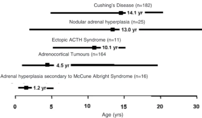
Pediatric CS is divided into ACTH-dependent and ACTH-independent forms. These are listed in table 1. Causes can be further classified according to age of onset (figure 2). For example, CS in infancy is usually associated with McCune-Albright syndrome, adreno-cortical tumours most commonly occur in children under four years of age and Cushing’s disease is the commonest cause of CS after five years of age. These conditions are discussed below.
Figure 1.Differential diagnosis of pediatric Cushing’s syn-drome.
Figure 2.Review of 398 pediatric CS cases from the literature showing ages of peak incidence, represented by the boxes.
Adrenal hyperplasia secondary to McCune Albright Syndrome (n=16) Cushing’s Disease (n=182)
Nodular adrenal hyperplasia (n=25)
Ectopic ACTH Syndrome (n=11)
Adrenocortical Tumours (n=164
CLINICAL PRESENTATION
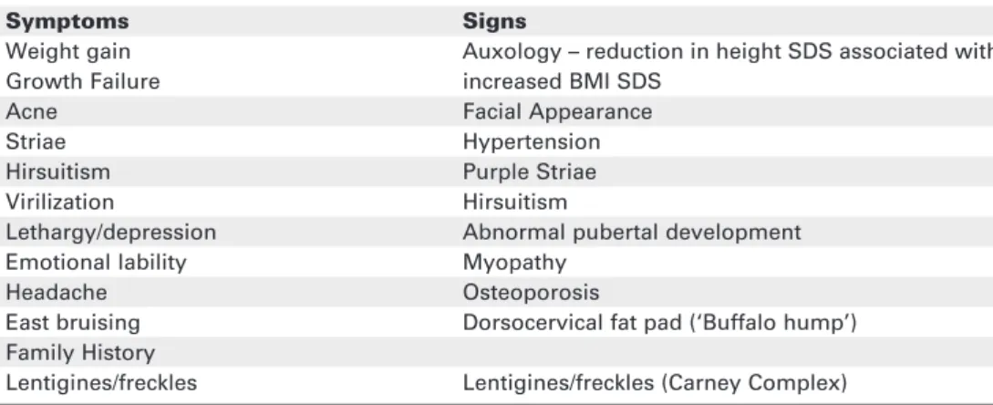
Children with CS can present with a number of symp-toms and signs. These can vary depending on the age of the child and cause of the CS. The key symptoms and clinical findings are summarised in table 2. In compari-son to adult CS, growth failure with associated weight gain is one of the most reliable indicators of hypercorti-solaemia in pediatric CS. Clinical features may occur gradually over a period of time and go unrecognised by parents and carers. For example, the change in facial appearance, which is almost always present, can often be missed. In our own series of 33 Cushing’s disease (CD) patients, the mean time from symptoms appearing to diagnosis was 2.5 yr (range 0.5–6.6 yr). Accurate auxo-logical assessment is vital and serial photographs can be helpful to document physical changes. More recently the comparison of height and BMI SDS in 29 pediatric CD patients and 44 patients with simple obesity showed that height was increased in simple obesity and decreased in CD. This confirmed that height and BMI SDS measurements provided a sensitive diagnostic dis-criminator in pediatric patients with CD and those with simple obesity (10).
ACTH-DEPENDENT CS
Cushing’s disease
Cushing’s disease (CD) is the most common cause of endogenous CS in childhood and adolescents after the age of 5 years (7,13) (figure 2). Defined as hypercortiso-laemia caused by an ACTH-secreting corticotroph ade-noma, pediatric CD accounts for approximately 75–80% of all pediatric CS cases (7,11,12). This compares to 49–71% in adult CS (11,12). The median age of presen-tation is 14.1 yrs (141 cases taken from the literature), with the youngest reported case being 6.2 years (14). Despite advances in the diagnosis and management of CD, the condition still causes significant morbidity and mortality (9,14,15). Unlike adult CD where the number of female patients predominates, pediatric CD is charac-terized by significant male preponderance in prepubertal years (4). This apparent lack of prepubertal female CD patients is shown in figure 3. This phenomenon was also seen in a large NIH series (12). Towards puberty the sex distribution of CD equalizes and the trend is reversed with female preponderance in adulthood. The explana-tion for this is unclear, but is perhaps due to the oestro-genic milieu during puberty in females (4).
Table 2.Key clinical symptoms and signs of Cushing’s syndrome.
Symptoms Signs
Weight gain Auxology – reduction in height SDS associated with
Growth Failure increased BMI SDS
Acne Facial Appearance
Striae Hypertension
Hirsuitism Purple Striae
Virilization Hirsuitism
Lethargy/depression Abnormal pubertal development
Emotional lability Myopathy
Headache Osteoporosis
East bruising Dorsocervical fat pad (‘Buffalo hump’) Family History
Lentigines/freckles Lentigines/freckles (Carney Complex)
SDS: standard deviation score
Table 1.Etiology of pediatric Cushing’s syndrome.
ACTH-dependent CS ACTH-independent CS
1. Cushing’s disease 1. Exogenous glucocorticoid administration (ACTH-secreting pituitary adenoma)
2. Ectopic ACTH syndrome 2. Adrenocortical tumour (adenoma or carcinoma) 3. Primary adrenocortical hyperplasia
The vast majority of pediatric CD is caused by ACTH-producing pituitary microadenomas. Macroadenomas are extremely rare although these have been reported in the literature (6,16,17). Most children and adolescents with CD have a typical cushingoid appearance. Subtle, subclinical or cyclical features are uncommon. In our patient series, all 33 CD patients experienced facial changes and weight gain. The frequency of the following features was: hir-suitism 60%, striae 51%, hypertension 45%, emotional lability 51% and fatigue 60%. Muscle weakness and easy bruising were uncommon. Young children are more likely to present with poor growth and obesity without the classical features of hisutism, plethora, acne and striae.
Growth and puberty in Cushing’s disease
Height SDS is almost always reduced and associated with increased BMI SDS (figure 4). Furthermore, bone age is typically delayed by a mean of 2 yr (range -0.5 to 4.1 yr) (18). Pubertal development in CD has recently been analyzed in 27 pediatric CD patients (19). Thirteen patients had excessive virilization, defined as inappropriate pubic hair development for stage of breast development or testicular size. Virilized patients had significantly higher serum androstene-dione, DHEA-S, and testosterone levels, accompanied by lower sex hormone binding globulin levels. Puber-tal CD patients also had low basal LH and FSH levels, suggesting impaired function of the hypothalamic-pituitary-gonadal axis secondary to long-standing hypercortisolaemia.
Ectopic ACTH syndrome
Ectopic ACTH syndrome (EAS) is extremely rare in the pediatric age group. This contrasts with adults where EAS accounts for approximately 15% of adult ACTH-dependent CS (20). The majority of pediatric cases result from carcinoid tumours of bronchial or thymic origin (21). However, carcinoid tumours of duodenal and renal origin have also been reported (22,23). EAS has been described in pediatric cases of clear cell sarcoma, malignant neuroendocrine tumours of the pancreas, Wilms’ tumour and adrenal neurob-lastoma (24-27). The median age of presentation is 9.5 years, with a female predominance.
ACTH-INDEPENDENT CS
Adrenocortical tumours (adenoma or carci-noma)
Adrenocortical tumours (ACTs) only account for 0.3–0.4% of all neoplasms in childhood. However there is a geographical variation with the highest inci-dence in southern Brazil, where the inciinci-dence is reported as 3.4–4.2 per million children (28,29). Much of what is known about ACTs in childhood has come from large Brazilian pediatric cohorts, pro-viding the basis of many comprehensive reviews on this topic (3,29). ACTs occur most commonly in
Figure 3. Preponderance of prepubertal male patients in pediatric CD as illustrated in our own case series (n = 33).
children under 4 years of age. In Southern Brazil, there is an association with Li-Fraumeni syndrome and a germline point mutation of the p53 tumour
suppressor gene (TP53). This mutation results in a
single amino acid change from Arginine to Histidine at amino acid position 337 (28). In comparison, older children and young adults with ACT do not appear to carry germline mutations in TP53. The presence of this mutation has no influence on prog-nosis (29).
In other syndromes such as Beckwith-Weide-mann syndrome ACT affects almost 15% of patients (3). ACTs associated with MEN type 1 are unlikely to present in childhood (3). In isolated hemihypertrophy approximately 20% of tumours are ACTs (3). A review of 254 children on the International Pediatric Adreno-cortical Tumour Registry identified virilization as the most common manifestation (29). At presentation, only 10% of pediatric ACT patients had no clinical evi-dence of an endocrine syndrome (non-functional tumours) and isolated overproduction of glucocorti-coids accounted for only 5.5% of patients. This con-trasts with the adult population where the majority of adrenocortical tumours are cortisol-secreting or non-functional at presentation (29). Approximately one third of pediatric patients present with hypertenision. At presentation, the majority of patients (192/254) had localized disease and metastatic disease was found in less than 5% of cases. Older children with CS or mixed androgen and cortisol secreting adrenocortical tumors had a worse prognosis compared to younger children (29).
Primary pigmented adrenocortical disease/ ‘micronodular adrenal disease’
Primary pigmented adrenocortical disease (PPNAD) also known as ‘micronodular adrenal disease’ is a his-tological diagnosis characterized by the presence of multiple small adrenocortical nodules, with associat-ed internodular cortical atrophy (30,31). The nod-ules are generally pigmented hence the term PPNAD. However, internodular cortical atrophy resulting from ACTH suppression is the defining fea-ture (32,33). The majority of PPNAD cases are asso-ciated with the Carney Complex (95%), an autosomal dominant multiple endocrine neoplasia, with features of lentigines, cardiac myxomas, endocrine and non-endocrine tumours (31). CS secondary to PPNAD is the most frequent presentation of the Carney Com-plex (CNC) in children and young adults (33). Hypertension and virilization are common present-ing features (32). Cyclical or periodic pediatric CS
and the absence of classical symptoms such as growth failure have been described in some patients (33). The ‘Atypical’ Cushing‘s syndrome phenotype is almost uniquely associated with PPNAD but rare in children (34). Patients have a thin body habitus with short stature, muscle and skin wasting and osteo-porosis (35).
Inactivating germline mutations of the protein kinase A regulatory subunit 1-alpha gene (PRKAR1A)have been identified in 40–50% of fami-lies with CNC (36). Somatic and germline mutations in PRKAR1A have also been described in isolated PPNAD, not associated with CNC (37,38).
ACTH-independent macronodular adrenal hyperplasia (AIMAH)
ACTH-independent macronodular adrenal hyperpla-sia (AIMAH) is a very rare condition, particularly in the pediatric age range. The etiology is unknown (39). Unlike PPNAD, AIMAH usually leads to frank CS (34). Hypogonadism and gynaecomastia in males and hirsutism in females are additional features that have been reported (34,40). There is massive enlargement of the adrenal glands (10 to 100 times their normal weight). Histologically, the adrenocortical nodules are not pigmented but are composed of two different cell types: lipid-rich (with clear cytoplasm) and lipid-poor (with compact cytoplasm) (39,40).
McCune-Albright syndrome causing nodu-lar adrenocortical hyperplasia
McCune-Albright syndrome (MAS) is a sporadic con-dition, which is more common in females. It is caused
by somatic activating mutations of the GNAS1 gene
which encodes the Gs alpha protein (41). Cellular mosaicism accounts for the phenotypic heterogeneity seen in this disorder. The syndrome is classically char-acterized by polyostotic fibrous dysplasia, cafe-au-lait skin pigmentation, and peripheral precocious puberty. However, a wide spectrum of associated endocrine and non-endocrine features are described. This includes CS secondary to nodular adrenal hyperplasia (42,43). CS in infancy, almost always associated with MAS, is often aggressive in its course and requires bilateral adrenalectomy. However, spontaneous resolution has been known to occur (42).
INVESTIGATION OF PEDIATRIC CS
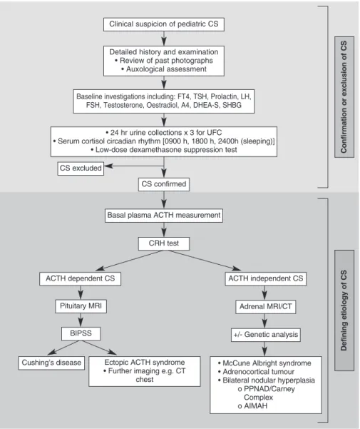
units (5,15). Guidelines for investigating pediatric CS have previously been published (7). The aim is firstly to confirm or exclude a diagnosis of CS, and secondly to determine the etiology. The general protocol that is used at our centre is summarized in figure 5.
Demonstration of Cushing’s syndrome Cushing’s syndrome is characterized biochemically by the loss of normal feedback suppression of the hypo-thalamic-pituitary-adrenal axis and loss of the normal circadian rhythm of cortisol secretion. Therefore inves-tigations test for disruption of the feedback mecha-nism and circadian rhythm. The following tests, par-ticularly when combined, have high sensitivity and specificity.
Urinary Free Cortisol Measurement
Three consecutive 24-hour urine collections for uri-nary free cortisol levels (UFC) are usually the first line investigation. UFC measurements have high sensitivi-ty but relatively low specificisensitivi-ty. If repeated UFC excre-tion is normal then CS is unlikely. Some patients with ‘atypical’ CS may have normal or minimally raised 24 hour UFC levels with the absence of normal circadian rhythm (15).
Serum cortisol circadian rhythm [0900 h, 1800 h, 2400 h(sleeping)]
The cortisol circadian rhythm is assessed at 3 time points, 0900 h, 1800 h and 2400 h. In the normal sleeping child the midnight serum cortisol level should be < 50 nmol/l. In our experience, an ele-vated sleeping midnight cortisol is the best
discrim-Figure 5.Protocol used in the investigation of pediatric CS.
CS excluded
CRH test
Defining etiology of CS
Confirmation or exclusion of CS
Cushing’s disease Ectopic ACTH syndrome • Further imaging e.g. CT
chest
Clinical suspicion of pediatric CS
Baseline investigations including: FT4, TSH, Prolactin, LH, FSH, Testosterone, Oestradiol, A4, DHEA-S, SHBG
Detailed history and examination • Review of past photographs
• Auxological assessment
• 24 hr urine collections x 3 for UFC
• Serum cortisol circadian rhythm [0900 h, 1800 h, 2400h (sleeping)] • Low-dose dexamethasone suppression test
CS confirmed
Basal plasma ACTH measurement
ACTH dependent CS
BIPSS
• McCune Albright syndrome • Adrenocortical tumour • Bilateral nodular hyperplasia
o PPNAD/Carney Complex o AIMAH +/- Genetic analysis
Adrenal MRI/CT ACTH independent CS
inator of CS. However, it is worth noting that some children may reach their cortisol nadir at a slightly earlier time than midnight.
Low-dose dexamethasone suppression test (LDDST)
A dexamethasone dosage of 0.5 mg 6 hourly given at 0900 h, 1500 h, 2100 h and 0300 h is generally used
in children weighing ≥ 40 kg. In those < 40 kg, a
dosage of 30 µg/kg/day is used as recommended by
NIH (7). Blood is taken for serum cortisol at 0 hr, 24 hr and 48 hr. Normal individuals will suppress their serum cortisol levels to < 50 nmol/l by 48 hours. A small proportion of patients with Cushing’s disease suppress normally during LDDST but patients with CS due to other etiologies tend not to.
Other screening tests
Midnight salivary cortisol measurements have been suggested as an alternative noninvasive screening test in the diagnosis of CS (44). It is not our practice to use this method in the screening of children due to the lack of pediatric normative data.
DEFINING THE CAUSE OF CS
Following confirmation of CS, the priority is to differ-entiate between ACTH-dependent and ACTH-inde-pendent forms of CS. This is done by accurate mea-surement of 0900 h plasma ACTH levels. In addition, all children at our centre with proven CS will undergo a corticotrophin-releasing hormone (CRH) test which may differentiate between CD and EAS. Only after ACTH-dependence or ACTH-independence has been established should further appropriate investigations be arranged.
Plasma ACTH levels
In our patients with adrenocortical tumours or nodu-lar adrenal hyperplasia (n = 8), 0900 h ACTH was undetectable (< 10 ng/L) (32). In contrast, all patients with CD (n = 33) had detectable ACTH lev-els ranging from 12 to 128 ng/L (N.R. 10–50 ng/L).
Corticotrophin-releasing hormone (CRH) test
In CD, the administration of CRH (1 µg/kg or 100
µg iv) will induce an exaggerated cortisol response
compared with normal subjects who usually have an increase of < 20%. In all 27 CD patients studied in our
unit, serum cortisol increased by > 100% from baseline (range 106–554) (45).
The High Dose Dexamethasone suppression test (HDDST)
HDDST i.e. dexamethasone administered at a dose of 2 mg 6 hourly over 48 hrs, is no longer routinely per-formed at our centre. This follows the analysis of 24 patients with CD where the reduction of cortisol ing the LDDST predicted the response observed dur-ing a HDDST (p < 0.05). Two thirds of CD patients suppressed their cortisol levels to > 30% during the LDDST. Therefore, the LDDST alone can largely dis-criminate between CD and other CS etiologies (46). However, in patients with PPNAD, HDDSTs have been shown to result in a paradoxical increase of UFC during the second phase of the test. This appears to be a specific feature of PPNAD (34).
GENETIC ANALYSIS IN CS PATIENTS
Genetic analysis may be helpful in certain situations,
for example, analysis of the PRKAR1Agene to help
in the diagnosis of PPNAD as part of the CNC. How-ever, genetic mutations only account for a proportion of cases and negative testing does not exclude the condition.
RADIOLOGICAL INVESTIGATIONS
Adrenal imaging
Once ACTH-independent CS is established, adrenal imaging in the form of adrenal CT or MRI is essential to differentiate between an adrenocortical tumour and primary nodular adrenal hyperplasia. Most adrenal tumours are visible by CT/MRI in contrast to PPNAD where the adrenals are usually normal sizes, although occasionally adrenocortical nodules (< 6 mm in size) are visible on imaging (47).
Pituitary imaging
Further imaging
Additional imaging maybe required for rare cases of EAS. For example, a CT scan of the chest using 0.5 cm cuts would help exclude a carcinoid tumor of bronchial origin.
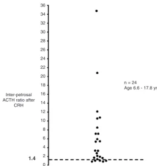
Bilateral Inferior Petrosal Sinus Sampling (BIPSS)
Due to the rarity of EAS in pediatric practice the pri-mary purpose of BIPSS is to attempt to demonstrate lat-eralisation of ACTH secretion. In experienced hands BIPSS has a predictive value of lateralisation of 75–80% in pediatric patients (12,13). BIPSS can be technically challenging and requires a highly specialised radiologist who regularly performs this procedure on adult patients. General anaesthesia may be required in very young chil-dren, which in turn may alter ACTH secretion. BIPSS has been used at our centre since 1987 and 24 patients have undergone this procedure without complications. Lateralisation, defined as an interpetrosal sinus ACTH
ratio of ≥ 1.4 after CRH, was observed in 79% of our
patients (figure 6). In contrast, a recent review of 94 patients found concordance between BIPSS lateralisa-tion and surgical finding in only 58% of patients, there-fore suggesting that the technique was non-essential (48). However, correlation increased to 70% following exclusion of 18 centrally located and 4 bilateral lesions.
TREATMENT
Primary adrenal lesions
Surgical excision is the first-line therapy for a cortical-secreting ACT. The optimum management of adreno-cortical carcinoma with metastasis is less clear. Mitotane therapy appears to be the treatment of choice. The benefits of additional cytotoxic agents have not been proven. Glucocorticoid replacement pre- and post surgery is required in view of inevitable contralateral adrenocortical suppression.
Definitive treatment of PPNAD or AIMAH is open or laparoscopic bilateral adrenelectomy. The aim of treatment is to prevent the detrimental effects of prolonged exposure to hypercortisolaemia. Metyrapone can be used as a temporary measure to normalise serum cortisol prior to surgery. Patients will require long-term glucocorticoid and mineralocorticoid replacement together with life-long endocrine follow-up.
Cushing’s disease
Medical therapies such as Metyrapone and Ketocona-zole to lower serum cortisol levels can be used as a short-term measure, but cannot be recommended as long-term therapy.
Transsphenoidal surgery with selective microadenomectomy
Transsphenoidal surgery with selective microade-nomectomy is now the accepted first line treatment of both adult and pediatric CD (49). The aim of the pro-cedure is selective removal of the microadenoma whilst preserving normal pituitary tissue. This is essential for the future development of the pediatric patient. Although a safe and effective procedure in pediatric CD, hypopituitarism is a known complication (50). At our centre, cure in adult CD is defined as a post-oper-ative serum cortisol level of < 50 nmol/L. Using this criteria, recurrence of CD following TSS is extremely uncommon (51). We use the same definition of cure for our pediatric CD patients. TSS can be technically difficult in children and even in the most experienced hands a certain number will fail to be cured and require second-line treatment. BIPSS performed pre-operatively can be beneficial. Since the introduction of BIPSS at our centre in 1987 the cure rate has improved to a current level of 72% (6,13).
Pituitary radiotherapy
Pituitary radiotherapy is an effective second-line treat-ment of pediatric CD following unsuccessful TSS and is the preferred option at our centre (52). Compared
Figure 6.Inter-petrosal ACTH ratio during BIPSS in 24 chil-dren. A ratio of ≥1.4 indicates lateralization and is seen in 79% of patients.
Inter-petrosal ACTH ratio after
CRH
n = 24 Age 6.6 - 17.8 yrs. 36
34 32 30 28 26 24 22 20 18 16 14 12 10 8 6 4 2
1.4
to adults, children respond more rapidly with a mean time of 0.8–1.0 yr when compared to 1.5–4 yr in adults (52). A decision is generally made within 2–4 weeks post TSS. At our centre, RT is administered in 25 fractions over 35 days delivering a total of 45 Gy. We have treated 12 patients in the last 25 yr with a cure rate of 92%, occurring at a mean interval of 0.83 yr (range 0.13–2.86 yr) following completion of RT. Long-term anterior pituitary function is often pre-served in children treated with pituitary RT (53).
Bilateral adrenelectomy and second TSS Bilateral adrenalectomy and repeat TSS can be used as secondary treatment options, but carry significant risks of Nelson’s syndrome and permanent pituitary defi-ciencies respectively (54,55). We have however used bilateral adrenalectomy as a last resort in a child too unwell to undergo TSS (14).
GROWTH IN PEDIATRIC CD FOLLOWING CURE BY TSS OR PITUITARY
RADIOTHERAPY
Pediatric CD is associated with a subnormal growth rate and short stature. Previous studies have shown poor catch-up growth and compromised final height, despite cure (56). This could be explained by GH defi-ciency (GHD) or the effects chronic hypercortiso-laemia on the growth plate (57). Our current approach is to test for GHD 3 months after TSS or completion of RT. If GHD is demonstrated (defined as a peak GH level of < 20 mU/L), GH therapy is ini-tiated a dose of 0.025 mg/kg/day. GnRH analogues maybe used in conjunction to delay epiphyseal fusion in pubertal patients. In this manner, acceptable final height close to or within the target range can be attained (58). GHD may persist for years after adult height is achieved, but is rarely severe enough to require treatment in adulthood (59).
BODY COMPOSITION AND BONE HEALTH IN PEDIATRIC CD
Body composition is more difficult to normalise fol-lowing cure. Many patients remain obese with high BMI SDS 3.9 yrs post cure (58). A long-term follow-up study of childhood and adolescent CD patients showed that total body fat was abnormally high 7 years after cure, with an elevated ratio of visceral to subcu-taneous fat (60). Bone mineral density (BMD) is often
but not universally reduced in children and adoles-cents with CD (61). We have reported that by nor-malizing pituitary function, a near normal bone min-eral density post-cure is attained.
COGNITIVE AND PSYCHOLOGICAL FOLLOW-UP IN PEDIATRIC CD
In adult CD there is a consensus that despite bio-chemical cure ‘quality of life’ may be impaired (15,62-64). Furthermore, chronic glucocorticoid exposure has been shown to result in loss of brain volume on radiological imaging. This appears to normalise with cure (64,65). Although, there is no evidence of long-term cognitive or psychological deficit in pediatric CD post cure, this has not been formally assessed.
SUMMARY
Pediatric CS is a challenging condition to manage and requires a multidisciplinary team of surgeons, endocri-nologists, biochemists, radiologists, oncologists and radiotherapists. Close collaboration with adult endocrine colleagues is vital. Weight gain with associat-ed growth failure is always abnormal in childhood and requires investigation. Even though, procedures such as BIPSS and TSS are technically more difficult in chil-dren, the overall prognosis of CD is good. Optimisa-tion of growth, puberty and body composiOptimisa-tion after cure of CS are important aspects to pediatric care. Pos-sible long-term complications in reproductive and neu-ropsychiatric function warrant further investigation.
REFERENCES
1. Pessanha TM, Campos JM, Barros AC, Pone MV, Garrido JR, Pone SM. Iatrogenic Cushing’s syndrome in a adolescent with AIDSs on Ritonavir and inhaled Fluticasone. Case report and literature review. AIDS 2007;21(4):529-32.
2. Volkl TM, Dorr HG. McCune-Albright syndrome: Clinical pic-ture and natural history in children and adolescents. J Pedi-atr Endocrinol Metab 2006;199(suppl 2):551-9.
3. Ribeiro RC, Figueiredo B. Childhood adrenocortical tumours.
Eur J Cancer 2004;40(8):1117-26.
4. Storr HL, Isidori AM, Monson JP, Besser GM, Grossman AB, Savage MO. Prepubertal Cushing’s disease is more common in males, but there is no increase in severity at diagnosis. J Clin Endocrinol Metab 2004;89(8):3818-20.
5. Newell-Price J, Trainer P, Besser M, Grossman A. The diag-nosis and differential diagdiag-nosis of Cushing’s syndrome and pseudo-Cushing’s states. Endocr Rev 1998;19(5):647-72. 6. Storr HL, Afshar F, Matson M, Sabin I, Davies KM, Evanson J,
7. Magiakou MA, Chrousos GP. Cushing’s syndrome in children and adolescents: Current diagnostic and therapeutic strate-gies. J Endocrinol Invest 2002;25(2):181-94.
8. Storr HL, Plowman PN, Carroll PV, Francois I, Krassas GE, Afshar F, et al. Clinical and endocrine responses to pituitary radiotherapy in pediatric Cushing’s disease: an Effective sec-ond-line treatment. J Clin Endocrinol Metab 2003; 88(1):34-7.
9. Newell-Price J, Bertagna X, Grossman AB, Nieman LK. Cush-ing’s syndrome. Lancet 2006;367(9522):1605-17.
10. Greening JE, Storr HL, McKenzie SA, Davies KM, Martin L, Grossman AB, et al. Linear growth and body mass index in pediatric patients with Cushing’s disease or simple obesity. J Endocrinol Invest 2006;29(10):885-7.
11. Weber A, Trainer PJ, Grossman AB, Afshar F, Medbak S, Perry LA, et al. Investigation, management and therapeutic outcome in 12 cases of childhood and adolescent Cushing’s syndrome. Clin Endocrinol (Oxf) 1995;43(1):19-28. 12. Magiakou MA, Mastorakos G, Oldfield EH, Gomez MT,
Dopp-man JL, Cutler GB, et al. Cushing’s syndrome in children and adolescents. Presentation, diagnosis, and therapy. N Engl J Med 1994;331(10):629-36.
13. Storr HL, Chan LF, Grossman AB, Savage MO. Paediatric Cushing’s syndrome: Epidemiology, investigation and thera-peutic advances. Trends Endocrinol Metab 2007; 18(4):167-74.
14. Greening JE, Brain CE, Perry LA, Mushtaq I, Sales Marques J, Grossman AB, et al. Efficient short-term control of hypercor-tisolaemia by low-dose etomidate in severe paediatric Cush-ing’s disease. Horm Res 2005;64(3):140-3.
15. Arnaldi G, Angeli A, Atkinson AB, Bertagna X, Cavagnini F, Chrousos GP, et al. Diagnosis and complications of Cushing’s syndrome: a Consensus statement. J Clin Endocrinol Metab 2003;88(12):5593-602.
16. Damiani D, Aguiar CH, Crivellaro CE, Galvão JA, Dichtcheken-ian V, SetDichtcheken-ian N. Pituitary macroadenoma and Cushing’s dis-ease in pediatric patients: Patient report and review of the lit-erature. J Pediatr Endocrinol Metab 1998;11(5):665-9. 17. Stratakis CA, Schussheim DH, Freedman SM, Keil MF, Pack
SD, Agarwal A et al. Pituitary macroadenoma in a 5-year-old: an Early expression of Multiple Endocrine Neoplasia type 1. J Clin Endocrinol Metab 2000;85(12):4776-80.
18. Peters CJ, Ahmed ML, Storr HL, Davies KM, Martin LJ, All-grove J, et al. Factors influencing skeletal maturation at diag-nosis of paediatric Cushing’s disease. Horm Res 2007; 68(5):231-5.
19. Dupuis CC, Storr HL, Perry LA, Ho JT, Ahmed L, Ong KK, et al. Abnormal puberty in paediatric Cushing’s disease: Rela-tionship with adrenal androgen, sex hormone binding globu-lin and gonadotrophin concentrations. Clin Endocrinol (Oxf) 2007;66(6):838-43.
20. Isidori AM, Kaltsas GA, Pozza C, Frajese V, Newell-Price J, Reznek RH, et al. The ectopic adrenocorticotropin syndrome: Clinical features, diagnosis, management, and long-term fol-low-up. J Clin Endocrinol Metab 2006;91(2):371-7. 21. Ilias I, Torpy DJ, Pacak K, Mullen N, Wesley RA, Nieman LK.
Cushing’s syndrome due to ectopic corticotropin secretion: Twenty years’ experience at the National Institutes of Health.
J Clin Endocrinol Metab 2005;90(8):4955-62.
22. Hannah J, Lippe B, Lai-Goldman M, Bhuta S. Oncocytic carci-noid of the kidney associated with periodic Cushing’s syn-drome. Cancer 1988;61(10):2136-40.
23. Amano S, Hazama F, Haebara H, Tsurusawa M, Kaito H. Ectopic ACTH-MSH producing carcinoid tumor with multiple endocrine hyperplasia in a child. Acta Pathol Jpn 1978;28(5):721-30.
24. Espinasse-Holder M, Defachelles AS, Weill J, De Keyzer Y, de Lasalle EM, Nelken B. Paraneoplastic Cushing syndrome due to adrenal neuroblastoma. Med Pediatr Oncol 2000; 34(3):231-3.
25. Hsiao JC, Yang CP, Lin CJ, Chuen H. Ectopic ACTH syndrome due to clear cell sarcoma of the kidney. Child Nephrol Urol 1991;11(2):103-6.
26. Hinnie J, Gray CE, McNicol AM, Carter R, Thomson JA, White A, et al. Cushing’s syndrome in a 16 year old girl due to ectopic ACTH precursor production from a pancreatic tumour. Clin Endocrinol (Oxf) 2000;53(4):539-40. 27. Thomas RJ, Sen S, Zachariah N, Mammen KE, Raghupathy P,
Seshadri MS, et al. Wilms’ tumor presenting as Cushing’s syndrome. Pediatr Surg Int 1998;13(4):293-4.
28. Ribeiro RC, Sandrini F, Figueiredo B, Zambetti GP, Michalkiewicz E, Lafferty AR, et al. An inherited P53 mutation that contributes in a tissue-specific manner to pediatric adrenal cortical carcinoma. Proc Natl Acad Sci U S A 2001;98(16):9330-5.
29. Michalkiewicz E, Sandrini R, Figueiredo B, Miranda EC, Caran E, Oliveira-Filho AG, et al. Clinical and outcome characteris-tics of children with adrenocortical tumors: a Report from the International Pediatric Adrenocortical Tumor Registry. J Clin Oncol 2004;22(5):838-45.
30. Shenoy BV, Carpenter PC, Carney JA. Bilateral primary pig-mented nodular adrenocortical disease. Rare cause of the Cush-ing syndrome. Am J Surg Pathol 1984; 8(5):335-44. 31. Carney JA, Gordon H, Carpenter PC, Shenoy BV, Go VL. The
complex of myxomas, spotty pigmentation, and endocrine overactivity. Medicine (Baltimore) 1985;64(4):270-83. 32. Storr HL, Mitchell H, Swords FM, Main KM, Hindmarsh PC,
Betts PR, et al. Clinical features, diagnosis, treatment and molecular studies in paediatric Cushing’s syndrome due to primary nodular adrenocortical hyperplasia. Clin Endocrinol (Oxf) 2004;61(5):553-9.
33. Gunther DF, Bourdeau I, Matyakhina L, Cassarino D, Kleiner DE, Griffin K, et al. Cyclical Cushing syndrome presenting in infancy: An early form of primary pigmented nodular adreno-cortical disease, or a new entity? J Clin Endocrinol Metab 2004;89(7):3173-82.
34. Stratakis CA, Kirschner LS. Clinical and genetic analysis of primary bilateral adrenal diseases (micro- and macronodular disease) leading to Cushing syndrome. Horm Metab Res 1998;30(6-7):456-63.
35. Sarlis NJ, Chrousos GP, Doppman JL, Carney JA, Stratakis CA. Primary pigmented nodular adrenocortical disease: Reevaluation of a patient with Carney complex 27 years after unilateral adrenalectomy. J Clin Endocrinol Metab 1997; 82(4):1274-8.
36. Kirschner LS, Sandrini F, Monbo J, Lin JP, Carney JA, Stratakis CA. Genetic heterogeneity and spectrum of muta-tions of the PRKAR1A gene in patients with the Carney com-plex. Hum Mol Genet 2000;9(20):3037-46.
37. Groussin L, Jullian E, Perlemoine K, Louvel A, Leheup B, Luton JP, et al. Mutations of the PRKAR1A gene in Cushing’s syndrome due to sporadic primary pigmented nodular adrenocortical disease. J Clin Endocrinol Metab 2002; 87(9):4324-9.
38. Groussin L, Horvath A, Jullian E, Boikos S, Rene-Corail F, Lefebvre H, et al. A PRKAR1A mutation associated with primary pigmented nodular adrenocortical disease in 12 kindreds. J Clin Endocrinol Metab 2006; 91(5):1943-9.
39. Bourdeau I. Clinical and molecular genetic studies of bilater-al adrenbilater-al hyperplasias. Endocr Res 2004;30(4):575-83. 40. Lieberman SA, Eccleshall TR, Feldman D. ACTH-independent
massive bilateral adrenal disease (AIMBAD): a Subtype of Cushing’s syndrome with major diagnostic and therapeutic implications. Eur J Endocrinol 1994;131(1):67-73. 41. Lumbroso S, Paris F, Sultan C. McCune-Albright syndrome:
Molecular genetics. J Pediatr Endocrinol Metab 2002; 15(suppl 3):875-82.
42. Kirk JM, Brain CE, Carson DJ, Hyde JC, Grant DB. Cushing’s syndrome caused by nodular adrenal hyperplasia in children with McCune-Albright syndrome. J Pediatr 1999; 134(6):789-92.
44. Yaneva M, Mosnier-Pudar H, Dugue M, Grabar S, Fulla Y, Bertagna X. Midnight salivary cortisol for the initial diagnosis of Cushing’s syndrome of various causes. J Clin Endocrinol Metab 2004;89(7):3345-51.
45. Peters CJ, Storr HL, Grossman AB, Savage MO. The role of corticotrophin-releasing hormone in the diagnosis of Cush-ing’s syndrome. Eur J Endocrinol 2006;155(suppl 1):S93-8. 46. Dias R, Storr HL, Perry LA, Isidori AM, Grossman AB, Savage MO. The discriminatory value of the low-dose dexametha-sone suppression test in the investigation of paediatric Cush-ing’s syndrome. Horm Res 2006;65(3):159-62.
47. Doppman JL. Problems in endocrinologic imaging.
Endocrinol Metab Clin North Am 1997;26(4):973-91. 48. Batista D, Gennari M, Riar J, Chang R, Keil MF, Oldfield EH, et
al. An assessment of petrosal sinus sampling for localization of pituitary microadenomas in children with Cushing disease.
J Clin Endocrinol Metab 2006;91(1):221-4.
49. Joshi SM, Hewitt RJ, Storr HL, Rezajooi K, Ellamushi H, Grossman AB, et al. Cushing’s disease in children and ado-lescents: 20 Years of experience in a single neurosurgical center. Neurosurgery 2005;57(2):281-5.
50. Massoud AF, Powell M, Williams RA, Hindmarsh PC, Brook CG. Transsphenoidal surgery for pituitary tumours. Arch Dis Child 1997;76(5):398-404.
51. Trainer PJ, Lawrie HS, Verhelst J, Howlett TA, Lowe DG, Grossman AB, et al. Transsphenoidal resection in Cushing’s disease: Undetectable serum cortisol as the definition of suc-cessful treatment. Clin Endocrinol (Oxf) 1993;38(1):73-8. 52. Storr HL, Plowman PN, Carroll PV, Francois I, Krassas GE,
Afshar F, et al. Clinical and endocrine responses to pituitary radiotherapy in pediatric Cushing’s disease: an Effective sec-ond-line treatment. J Clin Endocrinol Metab 2003; 88(1):34-7.
53. Chan LF, Storr HL, Plowman PN, Perry LA, Besser GM, Gross-man AB, et al. Long-term anterior pituitary function in patients with paediatric Cushing’s disease treated with pitu-itary radiotherapy. Eur J Endocrinol 2007;156(4):477-82. 54. Leinung MC, Kane LA, Scheithauer BW, Carpenter PC, Laws
ER Jr, Zimmerman D. Long term follow-up of transsphe-noidal surgery for the treatment of Cushing’s disease in child-hood. J Clin Endocrinol Metab 1995;80(8):2475-9. 55. McArthur RG, Hayles AB, Salassa RM. Childhood Cushing
disease: Results of bilateral adrenalectomy. J Pediatr 1979; 95(2):214-9.
56. Magiakou MA, Mastorakos G, Chrousos GP. Final stature in patients with endogenous Cushing’s syndrome. J Clin Endocrinol Metab 1994;79(4):1082-5.
57. Lebrethon MC, Grossman AB, Afshar F, Plowman PN, Besser GM, et al. Linear growth and final height after treatment for Cushing’s disease in childhood. J Clin Endocrinol Metab 2000;85(9):3262-5.
58. Davies JH, Storr HL, Davies K, Monson JP, Besser GM, Afshar F, et al. Final adult height and body mass index after cure of paediatric Cushing’s disease. Clin Endocrinol (Oxf) 2005;62(4):466-72.
59. Carroll PV, Monson JP, Grossman AB, Besser GM, Plowman PN, Afshar F, et al. Successful treatment of childhood-onset Cushing’s disease is associated with persistent reduction in growth hormone secretion. Clin Endocrinol (Oxf) 2004;60(2):169-74.
60. Leong GM, Abad V, Charmandari E, Reynolds JC, Hill S, Chrousos GP, et al. Effects of child- and adolescent-onset endogenous Cushing syndrome on bone mass, body compo-sition, and growth: a 7-Year prospective study into young adulthood. J Bone Miner.Res 2007;22(1):110-8.
61. Scommegna S, Greening JP, Storr HL, Davies KM, Shaw NJ, Monson JP, et al. Bone mineral density at diagnosis and fol-lowing successful treatment of pediatric Cushing’s disease. J Endocrinol Invest 2005;28(3):231-5.
62. Dorn LD, Burgess ES, Friedman TC, Dubbert B, Gold PW, Chrousos GP. The longitudinal course of psychopathology in Cushing’s syndrome after correction of hypercortisolism. J Clin Endocrinol Metab 1997;82(3):912-9.
63. Sonino N, Fava GA. Psychiatric disorders associated with Cushing’s syndrome. Epidemiology, pathophysiology and treatment. CNS Drugs 2001;15(5):361-73.
64. Forget H, Lacroix A, Cohen H. Persistent cognitive impair-ment following surgical treatimpair-ment of Cushing’s syndrome.
Psychoneuroendocrinology 2002;27(3):367-83.
65. Bourdeau I, Bard C, Noel B, Leclerc I, Cordeau MP, Belair M, et al. Loss of brain volume in endogenous Cushing’s syn-drome and its reversibility after correction of hypercorti-solism. J Clin Endocrinol Metab 2002;87(5):1949-54.
Address for correspondence:
Li Chan