ABSTRACT
Adrenocorticotropin hormone (ACTH)-dependent Cushing’s syndrome is most often due to a pituitary corticotroph adenoma, with ectopic ACTH-secreting tumors representing approximately 15% of cases. Biochemical and radiological techniques have been established to help distinguish between these two enti-ties, and thus aid in the localization of the neoplastic lesion for surgical resec-tion. The test that offers the highest sensitivity and specificity is bilateral infe-rior petrosal sinus sampling (BIPSS). BIPSS is an interventional radiology pro-cedure in which ACTH levels obtained from venous drainage very near the pitu-itary gland are compared to peripheral blood levels before and after corti-cotropin hormone (CRH) stimulation. A gradient between these two locations indicates pituitary Cushing’s, whereas the absence of a gradient suggests ectopic Cushing’s. Accurate BIPSS results require hypercortisolemia to sup-press normal corticotroph ACTH production and hypercortisolemia at the time of the BIPSS to assure excessive ACTH secretion. In some cases, intrapituitary gradients from side-to-side can be helpful to localize small corticotroph adeno-mas within the sella. BIPSS has rare complications and is considered safe when performed at centers with experience in this specialized technique. (Arq Bras Endocrinol Metab 2007;51/8:1329-1338)
Keywords:IPSS; BIPSS; Cushing’s; Ectopic; Pituitary; Bilateral inferior petros-al sinus sampling
RESUMO
O Papel da Amostragem Bilateral do Seio Petroso Inferior no Diagnóstico da Síndrome de Cushing.
A síndrome de Cushing (SC) ACTH-dependente é mais freqüentemente devida a um adenoma corticotrófico da hipófise, com os tumores ectópicos secretores de ACTH representando aproximadamente 15% dos casos. Técnicas bioquí-micas e radiológicas foram estabelecidas para permitir a distinção entre essas duas entidades e, assim, auxiliar na localização da lesão neoplásica para ressecção cirúrgica. O teste que oferece a mais alta sensibilidade e espe-cificidade é a coleta bilateral de amostras de sangue do seio petroso inferior (BIPSS). BIPSS é um procedimento de intervenção radiológica no qual os ní-veis de ACTH obtidos da drenagem venosa bem próxima da hipófise são comparados com os níveis sanguíneos periféricos antes e após estímulo com corticorrelina (CRH). Um gradiente entre essas duas localizações indica SC hipofisário, enquanto a ausência de gradiente sugere SC ectópica. Resultados acurados na BIPSS requerem a presença de hipercortisolemia e que ela suprima normalmente a produção de ACTH pelo corticotrofos por ocasião da BIPSS para garantir a secreção excessiva de ACTH. Em alguns casos, os gra-dientes intra-hipofisários de um lado para outro podem ser úteis na localização de pequenos adenomas corticotróficos no interior da sela. A BIPSS raramente apresenta complicações, sendo considerada segura quando realizada em centros com experiência nessa técnica especializada. (Arq Bras Endocrinol Metab 2007;51/8:1329-1338)
Descritores: IPSS; BIPSS; Síndrome de Cushing; ACTH ectópico; Hipófise; Coleta bilateral do seio petroso inferior
Recebido em 15/09/07 Aceito em 22/09/07
atualização
Neuroendocrine Unit,
Massachusetts General Hospital, Boston, Massachusetts, USA. ANDREAUTZ
T
HE EVALUATION OF A PATIENT WITH Cushing’ssyndrome requires a systematic approach. Follow-ing the establishment of pathologic hypercortisolemia and adrenocorticotropin hormone (ACTH) depen-dence, the source of ACTH excess is determined. This is essential because cure depends on surgical removal of the tumor. Neoplastic causes of excess ACTH include, in descending order of prevalence, pituitary corticotroph adenomas, ectopic ACTH-secreting tumors, or very rarely ectopic corticotropin releasing hormone (CRH)-secreting tumors. Methods to distin-guish between these entities have evolved and will be discussed below. Table 1 lists the biochemical tests that have been most carefully examined for their use-fulness in distinguishing between pituitary and ectopic ACTH hypersecretion.
The tests shown in table 1 rely on the assump-tion that although they are autonomous neoplastic lesions, pituitary corticotroph adenomas retain at least partial responsiveness to suppression by elevated glu-cocorticoid levels or stimulation by CRH or desmo-pressin, and conversely that ectopic ACTH-producing tumors do not have such regulation. While this is gen-erally true, variability in the responsiveness of both types of tumors underlies the lack of 100% accuracy with these tests. For instance, some corticotroph ade-nomas, particularly macroadeade-nomas, are not sup-pressed by high-dose glucocorticoids (1), and some are not stimulated by CRH; whereas some ectopic ACTH-secreting tumors show suppression by gluco-corticoids and/or stimulation by CRH (2).
Pituitary imaging does have a role in determining the cause of ACTH hypersecretion; however, certain caveats must be considered. Incidental pituitary lesions are relatively common in the general population (3) and thus a small lesion does not assure a pituitary source of ACTH. Conversely, the absence of a lesion on pituitary imaging does not rule-out a pituitary source, as many corticotroph adenomas are very small and undetectable by MRI (4). The size at which a pituitary lesion is near-ly certain to be a corticotroph adenoma, rather than an “incidentaloma,” is a matter of controversy. While the presence of a pituitary macroadenoma (i.e.lesion ≥ 10
mm) in the setting of ACTH-dependent Cushing’s syn-drome likely represents pituitary Cushing’s, some sug-gest that lowering the lesion size cut-off to > 6 mm pre-vents unnecessary BIPSS procedures without a signifi-cant sacrifice in specificity (5).
The characteristics of the tests above have been previously well described in the differential diagnosis of Cushing’s syndrome. High dose dexamethasone sup-pression testing continues to be used frequently to dis-tinguish between ectopic and pituitary Cushing’s; how-ever reports of low sensitivity (65–100%) and specifici-ty (60–100%), make this test insufficient as a sole means to indicate surgery (2,6). The peripheral CRH stimulation test has been reported to have a sensitivity that ranges from 70–93% and a specificity of 95–100% (7). BIPSS series show the following ranges: sensitivity of 88–100% and specificity 67–100% (2,7,8).
The choice of testing protocol used to distin-guish pituitary from ectopic Cushing’s varies by institu-tion and depends on many factors including accessibili-ty of the specialized BIPSS interventional radiology technique, financial considerations, availability of CRH, and sensitive MRI radiology equipment. Some centers support a step-wise approach by performing the less invasive high dose dexamethasone suppression and peripheral CRH stimulation tests first. It has been reported that concordant positive results for both of these tests predicts pituitary Cushing’s with 98% accura-cy. However, 18 to 65% of pituitary Cushing’s patients do not have concordant results and additional testing, usually BIPSS, is the next step for diagnosis (9). Due to the relatively high percentage of patients that will not be accurately diagnosed with these less invasive procedures, some centers proceed directly to BIPSS after the estab-lishment of ACTH-dependent hypercortisolemia.
BIPSS RATIONALE
ACTH-dependent Cushing’s syndrome is defined as the presence of normal or elevated levels of ACTH in the setting of hypercortisolemia. The presence of detectable ACTH in the systemic circulation does not
Table 1.Biochemical testing in the differential diagnosis of ACTH-dependent Cushing’s syndrome. High dose two-day dexamethasone suppression test (HDDST)
Eight milligram overnight dexamethasone suppression Metyrapone stimulation
Peripheral CRH stimulation Desmopressin stimulation
provide any information on the location of the source of the excess ACTH secretion. As the pituitary sella is the most likely location for such a source, it was pos-tulated that the venous drainage from the pituitary, in the presence of a corticotroph adenoma, would likely have a higher ACTH concentration than the systemic circulation.
The first efforts to measure pituitary venous effluent were reported by Corrigan et al. (10) and sev-eral other groups contributed significantly to estab-lishing this technique in the differential diagnosis of Cushing’s disease (11,12). Initial investigations com-pared unilateral inferior petrosal ACTH levels to peripheral blood levels. However, it was later estab-lished that ACTH levels in venous drainage from the pituitary may be asymmetric, either due to corti-cotroph adenoma location or anatomical variation. Pituitary venous drainage is usually ipsilateral, so that venous effluent on the contralateral side, relative to the corticotroph adenoma, may not have high ACTH values. In addition, some patients have asymmetric venous anatomy. Thus, simultaneous measurements from both inferior petrosal sinuses are necessary to avoid a falsely negative evaluation in the setting of a pituitary corticotroph adenoma (12). The technique has therefore been termed bilateral inferior petrosal sinus sampling (BIPSS).
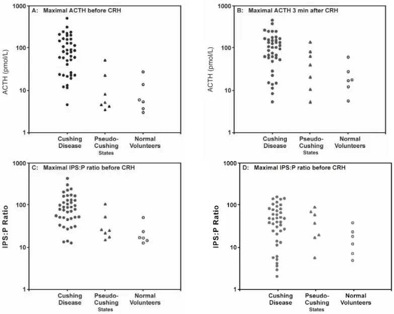
The success of BIPSS is dependent on the sup-pression of the normal corticotrophs in the pituitary gland by the longstanding hypercortisolemia associat-ed with Cushing’s syndrome. This prevents basal ACTH release or CRH-stimulated ACTH release by normal pituitary corticotrophs. Suppression of the normal cells ensures that any measurable plasma ACTH in the samples has been secreted by neoplastic tissue. The magnitude and duration of hypersolemia required to fully suppress normal corti-cotrophs is unknown. In normal individuals without hypercortisolemia, ACTH levels may be high due to normal pulsatile corticotroph ACTH release, and nor-mal corticotrophs respond to stimulation by CRH. This leads to absolute ACTH levels and central to peripheral ACTH ratios, obtained via BIPSS, in the same range as seen in true pituitary Cushing’s disease (figure 1) (13). Thus, in the absence of sustained hypercortisolemia (to suppress normal corticotroph function), a central to peripheral ACTH gradient, identical to that seen in pituitary Cushing’s, may occur. This means that the diagnosis of Cushing’s syn-drome (i.e.endogenous cortisol excess) must be estab-lished before performing BIPSS. In addition, the patient must be hypercortisolemic at the time of the
BIPSS and not controlled with medical therapy or adrenalectomy. Confirmation of hypercortisolemia with a 24-hour urine cortisol or late-night salivary cor-tisol measurement performed immediately prior to the BIPSS procedure is important.
BIPSS ANATOMY
Miller and Doppman (14) provide a detailed description of the venous anatomy and technique of bilateral inferior petrosal sinus sampling (figure 2). In the majority of individuals, each inferior petrosal sinus narrows to become a vein (approximately 2 mm in diameter) that empties directly into the ipsi-lateral internal jugular vein. In approximately 25% of people, the drainage of the inferior petrosal sinus is a plexus of channels emptying into the internal jugu-lar vein. In a small percentage of individuals (0.6–7%), there is no connection between the inferi-or petrosal sinus and the internal jugular vein, mak-ing standard samplmak-ing impossible (14,15). In approximately 60% of individuals, pituitary venous drainage is symmetric (16), with the majority of the venous effluent from each side of the pituitary drain-ing into the ipsilateral inferior petrosal sinus (14). Thus, the location of a corticotroph tumor within the sella can affect the levels of ACTH in pituitary venous drainage on that side. In some cases, this can assist in lateralization of the tumor and is the ratio-nale for calculating an interpetrosal gradient between left and right sides. More importantly, this explains the necessity to catheterize both inferior petrosal sinuses, as the contralateral side, relative to the tumor, may have ACTH levels similar to the periphery. This could mistakenly lead to misclassifi-cation as an ectopic ACTH source if only the con-tralateral side was sampled.
BIPSS PROCEDURE
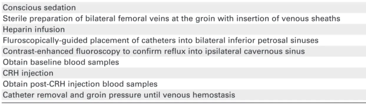
Table 2 outlines the basic procedure for performing BIPSS. The details of catheter choice and radiograph-ic technique are beyond the scope of the current review and interested readers are referred to the inter-ventional radiology literature (14).
Figure 1.Maximal ACTH concentrations (A)pre-CRH and (B)post-CRH; and central to peripheral ACTH ratios (IPS:P ratio) (C) pre-CRH and (D)post-CRH during BIPSS in patients with Cushing’s disease, pseudo-Cushing’s, and normal volunteers. ACTH (pg/ml) = ACTH (pmol/L) x 4.55. [Modified from (13) with permission]
Figure 2.Schematic views of the pituitary venous drainage. (A)Sagittal, (B)Coronal CS: cavernous sinus, IPS: inferior petros-al sinus, SPS: superior petrospetros-al sinus, JB: jugular bulb, SS: sigmoid sinus, IJV: internpetros-al jugular vein, FV: facipetros-al vein, PV: pha-ryngeal vein. [Reproduced from (14,37) with permission]
A B
AC
T
H
(p
mo
l/
L
)
AC
T
H
(p
mo
l/
L
)
States States
States
CS
JB
PV
FV
IJV SS
CAVERNOUS SINUS
JUGULAR VEIN CONFLUENT
PITUITARY VEINS
INFERIOR PETROSAL SINUS
IPS SPS
on the opposite side. Catheters are then advanced from the groin to each inferior petrosal sinus. Usually heparin is infused before catheters are advanced. Cor-rect catheter placement is confirmed by injection of contrast dye and fluoroscopic imaging assessment. Occasionally, it is not possible to perform BIPSS via a femoral approach (due to inferior vena cava filters, thrombosis, aberrant anatomy, or inability to cross the internal jugular valve) and other access, such direct internal jugular access is used.
After the correct placement of catheters, simulta-neous blood samples are obtained from each of the three ports (peripheral, left inferior petrosal sinus, and right inferior petrosal sinus). After obtaining baseline samples (usually 2 sets of baseline samples are obtained), CRH is injected as a bolus peripherally at 1 mcg/kg (maximum 100 mcg) and post-CRH samples are obtained from each port at 3, 5, 10, and 15 minutes. Blood samples are immediately placed into lavender-top EDTA-containing tubes and placed on ice. Processing of the blood, including centrifugation and plasma decantation should occur within one hour and samples are analyzed immediately or frozen until ACTH assay.
Many corticotroph adenomas are susceptible to stimulation by CRH, and this has been exploited to increase the sensitivity of BIPSS. It should be noted that two different forms of CRH, ovine CRH (oCRH) and human CRH (hCRH), have been used in the var-ious reported studies of CRH stimulation. A report by Nieman et al. compared the responses in 15 patients with Cushing’s disease following stimulation with ovine and human CRH and found the peripheral ACTH and cortisol responses to ovine CRH were sig-nificantly higher than with human CRH (17).
BIPSS DATA INTERPRETATION
Simultaneous measurements of ACTH levels in the systemic circulation (peripheral) and in each inferior
petrosal sinus (central) are used for calculations. Ratios are calculated at baseline and at each time point after CRH injection, between the left IPS and periphery and between the right IPS and periphery. Of note, the inferior petrosal sinus time point with the highest cen-tral to peripheral ACTH ratio is used for interpreta-tion. Based on the largest published series of BIPSS data, established criteria for a central (pituitary) source of excess ACTH are a pre-CRH stimulation central:peripheral ratio ≥2.0 OR a post-CRH stimula-tion ratio ≥ 3.0 (18). By convention, a “positive” BIPSS represents centralization, i.e. a pituitary source of ACTH excess; whereas a “negative” BIPSS repre-sents an ectopic source of ACTH. Examples of typical BIPSS data in pituitary and ectopic Cushing’s are shown in table 3.
BIPSS TESTING CHARACTERISTICS
The initial studies of BIPSS suggested near-perfect sensitivity and specificity. As further reports of BIPSS series have been published, diagnostic errors have surfaced. Many of these suggest that errors may be attributed to an inability to access the inferior petrosal sinus, either due to technique or individual anatomy. Reports of diagnostic accuracy may not include data from sampling that has been deemed inadequate or may exclude patients in whom the final diagnosis is unknown. This must be considered when interpreting the resulting data and will lead to lower actual sensitivity of BIPSS in clinical practice. As data continue to accumulate on the accuracy of BIPSS, most studies suggest a high degree of speci-ficity with lesser sensitivity. BIPSS series show the following ranges: sensitivity of 88–100% and speci-ficity 67–100% (2,7,8).
As the pre-test probability of a pituitary source of Cushing’s syndrome is high at approximately 70% (7), small decrements in sensitivity will significantly Table 2.Basic BIPSS procedure.
Conscious sedation
Sterile preparation of bilateral femoral veins at the groin with insertion of venous sheaths Heparin infusion
Fluroscopically-guided placement of catheters into bilateral inferior petrosal sinuses Contrast-enhanced fluoroscopy to confirm reflux into ipsilateral cavernous sinus Obtain baseline blood samples
CRH injection
Obtain post-CRH injection blood samples
increase the number of pituitary Cushing’s patients that are inappropriately classified as ectopic Cush-ing’s. A report by Swearingen et al. showed in 9 patients with negative BIPSS results, that transsphe-noidal surgery revealed an ACTH secreting corti-cotroph adenoma. Most had suggestive, but not definitive, pituitary MRI findings and no other source on detailed body imaging (8). Thus, following a neg-ative BIPSS and a comprehensive radiologic work-up that does not reveal a source of ectopic ACTH pro-duction, consideration should be given to transsphe-noidal exploration by a highly experienced pituitary surgeon, particularly in the setting of suggestive pitu-itary MRI findings.
One report highlighted two sources of false positive BIPSS results. The rare entity of ectopic CRH production may confound BIPSS testing, as the data will suggest a pituitary source due to pituitary corti-cotroph hyperplasia (8). A peripheral CRH level mea-surement may be helpful in patients with the appear-ance of hyperplasia on pituitary MRI or in those with persistent hypercortisolemia following transsphenoidal resection and no evidence of adenoma on pathologic assessment.
A second potential cause of decreased specifici-ty is performance of BIPSS in individuals with an adrenal source of cortisol excess. The presence of adrenal nodularity on abdominal imaging may be due to primary adrenal disease or stimulation by elevated ACTH levels. Although ACTH is usually completely suppressed in adrenal Cushing’s, levels in the low nor-mal range may be present in some patients with adren-al Cushing’s and this can incorrectly indicate that the hypercortisolemia is ACTH-dependent. Peripheral
CRH testing and high dose dexamethasone suppres-sion tests have been utilized in this situation. In the setting of adrenal Cushing’s there is a lack of ACTH or cortisol response to CRH and lack of cortisol sup-pression by dexamethasone (2,19). BIPSS in the set-ting of adrenal Cushing’s shows low absolute ACTH levels but may reach criteria for pituitary Cushing’s due to the low value in the denominator of the ratio calculation, producing a false positive result. For example, a maximum central ACTH value of 10 pg/mL, with a peripheral value of 1 pg/mL was noted in a patient with adrenal Cushing’s. This yielded a ratio of 10, thereby misclassifying the patient as having pituitary Cushing’s (8).
BIPSS FOR CORTICOTROPH ADENOMA LATERALIZATION
Since corticotroph adenomas may be small, tech-niques to pinpoint the lesion within the sella can be helpful for resection guidance. In some cases, MRI findings are unequivocal, such as in the case of macroadenomas. However, in the absence of an obvi-ous lesion on MRI or during surgical exploration, additional information gained from the BIPSS may be useful. Calculation of a side-to-side IPS ratio has been employed to predict the tumor site within the pitu-itary gland. An interpetrosal gradient between right and left inferior petrosal sinus ACTH levels before and/or after CRH stimulation can suggest the side where the adenoma might reside. Criteria for lateral-ization have been proposed, with a side-to-side gradi-ent ≥ 1.4 before or after CRH stimulation indicating Table 3.Examples of BIPSS results data represent ACTH levels in pg/ml units.
Peripheral Left IPS Right IPS Highest Lateralization IPS:peripheral ratio
ratio Pituitary Cushing’s
Basal 32 110 74 110/32 =3.44
3 min post-CRH 34 563 110 563/110 =5.12
5 min post-CRH 39 630 176
10 min post-CRH 43 725 210 725/43 =18.13
15 min post-CRH 54 304 280
Ectopic Cushing’s
Basal 33 41 38 41/33 =1.24
3 min post-CRH 34 51 35 51/34 =1.50
5 min post-CRH 32 46 36
10 min post-CRH 30 44 38
a lateral lesion and a gradient < 1.4 indicating a mid-line lesion, with approximately 70% accuracy. The highest side-to-side gradient is chosen for this calcu-lation (18). However, the usefulness of lateralization prediction has been questioned, with accuracies rang-ing from 50 to 100% (2). Incorrect lateralization may be due to asymmetric pituitary venous drainage, which may be found in up to 40% of individuals (16,20). However, if venous drainage has been shown to be symmetric with venous angiography and there is an interpetrosal ACTH gradient without an obvious lesion on MRI or during surgical resection, hemihy-pophysectomy on the side with higher ACTH may be indicated.
BIPSS CONFOUNDERS
As described above, an accurate BIPSS requires sup-pression of normal pituitary corticotroph ACTH pro-duction. Table 4 lists situations in which there may be inadequate suppression of ACTH from normal corti-cotrophs. The inability to sample adequately from the inferior petrosal sinus, either due to technical difficul-ty or due to aberrant venous drainage (21), can pre-vent the correct prediction that the ACTH-secreting tumor is in the pituitary gland (table 4).
BIPSS RISKS
Risks of BIPSS are uncommon; however, there are potential adverse events. The most common compli-cation is groin hematoma, occurring in 3–4% (14).
As the BIPSS procedure involves infusion of iodinat-ed contrast material, there is a risk of acute renal insufficiency that may be exacerbated by pre-existing renal insufficiency or hypovolemia, so measurement of BUN and creatinine is recommended prior to the procedure. Heparin is frequently infused following the insertion of sampling catheters to prophylax against cavernous sinus thrombosis. Thus, pre-pro-cedure assessment of the coagulation system, platelets, and hematocrit is suggested. Additional concerns include radiation exposure during fluo-roscopy, heparin-induced thrombocytopenia, and catheter-related infections. The placement of the catheter may cause discomfort, headache, ear pain, or hearing noises. Explaining to the patient in advance that these may occur can prevent anxiety during the procedure.
Very rare complications have included deep venous thrombosis and pulmonary thromboem-bolism (22,23), pontocerebellar junction stroke (24), brain stem injury (25), cranial nerve palsy (26), and venous subarachnoid hemorrhage and obstruc-tive hydrocephalus (27). Neurologic complications may be due to variant venous anatomy or specific catheter use and at least one report suggests that adverse sequelae may be minimized or averted if the BIPSS is aborted upon development of new neuro-logic signs or symptoms (25).
ALTERNATIVE BIPSS STRATEGIES
While sampling from the inferior petrosal sinus pro-vides the highest sensitivity, it is a technically chal-lenging procedure. At institutions without experience
Table 4.Pitfalls in BIPSS.
False positives (BIPSS predicts pituitary Cushing’s in the absence of a corticotroph adenoma):
Lack of suppression of normal corticotrophs: Cyclic Cushing’s syndrome
Cortisol blocking drugs (ketoconazole, metyrapone, mitotane, aminoglutethamide) Bilateral adrenalectomy
Normal individuals with factitious hypercortisolemia Pseudo-Cushing’s syndrome
Adrenal Cushing’s syndrome (with mild hypercortisolemia) Ectopic CRH-secreting tumor
False negative (BIPSS predicts non-pituitary Cushing’s in the presence of a corticotroph adenoma):
Aberrant pituitary venous drainage
in this procedure, it may be reasonable to proceed with advancing femoral catheters only as far as the internal jugular veins, known as jugular venous sampling (JVS). A study compared the two procedures, per-formed in the same subjects on separate days, in a pop-ulation of patients with Cushing’s syndrome (n = 74 for pituitary source and n = 11 with ectopic source). Different central to peripheral ACTH ratio cut-off val-ues were used. For BIPSS, ratios of > 1.6 pre-CRH and > 2.5 post-CRH were classified as pituitary Cush-ing’s. For JVS, ratios of > 1.7 pre-CRH and > 2.0 post-CRH were classified as pituitary Cushing’s. Using these criteria, the test was 100% specific for both tech-niques, 94% sensitive for BIPSS, and 83% sensitive for JVS (9). A similar study using standard cut-offs of ≥ 2.0 pre-CRH and ≥ 3.0 post-CRH showed sensitivi-ties of 94% and 81% for BIPSS and JVS, respectively (28). Thus JVS may be a reasonable first step in cen-ters without expertise in BIPSS. The lower sensitivity of JVS suggests that results indicating an ectopic source may be incorrect; therefore follow-up options include: referral for BIPSS or performance of a detailed radiologic assessment for an ectopic source. In the absence of a demonstrable ectopic source, exploratory pituitary surgery may be indicated if an expert pituitary surgeon is available.
Conversely some centers have reported improved accuracy and intra-sellar localization by sampling more proximally, from bilateral cavernous sinuses (29,30). However, increased risk of cranial nerve palsy with cavernous sinus sampling has been reported (20) and this procedure is not performed at most centers.
The most likely cause of a false-negative BIPSS is the inability to sample bilateral pituitary venous effluent adequately. In the setting of non-centralizing BIPSS results, verification of adequate sampling is valuable. This is generally obtained by reviewing the radiologic films to confirm appropriate catheter place-ment and standard venous anatomy via angiography. Biochemical confirmation of adequate sampling may be possible by assessing concurrent levels of other anterior pituitary hormones. For example, during BIPSS a central prolactin to peripheral prolactin ratio close to 1.0 may suggest inadequate sampling and if the ACTH results suggest an ectopic location, it may represent a false-negative result with consideration given to transsphenoidal exploration (31). However, previous studies of basal anterior pituitary hormone secretion did not suggest that normalization improved sensitivity of BIPSS testing (32). Further study of this approach would be valuable.
Methods for improving the sensitivity of BIPSS have been explored. Injection of desmopressin (10 mcg intravenous) alone (33) or concurrently with hCRH has been examined for its ability to increase the stimulation of ACTH release by corticotroph adeno-mas. Following hCRH and desmopressin injection, a post-stimulation central:peripheral ACTH ratio of > 2.0 produced a sensitivity of 97.9% and specificity of 100% for Cushing’s disease (34). Pre-treatment with metyrapone for 24 to 48 hours prior to BIPSS, to increase pituitary ACTH output, has been explored and may increase sensitivity when CRH is not available (35).
While it is appropriate to rule-out pseudo-Cushing’s prior to utilizing the BIPSS technique to distinguish between pituitary and ectopic Cushing’s, in occasional patients the distinction between true-Cushing’s and pseudo-true-Cushing’s may be difficult. In these instances, assessment of central CRH levels could theoretically be helpful in distinguishing between the two disorders. Unfortunately, CRH levels in the inferior petrosal sinuses were at or below the limits of detection of the CRH assay in normal indi-viduals, those with pseudo-Cushing’s, and Cushing’s disease, even when opiate and benzodiazepine seda-tion was withheld (36).
CONCLUSION
REFERENCES
1. Katznelson L, Bogan J, Trob J, Schoenfeld D, Hedley-Whyte E, Hsu D, et al. Biochemical assessment of Cushing’s disease in patients with corticotroph macroadenomas. J Clin Endocrinol Metab 1998;83:1619-23.
2. Newell-Price J, Trainer P, Besser M, Grossman A. The diag-nosis and differential diagdiag-nosis of Cushing’s syndrome and pseudo-Cushing’s states. Endocr Rev 1998;19:647-72. 3. Ezzat S, Asa S, Couldwell W, Barr C, Dodge W, Vance M, et al.
The prevalence of pituitary adenomas. Cancer 2004;101:613-9. 4. Patronas N, Bulakbasi N, Stratakis CA, Lafferty A, Oldfield E, Doppman J, et al. Spoiled gradient recalled acquisition in the steady state technique is superior to conventional postcon-trast spin echo technique for magnetic resonance imaging detection of adrenocorticotropin-secreting pituitary tumors. J Clin Endocrinol Metab 2003;88:1565-9.
5. Arnaldi G, Angeli A, Atkinson AB, Bertagna X, Cavagnini F, Chrousos G, et al. Diagnosis and complications of Cushing’s syndrome: A consensus statement. J Clin Endocrinol Metab 2003;88:5593-602.
6. Aron DC, Raff H, Findling JW. Effectiveness versus efficacy: The limited value in clinical practice of high dose dexam-ethasone suppression testing in the differential diagnosis of adrenocorticotropin-dependent Cushing’s syndrome. J Clin Endocrinol Metab 1997;82:1780-5.
7. Lindsay J, Nieman L. Differential diagnosis and imaging in Cushing’s syndrome. Endocrinol Metab Clin North Am 2005;34:403-21.
8. Swearingen B, Katznelson L, Miller K, Grinspoon S, Waltman A, Dorer DJ, et al. Diagnostic errors after inferior petrosal sinus sampling. J Clin Endocrinol Metab 2004;89:3752-63. 9. Ilias I, Chang R, Pacak K, Oldfield EH, Wesley R, Doppman J, et al. Jugular venous sampling: An alternative to petrosal sinus sampling for the diagnostic evaluation of adrenocorti-cotropic hormone-dependent Cushing’s syndrome. J Clin Endocrinol Metab 2004;89:3795-800.
10. Corrigan D, Schaff M, Whaley R, Czerwinski C, Earll J. Selec-tive venous sampling to differentiate ectopic ACTH secretion from pituitary Cushing’s syndrome. NEJM 1977;196:861-2. 11. Findling J, Aron D, Tyrrell J, Shinsako J, Fitzgerald P, Norman
D, et al. Selective venous sampling for ACTH in Cushing’s syndrome. Ann Intern Med 1981;94:647-52.
12. Doppman J, Oldfield E, Krudy A, Chrousos G, Schulte H, Schaaf M, et al. Petrosal sinus sampling for Cushing syn-drome: Anatomical and technical considerations. Work in progress. Radiology 1984;150:99-103.
13. Yanovski J, Cutler G, Jr, Doppman J, Miller D, Chrousos G, Oldfield E, et al. The limited ability of inferior petrosal sinus sampling with corticotropin-releasing hormone to distin-guish Cushing’s disease from pseudo-Cushing states or nor-mal physiology. J Clin Endocrinol Metab 1993;77:503-9. 14. Miller D, Doppman J. Petrosal sinus sampling: Technique and
rationale. Radiology 1991;178:37-47.
15. Shiu P, Hanafee W, Wilson G, RW R. Cavernous sinus venog-raphy. Am J Radiol 1968;104:57-62.
16. Mamelak A, Dowd C, Tyrrell J, McDonald J, Wilson C. Venous angiography is needed to interpret inferior petrosal sinus and cavernous sinus sampling data for lateralizing adrenocorti-cotropin-secreting adenomas. J Clin Endocrinol Metab 1996;81:475-81.
17. Nieman L, Cutler G, Jr, Oldfield E, Loriaux D, Chrousos G. The ovine corticotropin-releasing hormone (CRH) stimulation test is superior to the human CRH stimulation test for the diagno-sis of Cushing’s disease. J Clin Endocrinol Metab 1989; 69:165-9.
18. Oldfield E, Doppman J, Nieman L, Chrousos G, Miller D, Katz D, et al. Petrosal sinus sampling with and without corti-cotropin-releasing hormone for the differential diagnosis of Cushing’s syndrome. NEJM 1991;325:897-905.
19. Freda P, Wardlaw S, Jeffrey N, Post K, Goland R. Differential diagnosis in Cushing syndrome: Use of corticotropin-releas-ing hormone. Medicine 1995;74:74-82.
20. Lefournier V, Martinie M, Vasdev A, Bessou P, Passagia J-G, Labat-Moleur F, et al. Accuracy of bilateral inferior petrosal or cavernous sinuses sampling in predicting the lateralization of Cushing’s disease pituitary microadenoma: Influence of catheter position and anatomy of venous drainage. J Clin Endocrinol Metab 2003;88:196-203.
21. Doppman JL, Chang R, Oldfield EH, Chrousos G, Stratakis CA, Nieman LK. The hypoplastic inferior petrosal sinus: A potential source of false-negative results in petrosal sampling for Cushing’s disease. J Clin Endocrinol Metab 1999; 84:533-40.
22. Diez J, Iglesias P. Pulmonary thromboembolism after inferior petrosal sinus sampling in Cushing’s syndrome. Clin Endocrinol 1997;46:777.
23. Obuobie K, Davies J, Ogunko A, Scanlon M. Venous throm-bo-embolism following inferior petrosal sinus sampling in Cushing’s disease. J Endocrinol Invest 2000;23:542-4. 24. Sturrock N, Jeffcoate W. A neurological complication of
infe-rior petrosal sinus sampling during investigation for Cush-ing’s disease: A case report. J Neurol Neurosurg Psychia-try 1997;62:527-8.
25. Miller D, Doppman J, Peterman S, Nieman L, Oldfield E, Chang R. Neurologic complications of petrosal sinus sam-pling. Radiology 1992;185:143-7.
26. Lefournier V, Gatta B, Martinie M, Vasdev A, Tabarin A, Bessou P, et al. One transient neurological complication (sixth nerve palsy) in 166 consecutive inferior petrosal sinus samplings for the etiological diagnosis of Cushing’s syn-drome. J Clin Endocrinol Metab 1999;84:3401-2. 27. Bonelli FS, Huston III J, Meyer FB, Carpenter PC. Venous
sub-arachnoid hemorrhage after inferior petrosal sinus sampling for adrenocorticotropic hormone. AJNR Am J Neuroradiol 1999;20:306-7.
28. Erickson D, Huston J, Young W, Carpenter P, Wermers R, Bonelli F, et al. Internal jugular vein sampling in adrenocorti-cotropic hormone-dependent Cushing’s syndrome: A com-parison with inferior petrosal sinus sampling. Clin Endocrinol 2004;60:413-9.
29. Graham KE, Samuels MH, Nesbit GM, Cook DM, O’Neill OR, Barnwell SL, et al. Cavernous sinus sampling is highly accu-rate in distinguishing Cushing’s disease from the ectopic adrenocorticotropin syndrome and in predicting intrapitu-itary tumor location. J Clin Endocrinol Metab 1999; 84:1602-10.
30. Teramoto A, Yoshida Y, Sanno N, Nemoto. Cavernous sinus sampling in patients with adrenocorticotrophic hormone-dependent Cushing’s syndrome with emphasis on inter- and intracavernous adrenocorticotrophic hormone gradients. J Neurosurg 1998;89:890-3.
31. Findling JW, Kehoe ME, Raff H. Identification of patients with Cushing’s disease with negative pituitary adrenocorti-cotropin gradients during inferior petrosal sinus sampling: Prolactin as an index of pituitary venous effluent. J Clin Endocrinol Metab 2004;89:6005-9.
32. Zovickian J, Oldfield E, Doppman J, Cutler G, Jr, Loriaux D. Usefulness of inferior petrosal sinus venous endocrine mark-ers in Cushing’s disease. J Neurosurg 1988;68:205-10. 33. Machado MC, de Sa SV, Domenice S, Fragoso MCBV, Puglia
P, Pereira MAA, et al. The role of desmopressin in bilateral and simultaneous inferior petrosal sinus sampling for differ-ential diagnosis of ACTH-dependent Cushing’s syndrome. Clin Endocrinol 2007;66:136-42.
34. Tsagarakis S, Vassiliadi D, Kaskarelis IS, Komninos J, Sou-vatzoglou E, Thalassinos N. The application of the combined corticotropin-releasing hormone plus desmopressin stimula-tion during petrosal sinus sampling is both sensitive and spe-cific in differentiating patients with Cushing’s disease from patients with the occult ectopic adrenocorticotropin syn-drome. J Clin Endocrinol Metab 2007;92:2080-6. 35. Cuneo R, Lee W, Harper J, Mitchell K, Ward G, Atkinson R, et
36. Yanovski JA, Nieman LK, Doppman JL, Chrousos GP, Wilder RL, Gold PW, et al. Plasma levels of corticotropin-releasing hormone in the inferior petrosal sinuses of healthy volun-teers, patients with Cushing’s syndrome, and patients with pseudo-Cushing states. J Clin Endocrinol Metab 1998;83:1485-8.
37. Oldfield E, Chrousos G, Schulte H, Schaaf M, McKeever P, Krudy A, et al. Preoperative lateralization of ACTH-secreting pituitary microadenomas by bilateral and simultaneous infe-rior petrosal venous sinus sampling. NEJM 1985;312:100-3.
Address for correspondence:
Beverly M.K. Biller
Massachusetts General Hospital Bulfinch 457
55 Fruit Street