ISSN/$–see front matte © 2013 Sociedade Brasileira de Ortopedia e Traumatologia. Published by Elsevier Editora Ltda. All rights reserved. doi: 10.1016/j.rboe.2012.07.001
www.rbo.org.br/
*Corresponding author at: Rua Herculano Carlos Franco de Souza, 438, Água Verde, Curitiba, PR, Brazil. CEP 80240-290. Phone: (+55 41) 3044-2940. Fax: (+55 41) 3044-2941
E-mail: [email protected] article info
Article history:
Received December 4 2011 Approved July 17 2012
Keywords: Biomechanics Transplants
Anterior cruciate ligament Suture techniques
Original Article
Knee ligament injuries: biomechanics comparative study of
two suturetechnique in tendon – analysis “in vitro” tendon of
bovine
Elias Marcelo Batista da Silva,
1*Mauro Batista Albano,
2Hermes Augusto Agottani Alberti,
3Francisco Assis Pereira Filho,
4Mario Massatomo Namba,
5João Luiz Viera da Silva,
6Luiz Antônio Munhoz da Cunha
71Master’s Student on Surgical Clinical Medicine, Universidade Federal do Paraná (UFPR). Professor of the Specialization Course on Sports
Traumatology and Arthroscopy at UFPR and at the Workers’ Hospital (HT), Curitiba, PR, Brazil.
2Doctoral Student on Surgical Clinical Medicine, UFPR. Professor of the Specialization Course on Sports Traumatology and Arthroscopy at
UFPR/HT, Curitiba, PR, Brazil.
3Master’s Student on Biomedicine, Universidade Tecnológica Federal do Paraná (UTFPR). Professor of the Specialization Course on Sports
Traumatology and Arthroscopy at UFPR/HT, Curitiba, PR, Brazil.
4Professor of the Specialization Course on Sports Traumatology and Arthroscopy at UFPR/HT, Curitiba, PR, Brazil.
5MSc on Surgical Clinical Medicine, UFPR; Professor and Coordinator of the Specialization Course on Sports Traumatology and Arthroscopy
at UFPR/HT, Curitiba, PR, Brazil.
6Titular Professor of Orthopedics and Traumatology, Universidade Positive (UP), and Professor of the Specialization Course on Sports
Traumatology and Arthroscopy at UFPR/HT, Curitiba, PR, Brazil.
7Titular Professor of Orthopedics and Traumatology, UFPR, and Head of the Orthopedics and Traumatology Service, UFPR, Curitiba, PR, Brazil.
Work performed within the Stricto Sensu Postgraduate Course on Surgical Clinical Medicine, Health Sciences Sector, UFPR.
a b s t r a c t
Introduction
Ligament injuries occur very commonly in humans, particularly at knee level, where the anterior cruciate ligament (ACL) is one of the most frequently injured ligaments.1
Cruciate ligament reconstruction surgery is based on two well-established concepts: a) use of biological grafts with biomechanical characteristics similar to those of the ACL; b) graft fixation as rigidly as possible and as close as possible to the ligament exit point in the joint. The grafts most used for reconstructing knee ligaments come from the central third of the patellar tendon, with its bone insertions, and the tendons of the hamstring muscles or the flexor tendons in a quadruple configuration.2 Independent of the type of
tendon graft obtained, one of the problems for surgeons consists of adequate preparation of the tendon. Suitably resistant suturing at the time of fixation enables tension levels that are sufficient for promoting the best conditions for graft incorporation to the host bone.
However, there is no standard preparation method, or any consensus regarding the best technique. There are probably as many techniques for graft preparation as there are surgeons performing ACL reconstruction surgery.3 Stitches
such as the whipstitch, whipknot, Prusik knot, Kessler, crisscross, Bunnell, baseball stitch, prefabricated “loop” stitch (Fiber loop) with and without locking and Krackow have been used and described as techniques.3,4
The aim of the present study was to evaluate and compare the biomechanical behavior of two different suture configurations that were subjected to tests on a traction machine: 1) “X” stitches; 2) “loop” stitches. These were prepared on bovine common digital extensor tendons, which can replace the human flexor tendons in experimental traction-test studies.5
Material and method
Ten common digital extensor tendons from cattle of Nellore breed were acquired fresh from a specialist beef slaughter and trading company. The distal part of the anterior limb of
the cattle was obtained for extraction. The animals’ mean age was two years.
Each tendon was divided, thus forming a total of 20 paired tendons that simulated the flexor, gracilis and semitendinosus tendons of the human knee.5 The pairs
were divided into two groups of ten tendons and were all cut to the length of 20 cm.
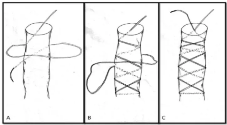
The first group, defined as the “X” configuration, was prepared using synthetic polyester Ethibond® No. 5 braided thread on a needle. The suturing was started in the distal portion of the graft, on one of the margins, using transfixing stitches across the entire substance of the tendon, with spacing of 7.5 mm between the stitches, until reaching 3 cm distally to the start of the suture. The suturing then returned along the line in the same manner, from the same margin as at the start of the suture, transfixing the tendon in the open spaces, intercalating the stitches and crossing the suture line in an “X” configuration. The same procedure was followed at the other end of the tendon (Figs. 1 and 2).
The second group, defined as stitches in a “loop” configuration, was prepared with the same type of synthetic Result: The Maximum Force of Rupture suture in “Loop” was 444.45 N and the suture in “X” was 407.59 N with statistical significance (p = 0.030). The average Tension obtained at the suture in “Loop” was 27.67 MPa and at the suture in “X” was 25.73 MPa with a statistically significant difference (p = 0.036). The stiffness showed no statistical differences (p = 0.350) at 11.804 N / mm at the point where “Loop” and 11.570 N / mm at the suture “X”. Conclusion: The suture in “Loop” had a higher biomechanical behavior to the suture “X”, considering the Maximum Force and Tension.
© 2013 Sociedade Brasileira de Ortopedia e Traumatologia. Published by Elsevier Editora Ltda. All rights reserved.
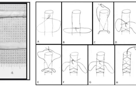
polyester Ethibond® No. 5 braided thread on a needle. However, the needle was removed to use only the thread, which was mounted in a double manner on a single needle, to form a loop (Fig. 3). The end of the graft was introduced into the loop, with the needle positioned superiorly. The stitches were started by transfixing the graft from the upper face to the lower face in each stitch. The suturing was started 3 cm from the end of the graft, by means of loops and transfixing stitches across the entire substance of the tendon, with spacing between the stitches of 7.5 mm (Figs. 4 and 5). This was also done at the other end.
All the tendons that had previously been prepared (Fig. 6) were folded over in the middle, over an Ethibond® No. 5 thread, in horse-rider style. In this double configuration, the
Fig. 2 - Tendons prepared using “X” stitches”.
Fig. 3 - (A) Synthetic polyester No. 5 braided thread on needle. (B) Needle separated from the thread. (C) Synthetic polyester No. 5 braided thread, threaded doubly on a single needle.
Fig. 4 - (A) Start of preparation of stitches in “loop” configuration, looping around the tendon 3 cm proximally to the end of the graft. (B) First stitch transfixing the graft in this region (C, D, E, F, G), looping around the tendon, and stitch transfixing the tendon with spacing of 7.5 mm. (F) Stitch in “loop” configuration completed.
Fig. 5 - Tendon prepared with “loop” stitches.
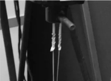
tendons were named “test bodies”, which were of length 10 cm and were kept under tension of 20 N (Fig. 7).
Each test body was coated with an alginate paste (Jeltrade alginate type II, regular set), which developed the consistency of rubber after a few seconds. The tendon was then removed, leaving an impression of the test body in the alginate, like in a mold. This mold was then sectioned transversally.
The sections generated from the alginate mold were then digitized with a resolution of 600 dpi, by means of an HP J5780® digitizer.
The cross-sectional areas of the molds were measured with the aid of the Image-Pro Plus® software, which had the capacity to supply measurements of the cross-sectional area from the digitized images.6
The maximum strength and the force-versus-displacement results were obtained using a load cell (model 661.19 F-02; MTS Systems Corporation) with a capacity of 10 KN and test velocity of 20 mm/min.
East test body was installed in the machine in horse-rider style, on a steel bar of diameter 6.35 mm at the top of the machine. The thread used for suturing the ends of each test body were fixed at the base of the machine with the aid of surgical hemostatic forceps, in the same way as done in ACL reconstruction surgery to maintain the tension when the graft is fixed in the tibial tunnel. The same length of thread (15 cm) was left between the end of the tendon and the forceps, in all the tests (Figs. 8 and 9).
In the statistical analysis, the normality of the data was tested using the Shapiro-Wilk test. Discrepant values or outliers were identified using a boxplot graph and then the t test for independent samples was performed, taking p < 0.05. The statistical analyses were performed using the SPSS 17 software for Windows.
Results
The “loop” stitches presented mean maximum strength of 444.45 N. The “X” stitches presented mean maximum strength of 407.59 N. The Shapiro-Wilk normality test demonstrated that the groups of “loop” and “X” stitches had distributions compatible with normal in relation to maximum strength. The t test for independent samples showed that after three outliers had been removed, the difference in maximum strength between the groups presented a statistically significant difference, with p = 0.030.
The mean cross-sectional area was 16.2 mm2 in the “loop”
stitches and 16.08 mm2 in the “X” stitches. There was no
statistically significant difference between the two groups, with p = 0.283.
The mean rigidity of the “loop” stitches was 11.570 N/mm and of the “X” stitches, 11.804 N/mm. The difference between the two groups was not significant, with p = 0.350.
The Shapiro-Wilk normality test demonstrated that the groups with “loop” stitches and with “X” stitches had distribution compatible with normal in relation to tension. The mean tension obtained in the “loop” stitches was 27.67 MPa and in the “X” stitches, 25.73 MPa. The statistical difference between the two groups after removal of an outlier was significant, with p = 0.036. The data obtained from the test bodies with “X” stitches are in Table 1. The data obtained from the test bodies with “loop” stitches are in Table 2.
Fig. 6 - “X” and “loop” stitches.
Fig. 7 - Test body.
Fig. 8 - Test body positioned in traction machine, in horse-rider position.
The failures in the maximum strength test occurred in the proximal portion of the tendon-thread complex (Fig. 10).
Discussion
ACL reconstruction is one of the surgical procedures most performed on the knee. Over recent decades, there have been advances in knowledge and refinement of the techniques and materials. The great majority of studies on refinements have been based on improvements to the methods for fixing grafts, choosing grafts and developing reconstruction techniques that allow restructuring of the biomechanics prior to the ACL injury in the knee.
Many details within the reconstruction still need to be evaluated and refined. Little is known about the best way of preparing grafts and how this preparation might influence the final results from the reconstruction.
Preparation of grafts from flexor tendons using “X” and “loop” stitches is very frequently done. However, there is little in the worldwide literature on which this practice can be based. Some authors have advocated Krackow stitches and prefabricated “loop” stitches (fiber loop) with locking.4
Others have preferred the Whipstitch and crisscross suturing.7
“X” stitches resemble crisscross stitches but suture the graft without joining the stitches. Each graft stitch is prepared individually, thus differing from crisscross suturing. “X” stitches suture the graft in a manner that is easy to perform and reproduce and which rarely produces cuts in the region where applied or divisions at the edge of the graft. “Loop” stitches, which were developed to simulate a prefabricated loop stitch (fiber loop), are used because they are economical, use materials that are easy to obtain and acquire, both in public and in private healthcare clinics, and enable use of the same principles as in fiber loops, i.e. easy and fast application in graft preparation.
We chose to use bovine common digital extensor tendons for this experiment based on studies that compared properties between these and the flexor tendons of the human knee.5 These demonstrated that grafts using bovine
common digital extensor tendons can replace the flexor tendons in traction tests. Another factor in making this choice was the possibility of obtaining fresh tendons and performing the traction tests in the same period of the day of their extraction, thereby avoiding changes such as those that occur to the modulus of elasticity of the tendons when they are stored in frozen condition8 or in formalin, which
harden the tissue.9
The data on maximum rupture strength and tension obtained from the test bodies were displayed on boxplot graphs to show the presence of any outlying discrepant values, which were then eliminated in accordance with the statistical analysis. The boxplot method for determining discrepant data was used because it is greatly used, easy to use and has great precision for detecting truly atypical observations.
The “loop” stitches presented a mean maximum rupture strength that was greater than that of the “X” stitches, with
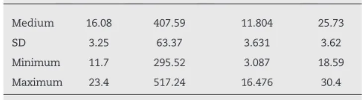
Area (mm2) Strength (N) Stiffness (N/mm) σ (MPa)
Medium 16.08 407.59 11.804 25.73
SD 3.25 63.37 3.631 3.62
Minimum 11.7 295.52 3.087 18.59
Maximum 23.4 517.24 16.476 30.4
N: # tests, σ: tension.
Area (mm2) Strength (N) Stiffness (N/mm) σ (MPa)
Medium 16.2 444.45 11.57 27.67
SD 2.69 94.06 3.287 5.17
Minimum 13.5 257.94 7.341 14.82
Maximum 22.3 600.68 18.372 33.47
N: # tests, σ: tension.
Table 1 - Test body results from stitches in “X” configuration (N = 10).
Table 2 - Test body results from stitches in “loop” configuration (N = 10).
a statistically significant difference (p = 0.030). When the choice for preparing grafts using Ethibond® No. 5 thread is between using “loop” or “X” stitches, taking only the mean maximum rupture strength into consideration, use of “loop” stitches is more recommended. However, taking only the tension strength of 60 to 140 N that is needed in the graft at the time of its fixation,10 both types of preparation
are adequate, even considering the minimum value for the maximum rupture strength, which was 295.92 N for the “X” stitches and 257.94 N for the “loop” stitches. Both stitch types are sufficient for supporting the recommended tension forces during graft fixation in the receptor regions of the femur and tibia.
With advances in the technique of double-band reconstruction, the preparation of flexor tendons has gained an important mechanical characteristic. When reconstruction is done using a single band, flexor grafts are used in double form for the semitendinosus and gracilis, which together form a quadruple arrangement in which they are positioned in a single tunnel in the femur and tibia. In addition, they are fixed in this position as a single structure, and not separately for each tendon. However, when the double-band technique is performed, grafts from the semitendinosus are used in double form to simulate one of the bands and the gracilis is used in double form to simulate the other band, such that two tunnels are needed in the femur and two in the tibia, with independent fixation for each graft in each tunnel. Through using the test body configuration in double form in the present study, we were able to show that separately, the mean maximum rupture strengths of the two types of stitches in preparing the tendons (444.45 N for the “loop” and 407.59 N for the “X”) were insufficient to enable modern rehabilitation protocols subsequent to ACL surgical reconstruction, when only indirect or post fixation is used, i.e. in which the preparation suture threads are used to fix the graft. It has to be borne in mind that during the first six weeks after the operation, the graft fixation has to withstand mechanical loads from day-to-day activities that are estimated to be up to 454 N.11
The mean values for the cross-sectional area in the test body groups with “loop” and “X” stitches did not present statistical differences, which enabled better comparison between the two groups.
The mean maximum tension obtained before the rupture accompanied the mean maximum rupture strength. The mean tension values for the “loop” stitches before the rupture were 7.01% greater than those for the “X” stitches, with a statistically significant difference (p = 0.036). In reconstructing the ACL, choosing the quadruple configuration for flexor grafts increases the cross-sectional area of the test bodies and the capacity to withstand the tension.
The stiffness obtained in the two types of graft preparation did not show any statistical difference, with 11.804 N/mm for the “X” stitches and 11.57 N/mm for the “loop” stitches, in comparison with the stiffness of the ACL of 242 N/mm. Both preparations were well below the stiffness of this structure. Compared with the stiffness of a system configured as a single fold of the gracilis (336 N/mm) and a
single fold of the semitendinosus (469 N/m), the stiffness of the test bodies was 30 to 40 times lower. This demonstrates that the preparation or the thread, suture and graft complex is the link that weakens the graft stiffness, as also observed by Hamner et al.12
Conclusion
Based on the experimental model used, the “loop” suture configuration presented biomechanical behavior that was superior to that of the “X” configuration, taking the maximum strength and tension into consideration. Both configurations were valid for supporting the recommended graft tensions at the time of fixation in ACL ligament reconstruction.
Conflicts of interest
The authors declare that there was no conflict of interests in conducting this study.
R E F E R E N C E S
1. Beynnon BD, Johnson RJ, Abate JA, Fleming BC, Nichols CE. Treatment of anterior cruciate ligament injuries. Part I. Am J Sports Medi. 2005;33(10):1579-602.
2. Fu FH, Bennett CH, Ma CB, Menetrey J, Lattermann C. Current trends in anterior cruciate ligament
reconstruction. Part II. Operative procedures and clinical correlations. Am J Sports Med. 2000;28(1):124-30.
3. Charlick DA, Caborn DN. Technical note: alternative softtissue graft preparation technique for cruciate ligament reconstruction. Arthroscopy. 2000;16(8):E20. 4. White KL, Camire LM, Parks BG, Corey WS, Hinton RY.
Krackow locking stitch versus locking premanufactured loop stitch for soft-tissue fixation: a biomechanical study. Arthroscopy. 2010;26(12):1662-6.
5. Donahue TLH, Gregersen C, Hull ML, Howell SM. Comparison of viscoelastic, structural, and material properties of doublelooped anterior cruciate ligament grafts made from bovine digital extensor and human hamstring tendons. J Biomech Eng. 2001;123(2):162. 6. Stieven Filho E, Malafaia O, Ribas-Filho JM, Diniz OE dos
S, Borges PC, Albano M, et al. Análise biomecânica da solidarização de tendões para reconstrução do ligamento cruzado anterior. Rev Col Bras Cir. 2010;37(1):52-7. 7. Howell SM, Goi-flieb JE. Endoscopic fixation of a
doublelooped semitendinosus and gracilis anterior cruciate ligament graft using bone mulch screw. Oper Tech Orthop. 1996;6(3):152-160.
8. Matthews LS, Ellis D. Viscoelastic properties of cat tendon: effects of time after death and preservation by freezing. J Biomech. 1968;1(2):65-71.
10. Fleming BC, Abate JA, Peura GD, Beynnon BD. The relationship between graft tensioning and the anterior-posterior laxity in the anterior cruciate ligament reconstructed goat knee. J Orthop Res. 2001;19(5):841-4. 11. Noyes FR, Butler DL, Grood ES, Zernicke RF, Hefzy MS.
Biomechanical analysis of human ligament grafts used in knee-ligament repairs and reconstructions. J Bone Joint Surg Am. 1984;66(3):344-52.