www.rbo.org.br
0102-3616/$–see front matter © 2013 Sociedade Brasileira de Ortopedia e Traumatologia. Publicado pela Elsevier Editora Ltda. Todos os direitos reservados. Work performed at the Knee Surgery Center, Instituto Nacional de Traumatologia e Ortopedia (INTO), Rio de Janeiro, RJ, Brazil *Corresponding author at: Praia do Flamengo, 66, Bloco B, Sala 1313, Rio de Janeiro, RJ. CEP: 22210-030.
E-mail: [email protected] (A.P. Mozella).
Original Article
Homologous structural graft for treatment of bone defect
during knee revision arthroplasty
Hugo Alexandre de Araújo Barros Cobra,
aMario Corrêa Netto Pacheco Junior,
band Alan de Paula Mozella
c,*
aOrthopedist and Head of the Knee Surgery Center, Instituto Nacional de Traumatologia e Ortopedia (INTO), Rio de Janeiro, RJ, Brazil
bTrainee Physician in the Knee Surgery Center, INTO, Rio de Janeiro, RJ, Brazil cOrthopedist in the Knee Surgery Center, INTO, Rio de Janeiro, RJ, Brazil.
doi: 10.1016/j.rbo.2012.08.007 A RT I C L E I N F O
Article history:
Received on May 7, 2012 Accepted on August 20, 2012
Keywords:
Arthroplasty, replacement, knee Bone transplantation
Review
a b s t r a c t
Objective: Obtaining stable bone-implant interface, correct alignment of the components, proper balance of soft tissues’ tension, maintenance of proper joint interline are fundamental principles for success in surgical revision total knee arthroplasty, which are only obtained with management bone deficiency. However, proper treatment of large defects remains unclear. The aim of this study was to evaluate the clinical and radiographic results of patients that had underwent revision surgery for total knee arthroplasty with use of structural grafts of musculoskeletal tissue bank in the period between January 2002 to December 2010 by the Knee Surgery Center of National Institute of Traumatology and Orthopaedics (INTO). The study included 26 revision arthroplasties with homologous structural bone grafting in 25 patients. Thirty-four structural bone grafts were used during the 26 revision total knee arthroplasty surgeries studied. The proximal tibia and distal femur were the grafts most frequently used. Six patients developed deep infection and in one of them with damage to the extensor mechanism associated. The average score on the WOMAC was 24,9. In the assessment of functional capacity in the SF-36, the average was 52.5. In radiographic evaluation, resorption of the graft occurred in three patients and no cases were observed of osteolysis, fracture of the graft, migration or subsidence of the components. Bone grafting of a musculoskeletal tissue bank is a satisfactory option to the handling of the bone defect in the setting of revision surgery for total knee arthroplasty.
Enxerto homólogo estrutural para tratamento do defeito ósseo durante artroplastia de revisão do joelho
Introduction
Today, statistical data show that life expectancy is increasing among the population worldwide, with growing demands for improvements in quality of life. This has led to increases in the number of total knee arthroplasty (TKA) procedures and consequently greater numbers of revision operations.1 Kurtz et al.2 estimated that the number of revision surgery procedures in the United States will increase by 600% by 2030.
TKA revision involves complex procedures with high technical demands, in which appropriate bone deficiency management becomes imperative in order to obtain satisfactory clinical results.3,4
Obtaining a stable bone-implant interface, correct alignment of the components, proper balance of soft tissue tensions and maintenance of proper joint interline level are fundamental principles for success in these surgical procedures, which is only achieved through management of the bone deficiency.5-7 Bone defects may result from primary disease, the technique and implants used, the failure mechanism of the TKA or the difficulty in extracting fixed implants. The classification system of the Anderson Orthopedics Research Institute (AORI), as described by Engh and Parks,8 is the system currently most used: type I presents intact metaphyseal bone; type II presents moderate metaphyseal deficiency in one (IIA) or two (IIB) femoral or tibial condyles; and type III has severe metaphyseal deficiency, with possible detachment of the collateral ligaments or the patellar ligament.8,9
Bone defects cam be filled with methyl methacrylate or be managed by using modular metallic expanders, thicker polyethylene components or unconventional prostheses. In addition, autologous or homologous bone grafts, which may be spongy or structural, can also be used.8-12 Nonetheless, the correct treatment for large defects remains undefined.
Structural bone grafts offer many advantages, which include biocompatibility, restoration of the bone stock and the potential for ligament reinsertion.7,13,14 The main disadvantages include the possibility of bone reabsorption, probably secondary to the immune response;14 the risk of fracturing or pseudarthrosis;14,15 and the possibility of disease transmission.16
The objective of the present study was to evaluate the clinical and radiographic results from patients who underwent TKA revision surgery using structural grafts.
Material and methods
Patients who underwent TKA revision surgery with use of structural grafts from a musculoskeletal tissue bank, at the Knee Surgery Center of the National Institute of Traumatology and Orthopedics (INTO), between January 2002 and December 2010, were evaluated.
Structural grafts were used in surgical procedures in which the bone deficiency presented could not be adequately treated by means of metallic expanders and were defined in accordance with concepts currently used in the literature. r e s u m o
Objetivo: A obtenção de estável interface osso-implante, o correto alinhamento dos componentes, o apropriado equilíbrio das tensões de partes moles, a manutenção de adequada altura da interlinha articular são princípios fundamentais para êxito nas cirurgias de revisão de artroplastia total de joelho, os quais somente são obtidos com manejo da deficiência óssea. Contudo, o correto tratamento de grandes defeitos permanece indefinido. O objetivo deste estudo foi avaliar os resultados clínicos e radiográficos dos pacientes submetidos à cirurgia de revisão de artroplastia total do joelho com uso de enxerto estrutural de Banco de Tecidos Músculos-Esqueléticos, entre janeiro de 2002 e dezembro de 2010, no Centro de Cirurgia do Joelho do Instituto Nacional de Traumatologia e Ortopedia (INTO). Foram incluídos no estudo 26 artroplastias de revisão com enxertia óssea homóloga estrutural em 25 pacientes. Foram usadas 34 peças estruturais para enxertia homóloga durante as 26 cirurgias de revisão de artroplastia total de joelho. O terço proximal da tíbia e o terço distal do fêmur foram as peças mais frequentemente usadas. Seis pacientes evoluíram com infecção profunda, em um desses casos associada à lesão do mecanismo extensor. O valor médio da pontuação obtida no questionário WOMAC foi de 24,9. Na avaliação da capacidade funcional no SF-36, o valor médio foi de 52,5. Na avaliação radiográfica, a reabsorção do enxerto ocorreu em três pacientes e não foram observados casos de osteólise, fratura do enxerto, migração ou afundamento dos componentes. Enxerto ósseo de Banco de Tecidos Músculo-Esqueléticos representa satisfatória opção ao manejo da falha óssea no cenário da cirurgia de revisão de artroplastia total de joelho.
© 2013 Sociedade Brasileira de Ortopedia e Traumatologia. Publicado pela Elsevier Editora Ltda. Todos os direitos reservados. Palavras-chave:
Artroplastia do joelho Revisão
Grafts were taken from the femoral or humeral head, the proximal third of the tibia or the distal third of the femur. All the specimens were conserved by means of ultra-freezing at -80º Celsius in the tissue bank of INTO.
All patients who underwent TKA revision surgery during the study period, in whom one or more specimens of homologous structural graft were used, were included in this study. The minimum postoperative follow-up period was 12 months.
Patients who received homologous structural grafting during surgical procedures other than TKA revision, and those in whom the bone grafting was done using autologous tissues or fragmented homologous tissues, were excluded. Likewise, patients who did not attend clinical and radiographic assessments, or for whom insufficient medical documentation was available, or who had not been followed up for a minimum of 12 months, were also excluded.
Through analysis on the medical documentation, data were gathered with regard to the location and AORI classification of the bone defect, type of homologous graft used and, if applicable, the fixation performed, along with data on the prosthetic implants used.
Postoperative clinical evaluations were conducted 15 days, one month, three months and one year after the procedure, and annually thereafter. Demographic data and information on the cause of the primary TKA, duration of the arthroplasty, failure mechanism of the TKA and presence of complications during the postoperative period were gathered.
Radiographic assessments were conducted through sequential examinations in the anteroposterior and lateral views with the knee flexed at 30°. The number, location and width of radiolucency lines at the bone-prosthesis interface were measured in accordance with the criteria of the Knee Society.17 The existence of osteolysis, pseudarthrosis, fractures, graft reabsorption and subsidence or migration of the prosthetic components was documented.
Absorption of part or all of the graft was evaluated in accordance with the criteria defined by Clatworthy et al.:7 mild reabsorption – partial loss of thickness of one cortex with a length of less than 1 cm; moderate – partial loss of thickness of one cortex with a length of more than 1 cm; and severe – complete loss of thickness of one cortex, of any length.
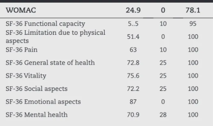
To evaluate quality of life and functional capacity, the previously validated Short Form 36 (SF-36)18 and Western Ontario and McMaster Universities (WOMAC)19 questionnaires were used, respectively.
The SF-3618 analysis was divided into the following items: functional capacity, limitation due to physical aspects, pain, general state of health, vitality, social aspects, emotional aspects and mental health.
This study was submitted for appraisal to and approved by the Research Ethics Committee of the National Institute of Traumatology and Orthopedics and was conducted at INTO’s Knee Surgery Center.
Results
Twenty-six cases of revision arthroplasty using homologous structural bone grafting, in 25 patients, were included in this study.
Clinical evaluation
Eighteen patients were female and nine were male. Their ages ranged from 46 to 83 years, with a mean of 70. The surgery was performed on the right side in 14 patients and on the left side in 12. Knee osteoarthrosis was the most prevalent diagnosis for performing the primary surgery, and this accounted for 85% of the cases. Rheumatoid arthritis was the diagnosis for 7.5% and post-traumatic arthrosis for 7.5%.
The mean length of postoperative follow-up was 55 months, with a minimum of 18 and maximum of 114 months.
Failure of the primary arthroplasty occurred after a mean of 7.8 years and ranged from 5 months to 13 years.
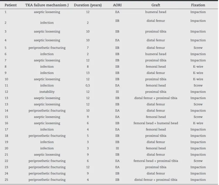
Aseptic loosening was the commonest failure mechanism, and this occurred in 11 cases (42%). Infection was the cause of the revision with homologous grafting in eight patients (31%). Periprosthetic fracturing occurred in six cases (23%). Post-prosthesis instability was the cause of revision in one patient (4%) (Table 1).
In 25 patients, revision implants from the Total Condylar 3® (TC 3) system (DePuy Inc., Warsaw, IN, USA) were used. In one patient, revision implants from the Coordinate® system (DePuy Inc.) were used. In all the cases, intramedullary nails were used, with the hybrid cementation technique. In 23 procedures, metallic expansion wedges were used in the femur, while in 11 patients, metallic wedges were used in the tibia.
During the 24 revision surgery procedures, we noted the presence of tibial bone defects: in 22 cases in the medial plateau and in 16 cases, lateral defects.
Bone defects in the femur were observed in 24 patients. In 23 procedures, bone deficiencies were seen in the posterior region of the condyles and in 16 cases there were bone defects in the lateral condyle and in 14 in the medial condyle.
The bone defects observed were classified (AORI) as II B in 16 patients (61.5%), II A in eight cases (30.5%) and III in two individuals (8%).
Thirty-four structural specimens were used for homologous grafting during the 26 surgical procedures studied (Fig. 1). The proximal third of the tibia and the distal third of the femur were the specimens most frequently used, and were implanted in 11 procedures each (32%). The bone defects were managed using homologous femoral head grafts in nine revisions (27%). Humeral head grafts were necessary in three cases (9%).
The graft fixation was achieved by means of bone impaction in 17 patients (65%), without the need for supplementary fixation. Cortical screws were used in five patients (19%) and Kirschner wires were necessary in four revisions (16%).
Six patients developed deep infection, which in one cases was associated with lesions of the extensor mechanism. Only one of these patients was characterized as a case of recurrence of infection (reinfection).
Arthrodesis, as a limb salvage measure, was performed with satisfactory results in two cases of acute infection that was refractory to venous antimicrobial treatment and serial dressings.
In two patients, transfemoral amputation was necessary. In one patient, resection arthroplasty was performed. In another case, after two surgical dressings and suppression using venous
Patient TKA failure mechanism J Duration (years) AORI Graft Fixation
1 aseptic loosening 12 IIA humeral head Impaction
2 infection 2 IIB distal femur Impaction
3 aseptic loosening 10 IIB proximal tibia Impaction
4 aseptic loosening 10 IIA distal femur Impaction
5 periprosthetic fracturing 7 IIB distal femur Screw
6 infection 2 IIB humeral head Impaction
7 aseptic loosening 12 IIB proximal tibia Impaction
8 infection 8 IIB femoral head K-wire
9 infection 13 IIB distal femur K-wire
10 aseptic loosening 12 IIB proximal tibia K-wire
11 infection 0,5 IIA femoral head Screw
12 instability 12 III proximal tibia Impaction
13 aseptic loosening 12 IIB distal femur + proximal tibia Impaction
13 aseptic loosening 12 IIB distal femur Screw
14 periprosthetic fracturing 10 IIA distal femur Impaction
15 aseptic loosening 9 IIA femoral head Screw
16 aseptic loosening 6 IIB femoral head + humeral head K-wire
17 infection 4 IIA femoral head Impaction
18 periprosthetic fracturing 5 IIB proximal tibia Impaction
19 infection 3 IIB distal femur Impaction
20 infection 3 III femoral head Impaction
21 aseptic loosening 9 IIB distal femur Impaction
22 periprosthetic fracturing 6 IIA femoral head + proximal tibia Screw
23 periprosthetic fracturing 12 IIA proximal tibia Impaction
24 periprosthetic fracturing 9 IIB distal femur Impaction
25 periprosthetic fracturing 4 IIB distal femur + proximal tibia Impaction
AORI, classification system of the Anderson Orthopedics Research Institute; TKA, total knee arthroplasty.
Table 1 - Evaluation of the surgical data on patients who underwent homologous grafting within knee revision arthroplasty.
antibiotic therapy, the patient then evolved without symptoms up to the present time.
One patient presented extensive cutaneous necrosis with the need to provide coverage, using a muscle flap. This was done without development of infection.
Another patient presented a voluminous hematoma that required a surgical approach for drainage.
So far, none of the patients has required any surgery for revision of the components.
WOMAC 24.9 0 78.1
SF-36 Functional capacity 5..5 10 95
SF-36 Limitation due to physical
aspects 51.4 0 100
SF-36 Pain 63 10 100
SF-36 General state of health 72.8 25 100
SF-36 Vitality 75.6 25 100
SF-36 Social aspects 72.2 25 100
SF-36 Emotional aspects 87 0 100
SF-36 Mental health 70.9 28 100
Table 2 - Mean functional evaluation on 20 of the 26 patients who underwent homologous grafting for knee revision arthroplasty.
Figure 2 - Before and after TKA revision procedure using structural graft.
condyle, where a graft originating from the proximal third of the humerus had been used. In one graft in the medial tibial plateau, we observed mild absorption; this case received a homologous graft from the distal femur.
In the series analyzed here, no cases of osteolysis or component migration of subsidence were observed. In one case, a segmental graft from the distal femur in a patient with rheumatoid arthritis evolved with nonunion, but without fracturing or loss of weight-bearing capacity (Fig. 2).
Discussion
Bone deficiency within the scenario of complex TKA revision surgery represents an enormous technical challenge with regard to obtaining a stable support surface for implanting prosthetic components and, consequently with regard to the durability and results of the surgery.
Use of structural grafts from tissue banks is an option for managing bone deficiencies in joint reconstruction surgery. However, the effectiveness of maintaining this structure as support over the long term has been questioned, along with the possibility of high rates of infection.
In 1992, Tsahakis et al.20 published a study that demonstrated three cases of mechanical failure after using segmental structural grafts. On the other hand, they highlighted that the bone stock was improved. In 1994, the same authors21 studied 21 patients who received structural grafts, with a mean Quality-of-life assessment
The WOMAC and SF-36 questionnaires were used to evaluate 20 of the procedures. Patients whose implants and grafts were removed because of infection and one patient who died from causes unrelated to the surgery (14 months after the grafting) were excluded from this analysis. The results are presented in Table 2.
Radiographic evaluation
Radiographic evaluations were performed on 21 TKA revision procedures, among which one patient underwent bilateral surgery. Five patients were excluded from the radiographic analysis because of postoperative complications (infection) that gave rise to loss of the implants and grafts.
Radiolucency lines were observed in eight patients (31%). Concordant with the criteria of the Knee Society,17 the commonest location was the medial tibial plateau (zone 1), which was found in five patients. Radiolucency lines were also found in the lateral tibial plateau (zone 3) in three patients; at the tip of the tibial nail (zone 6) in one patient; and at the medial tibial nail (zone 5) in one patient. In the femur, only one patient presented radiolucency lines, which occurred around the femoral nail. None of the cases showed the criteria of implant loosening.
follow-up of 42 months, and they noted that incorporation occurred in all the cases.
In the series reported by Mnaymneh et al.,14 14 specimens of homologous structural graft were used in 10 patients, with a mean follow-up of 40 months, and they found that graft incorporation occurred in 86% of the cases. When analyzed in more detail, the grafts used in tibial defects were found to have become incorporated in all the cases, but only 70% of the femoral grafts presented consolidation.
Dorr et al.22 studied 24 cases of revisions of primary prostheses using structural grafts, and found that union without collapse occurred in 92% of the patients. They therefore recommended that structural grafts should be used preferentially when there are defects larger than 50% of the femoral plateau or condyle. Dennis13 evaluated 30 patients who received segmental structural grafts, with 50 months of follow-up and found results classified as good or excellent in 86% of the cases. These data were corroborated by the series of Engh et al.,6 in which the structural graft most used was the femoral head; these authors presented good or excellent results corresponding to 87% of the patients. Ghazavi et al.23 presented a more modest success rate of around 77%, in a medium-term analysis.
In the largest series published, Clatworthy et al.7 evaluated 66 graft specimens that were used in 52 TKA revision surgical procedures and reported a five-year success rate of 92% and a 10-year rate of 72%. However, they highlighted that revision was needed for 23% of the cases after 71 months, among which 8% were because of infection and 8% because of failure due to graft reabsorption.
In our series, five patients evolved with failure and necessity to remove the implants and grafts because of infection. In none of these cases was recurrence of previous infection characterized. In another case, the patient evolved with the criteria of cured infection, and the implants and graft were kept in place after treatment with venous antibiotic therapy and serial dressings.
One patient who received a segmental distal femoral graft evolved with nonunion, but no reabsorption, collapse or fracturing of the graft was observed, and also there were no mechanical complications. In two other cases, we observed moderate reabsorption of the grafting in the distal femur and in another patient, we noted mild reabsorption in a femoral graft placed in the tibia. These data are concordant with what was presented by Mnaymneh et al.,14 in which all the grafts placed in the tibia presented incorporation, while only 70% of the femoral grafts became consolidated.
Graft fracturing was described in two cases among the series of 23 patients of Stokley et al.24 In the study by Clatworthy et al.,7 8% of the revisions failed because of graft reabsorption. In our study, we did not identify these complications or failures of arthroplasty relating to the graft. All the complications were caused by deep infection. In the single case of non-incorporation, we noted that the support function of the graft was maintained.
Tsahakis et al.21 did not report any occurrences of infectious complications after using homologous grafts in 15 TKA revision procedures. Septic failure was observed in three cases.
Mnaymneh et al.14 found that the failure rate due to infection was 7%.
In the series analyzed by Stokley et al.,24 infection occurred in 13% of the patients and, of these, one patient evolved to limb amputation. In our sample, the infection rate was 23% and two patients evolved to transfemoral amputation, two to arthrodesis and one to resection arthroplasty.
The cases of amputation resulting from incurable infection occurred in patients who had received segmental structural grafts (complete specimens from the proximal tibia).
We also highlight that in our sample, we did not see any cases of recurrence of infection in patients who had received a graft during the second stage of septic revision, and this finding was shared by Lord et al.25
Final remarks
In our sample, we did not observe any mechanical failures. There was only one case in which incorporation did not occur, and even so, the surgical result was not compromised. In all the cases, the grafts presented preserved weight-bearing function, without any occurrence of fracturing or loosening of components.
The main complication presented in the study group was infection, which occurred in 23% of the cases. Most of these cases evolved with implant loss and limited results.
Bone grafts from a musculoskeletal tissue bank form an option for managing bone failure within the scenario of TKA revision. However, studies with larger numbers of cases and longer follow-up are necessary.
Conflicts of interest
The authors declare no conflicts of interest.
R E F E R E N C E S
1. Harrison RJ Jr, Thacker MM, Pitcher JD, Temple HT, Scully SP. Distal femur replacement is useful in complex total knee arthroplasty revisions. Clin Orthop Relat Res. 2006;446:113-20. 2. Kurtz S, Ong K, Lau E, Mowat F, Halpern M. Projections of
primary and revision hip and knee arthroplasty in the United States from 2005 to 2030. J Bone Joint Surg Am. 2007;89(4):780-5.
3. Lavernia CJ, Guzman JF, Gachupin-Garcia A. Cost effectiveness and quality of life in knee arthroplasty. Clin Orthop Relat Res. 1997;(345):134-9.
4. Healy WL, Finn D. The hospital cost and the cost of the implant for total knee arthroplasty. A comparison between 1983 and 1991 for one hospital. J Bone Joint Surg Am. 1994;76(6):801-6.
5. Whittaker JP, Dharmarajan R, Toms AD. The management of bone loss in revision total knee replacement. J Bone Joint Surg Br. 2008;90(8):981-7.
7. Clatworthy MG, Ballance J, Brick GW, Chandler HP, Gross AE. The use of structural allograft for uncontained defects in revision total knee arthroplasty. A minimum five-year review. J Bone Joint Surg Am. 2001;83-A(3):404-11.
8. Engh GA, Parks NL. The use of a bone defect classification system in revision total knee arthroplasty. Orthop Trans. 1995;18:1136.
9. Bezwada HP, Shah AR, Zambito K, Cerynik DL, Johanson NA. Distal femoral allograft reconstruction for massive osteolytic bone loss in revision total knee arthroplasty. J Arthroplasty. 2006;21(2):242-8.
10. Windsor RE, Insall JN, Sculco TP. Bone grafting of tibial defects in primary and revision total knee arthroplasty. Clin Orthop Relat Res. 1986;(205):132-7.
11. Radnay CS, Scuderi GR. Management of bone loss: augments, cones, offset stems. Clin Orthop Relat Res. 2006;446:83-92. 12. Hockman DE, Ammeen D, Engh GA. Augments and allografts in
revision total knee arthroplasty: usage and outcome using one modular revision prosthesis. J Arthroplasty. 2005;20(1):35-41. 13. Dennis DA. The structural allograft composite in revision
total knee arthroplasty. J Arthroplasty. 2002;17(4Suppl 1):90-3.
14. Mnaymneh W, Emerson RH, Borja F, Head WC, Malinin TI. Massive allografts in salvage revisions of failed total knee arthroplasties. Clin Orthop Relat Res. 1990;(260):144-53. 15. Berrey BH Jr, Lord CF, Gebhardt MC, Mankin HJ. Fractures of
allografts. Frequency, treatment, and end-results. J Bone Joint Surg Am. 1990;72(6):825-33.
16. Buck BE, Malinin TI, Brown MD. Bone transplantation and human immunodeficiency virus. An estimate of risk of acquired immunodeficiency syndrome (AIDS). Clin Orthop Relat Res. 1989;(240):129-36.
17. Ewald FC. The Knee Society total knee arthroplasty
roentgenographic evaluation and scoring system. Clin Orthop Relat Res. 1989;(248):9-12.
18. Ware JE Jr, Sherbourne CD. The MOS 36-item short-form health survey (SF-36). I. Conceptual framework and item selection. Med Care. 1992;30(6):473-83.
19. Bellamy N, Kirwan J, Boers M, Brooks P, Strand V, Tugwell P, et al. Recommendations for a core set of outcome measures for future phase III clinical trials in knee, hip, and hand osteoarthritis. Consensus development at OMERACT III. J Rheumatol. 1997;24(4):799-802.
20. Tsahakis PJ, Brick GW, Sledge CB. The use of bulk allografts for uncontained defects in revision total knee arthroplasty. Orthop Trans. 1992;16:682.
21. Tsahakis PJ, Beaver WB, Brick GW. Technique and results of allograft reconstruction in revision total knee arthroplasty. Clin Orthop Relat Res. 1994;(303):86-94.
22. Dorr LD, Ranawat CS, Sculco TA, McKaskill B, Orisek BS. Bone graft for tibial defects in total knee arthroplasty. Clin Orthop Relat Res. 1986;(205):153-65.
23. Ghazavi MT, Stockley I, Yee G, Davis A, Gross AE. Reconstruction of massive bone defects with allograft in revision total knee arthroplasty. J Bone Joint Surg Am. 1997;79(1):17-25.
24. Stockley I, McAuley JP, Gross AE. Allograft reconstruction in total knee arthroplasty. J Bone Joint Surg Br. 1992;74(3):393-7. 25. Lord CF, Gebhardt MC, Tomford WW, Mankin HJ. Infection in