DM
February | 2018
Gonçalo Nuno Gouveia Martins
MASTER IN APPLIED BIOCHEMISTRYRelease of Resin-bound Ferulic Acid
During an
In Vitro
Digestion Simulation
and Antioxidant Activity Evaluation
Gonçalo Nuno Gouveia Martins
MASTER IN APPLIED BIOCHEMISTRY
During an
In Vitro
Digestion Simulation
and Antioxidant Activity Evaluation
MASTER DISSERTATIONORIENTADORA
RELEASE OF RESIN-BOUND FERULIC ACID DURING
AN IN VITRO DIGESTION SIMULATION AND
ANTIOXIDANT ACTIVITY EVALUATION
Esta dissertação foi desenvolvida no grupo de Produtos Naturais do Centro de Química da Madeira (CQM), sob a orientação da Professora Doutora Paula Cristina Machado Ferreira Castilho. Foi apresentada à Universidade da Madeira, para cumprimento dos requisitos necessários à obtenção do grau de Mestre em Bioquímica Aplicada.
Gonçalo Nuno Gouveia Martins
2018
“A química não se pode eliminar, pelo simples facto que se encontra em tudo
o que nos rodeia e em nós. Está à nossa volta nos fenómenos naturais
indispensáveis à vida, como a fotossíntese, e nos produtos artificiais de
importância primária para a nossa civilização […]. Está em nós porque o
homem "funciona" ou "não funciona" através de reacções químicas. A
concepção, o crescimento e a morte são processos químicos, ainda que muito
complexos. […] Portanto
, abolir a química quereria dizer não só abolir as
adulterações alimentares e poluição, mas também abolir os combustíveis, os
fármacos, os fertilizantes, as matérias plásticas, os semicondutores, os
detergentes, ou seja, todos os benefícios que, dum modo quase inconsciente,
usufruímos todos os dias; e quereria também dizer abolir as plantas, os
animais, e o próprio homem. Quereria dizer abolir tudo, porque tudo […] é
q
uímica.”
Vincenzo Balzani,
Boletim da Sociedade Portuguesa de Química,
I would like to thank the Madeira Chemistry Research Centre (CQM) and the University of Madeira for the opportunity to develop this work and present it at the 5th CQM Annual
Meeting, and for all the expertise and knowledge acquired during the Bachelor’s and, presently, the Master’s degree.
This work was supported by FCT-Fundação para a Ciência e a Tecnologia (project PEst-OE/QUI/UI0674/2013, CQM, Portuguese Government funds), and through Madeira 14-20 Program, project PROEQUIPRAM - Reforço do Investimento em Equipamentos e Infraestruturas Científicas na RAM (M1420-01-0145-FEDER-000008), the Portuguese National Mass Spectrometry Network (contract RNEMREDE/1508/REM2011), and by ARDITI-Agência Regional para o Desenvolvimento da Investigação Tecnologia e Inovação, through the project M1420-01-0145-FEDER-000005 - Centro de Química da Madeira - CQM+ (Madeira 14-20).
I thank my supervisor, Professor Paula C. Castilho, for the all challenges placed before me and for the trust, motivation, and advice provided through the years.
Acknowledgments also go to LANXESS, Germany, for providing the resin Lewatit® VP
OC 1064 MD PH, and to Farmácia Morna, in particular to Sara Morna, for kindly
providing the gelatine capsules, Capsulas 00 incoloras.
Also, I thank the Faculty of Exact Sciences and Engineering and Jorge Lopes for the production of the sample holder used in the ATR-FTIR analyses.
A big thank you goes to my lab colleagues, Vitor Spínola and Núria Fernandes, for their friendship, support, “advice”, and the paramount help throughout this work.
I am grateful to Carla Miguel and Radenka Whiffen for the help in the SEM and EDS analysis, Pedro Silva in the chromatography, João Serina with the bibliography, Cláudia Camacho in the Fluorescence Spectroscopy measurements, Professor Pedro Pires for the help with the kinetic data treatment, and my dear friend and colleague Natacha Antunes, for the support, advice and for drawing the molecular structures depicted in this dissertation.
I also acknowledge the help of lab technicians Paula Andrade and Paula Sousa for their essential assistance with reagents, materials, and equipment.
To my family, particularly my parents, Teresa Martins and Jorge Martins, and my brother, Ricardo Martins, I send a big hug and a meaningful “Thank you!” for all the support and patience throughout all the ups and downs of this journey.
Ferulic acid is one of the most abundant hydroxycinnamic acids in Nature, with impact in human health whereas an antioxidant protection might be implicated. It has uses in cosmetics and food industry. It can be found in various foodstuffs, but mostly present etherified to lignins or esterified to carbohydrates or sterols, which hinders its absorption by the organism during the digestive process. The free form of ferulic acid is rapidly absorbed from the stomach, jejunum and, in a much lesser extent, from the ileum, in a pH dependent process.
Since the ileum is particularly susceptible to inflammation and oxidative stress it is important to ensure that ferulic acid is able to exert its action in that part of the body without being absorbed from the stomach. A possible way to increase ferulic acid’s
bioavailability to the ileum is by immobilization in solid matrixes resistant to low pH but not neutral or basic conditions.
In the present work, the adsorption of ferulic acid onto the polystyrene adsorbent resin Lewatit® VP OC 1064 MD PH was studied, and a loading of 144 mg/g dry resin was obtained.
To evaluate the release of the resin-bound ferulic acid, an in vitro “digestion” was
performed with simulated gastrointestinal juices. The intestinal step was the most relevant with a release of 13-35 % of FA from the loaded resin.
After each step of the in vitro digestion simulation, the antioxidant activity of ferulic acid was evaluated using the DPPH radical scavenging assay and all samples successfully maintained antioxidant activity throughout the digestive process.
The confirmation of the incorporation of ferulic acid onto the resin was made by ATR-FTIR spectroscopy and morphological analysis was made by SEM. The quantification of ferulic acid in solution was performed by HPLC-DAD throughout the entire work. This work showed that the free form of ferulic acid can be delivered in the intestine, after immobilization of solid matrixes, maintaining its antioxidant activity. This study is probably the first on this subject with these materials and methods.
O ácido ferúlico é um dos ácidos hidroxicinâmicos mais abundantes na Natureza, podendo ter um impacto benéfico na saúde humana, em processos que poderão ter por base o seu potencial antioxidante. Actualmente as suas aplicações passam pelas indústrias cosmética e alimentar. Pode ser encontrado em várias fontes alimentares, onde a sua forma eterificada a ligninas ou esterificada a carbohidratos ou esteróis é muito comum, o que dificulta a sua absorção pelo organismo durante o processo digestivo. A forma livre, por outro lado, é rapidamente absorbida no estômago, jejuno e, em menor extensão, no íleo, num processo dependente do pH.
Visto que o íleo é particularmente suspectível a inflamação e stress oxidativo a entrega
da forma livre do ácido ferúlico neste segmento do tracto gastrointestinal deve ser assegurada, sem que seja absorvido no estômago.
Uma forma de aumentar a biodisponibilidade do ácido ferúlico é por imobilização em matrizes sólidas que sejam resistentes ao meio ácido do estômago, mas que garantam a sua entrega em meios neutros ou alcalinos. Neste trabalho, a imobilização do ácido ferúlico foi feita por adsorção na resina adsorvente de polistireno Lewatit® VP OC 1064 MD PH, tendo sido obtida uma incorporação de 144 mg/g resina seca.
Através de uma simulação in vitro da digestão, a libertação do ácido ferúlico da resina foi
avaliada. Efectivamente, o meio intestinal proporcionou a maior libertação de ácido ferúlico, sendo que 13-35 % do ácido ferúlico incorporado na resina foi libertado nestas condições.
Foram recolhidas amostras após cada passo da digestão e as suas actividades antioxidantes foram aferidas com recurso ao teste do DPPH, tendo sido observado que todas as amostras apresentaram actividade antioxidante após a digestão.
A confirmação da incorporação do ácido ferúlico na resina foi feita por espectroscopia ATR-FTIR e foram realizadas análises físicas e químicas por SEM. Durante o decorrer do trabalho, a quantificação do ácido ferúlico foi feita por HPLC-DAD.
Este trabalho demonstrou que a forma livre do ácido ferúlico pode ser entregue no intestino, após imobilização em matrizes sólidas, mantendo a sua actividade antioxidante. Este é possivelmente o primeiro estudo sobre este assunto com estes materiais e nestas condições.
List of Oral Communications
Martins, G, Castilho PC. Delivery of Ferulic Acid During an In Vitro Digestion
Simulation. 5th CQM Annual Meeting, Funchal, Madeira. 01-03 of February 2018. ISBN:
Acknowledgments ... v
Abstract ... vii
Resumo ... ix
List of Oral Communications ... xi
List of Figures ... xvii
List of Tables ... xxi
Abbreviations ... xxiii
I. Introduction ... 1
1. Secondary metabolites as bioactive compounds – the case of ferulic acid ... 3
1.1. Introduction – secondary metabolites ... 3
1.2. Hydroxycinnamic acids ... 4
1.2.1. Biosynthesis ... 4
1.2.2. Ferulic acid ... 5
1.2.2.1. Occurrence ... 6
1.2.2.2. Bioactivity, potential uses and applications ... 9
1.2.2.3. Intake and pharmacokinetics ... 14
2. Polymeric resins ... 19
2.1. Polymeric resins in medicine and biomedical applications ... 19
2.2. Adsorption of phenols by polymeric resins... 20
3. Simulated in vitro digestion ... 21
3.1. Absorption ... 21
3.2. Release - In vitro digestion simulation ... 22
II. Methods and Experimental Procedures ... 23
1. Immobilization in macroporous resin ... 25
1.1. Objective ... 25
1.2.1. Adsorption experiments ... 25
1.2.1.1. Selection and pre-treatment of the adsorbent ... 25
1.2.1.2. Studying the influence of experimental parameters in the adsorption process 26 1.2.1.3. Resin selectivity ... 29
1.2.2. HPLC-DAD compound quantification... 29
1.2.3. Physical and chemical characterization... 32
2. In vitro digestion simulation ... 35
2.1. Methods ... 35
2.1.1. Samples ... 35
2.1.2. In vitro digestion assays ... 35
3. Antioxidant activity evaluation ... 39
III. Results and Discussion ... 43
1. Immobilization in macroporous resin ... 45
1.1. Studying the influence of experimental parameters in the adsorption process 45 1.2. Resin selectivity ... 50
1.3. Physical and chemical characterization ... 52
1.3.1. ATR-FTIR Spectroscopy ... 52
1.3.2. SEM morphological and elemental analysis ... 53
2. In vitro digestion simulation ... 57
2.1. HPLC-DAD compound detection ... 57
2.2. Liquid samples’ digestion ... 57
2.3. Solid samples’ digestion ... 59
3. Antioxidant activity evaluation ... 67
3.1. Standard ferulic acid solutions ... 67
3.2. Supernatants of the Lewatit+FA digestions ... 68
IV. Conclusion and Future Perspectives ... 73
V. References... 77
List of Figures
Figure 1 - General structure of a hydroxycinnamic acid. R1, R2 = H; OH; OCH3. ... 4
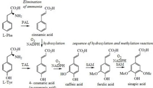
Figure 2 - The formation of p-coumaric, caffeic, ferulic and sinapic acids. Adapted from Dewick, M. (2002).[1] ... 4
Figure 3 – Structures of some chlorogenic acids.[5] ... 5
Figure 4 - Structure of ferulic acid. ... 6
Figure 5 – Structure of 8-8-diferulic acid. ... 8
Figure 6 - Bioactivities and applications of FA. ... 9
Figure 7 - Nitrogen-containing compounds found in the Alangiceae plant family bearing a feruloyl moiety. Adapted from Silva, E. and Batista, R. (2017).[11] ... 10
Figure 8 - Resonance stabilization of FA’s phenoxyl radical, during radical-scavenging. Adapted from Silva, O. and Batista, R. (2017). [11] ... 12
Figure 9 – Structure of curcumin. ... 12
Figure 10 - Types of drug transport across the intestinal epithelial cells. ... 16
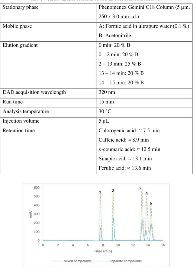
Figure 11 - Chromatograms of chlorogenic (1), caffeic (2), p-coumaric (3), sinapic (4), and ferulic (5) acids isolated in different solutions and in the same solution (dashed green line), determined by HPLC-DAD at 320 mn with the conditions described in Table 5. 30 Figure 12 - Chromatogram of FA as determined by HPLC-DAD at 320 nm, with the conditions described inTable 6. ... 31
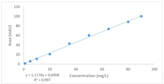
Figure 13 - Calibration curve obtained for FA by HPLC-DAD at 320 nm, using the conditions in Table 6. ... 32
Figure 14 – Specially made sample holder for ATR-FTIR spectroscopy. ... 33
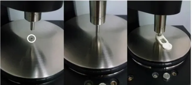
Figure 15 - Left: detail of the 1.5 mm IRE (the black spot inside the white circumference); centre: scanning without the sample holder; right: scanning with the sample holder. ... 33
Figure 16 - Schematics of the in vitro simulated digestion procedure. ... 37
Figure 17 – Encapsulated loaded resin. ... 38
Figure 18 - UV-Vis spectra of DPPH and DPPH + Trolox. ... 40
Figure 19 - Calibration curve for the absorbance at 550 nm versus the concentration of Trolox. ... 41
Figure 20 - Samples analysed in the DPPH assay. Circles and squares represent solid and liquid samples, respectively. ... 42
Figure 22 - Amount of FA adsorbed per gram of dry resin, using different initial concentrations. Values are presented as mean ± SD (n = 3). ... 47 Figure 23 - Amount of FA adsorbed per gram of dry resin, at different temperatures. Values are presented as mean ± SD (n = 3). ... 48 Figure 24 - Percentage of adsorption over time. Dots are the experimental results (average of three assays) and the dashed line is function that better fitted the present data. ... 49 Figure 25 - Amount of each HCAs studied in this work adsorbed to the Lewatit resin (mg/g dry weight). Values are presented as mean ± SD of three assays. ... 51 Figure 26 - ATR-FTIR spectra of the unloaded and loaded resin and FA. * = peaks attributed to FA. ... 52 Figure 27 – Scanning electron micrographs of unloaded and loaded Lewatit resin. Upper line: general view of intact and broken unloaded resins (left) and detail with size measurement (right). Bottom line: optical visualization (left) and electronic visualization details with size measurements (right) of the FA loaded resin... 54 Figure 28 – Structure of polystyrene. ... 55 Figure 29 - Chromatogram of FA obtained at 320 nm by HPLC-DAD after a full digestion with adjuncts. ... 57 Figure 30 - FA's concentration in the digestion media. The presented values are the average of three assays ± SD. In the case of the Prediction the values refer to theoretical calculations. ... 58 Figure 31 - Amount of resin-bound FA released in gastrointestinal conditions. The variant
“Capsule” refers to the full in vitro digestion using the gelatine capsule. The presented
values are the average ± SD of three assays. ... 59 Figure 32 - Digestion of the encapsulated resin-bound FA: A - before the addition of the salivary juice; B – middle of the gastric digestion; C – end of the gastric step; D –
intestinal stage. ... 61 Figure 33 - Loaded resin’s simulated digestion in the stomach (left) and the intestine
Figure 37 – DPPH inhibitions (%) obtained for the standard FA solution before and after each digestion step, the predicted values for each concentration, and for the simulated digestive juices. The values presented are the mean ± SD of several assays: n = 3 for the undigested solution and simulated digestive juices; and n = 9 for each of the digested samples. ... 67 Figure 38 - DPPH inhibitions (%) of the supernatants of each step of the Lewatit+FA
digestion, as well as the prediction based on each sample’s concentration. The values
presented are the mean ± SD of nine assays. ... 68 Figure 39 - Trolox eq. (mM/g/L FA) of the supernatants of the Lewatit+FA digestion. 69 Figure 40 - DPPH inhibitions of the undigested unloaded and loaded Lewatit resin and gelatine capsule; and of the digested Lewatit+FA after each step of the simulated in vitro
digestion. Values presented are the mean ± SD of three and nine assays for the undigested
samples and the digested samples, respectively. The sample “Gelatine” refers to the
List of Tables
Table 1 - FA (mg/100 g of fresh weight) present in different foodstuffs. Adapted from Zhao, Z. and Moghadasian, M. H. (2008). [7] ... 7 Table 2 - HCAs’ content in different grains (mg/kg of fresh weight). Legend: 1 = flour; 2
AA Antioxidant Activity
ATR-FTIR Attenuated Total Reflectance-Fourier Transform Infrared
BCS Biopharmaceutics Classification System
CAT Catalase
DPPH 2,2-diphenyl-1-picrylhydrazyl
DVB Divinylbenzene
EDS Energy-Dispersive X-ray Spectroscopy
FA Ferulic acid
HCA(s) Hydroxycinnamic acid(s)
HMG-CoA Hydroxymethylglutaryl coenzyme A
HPLC-DAD High Performance Liquid Chromatography with Diode-Array Detection
IRE Internal Reflection Element
IVIVC In vivo-in vitro correlation
Lewatit Lewatit® VP OC 1064 MD PH
L-Phe L-phenylalanine
L-Tyr L-tyrosine
qe Amount of compound adsorbed for unit mass of adsorbent (mg/g)
qr Amount of ferulic acid released from the resin (mg/g)
ROS Reactive oxygen species
SD Standard Deviation
SEM Scanning Electron Microscopy
SOD Superoxide dismutase
UV Ultraviolet
1. Secondary metabolites as bioactive compounds
–
the case
of ferulic acid
1.1. Introduction – secondary metabolites
In order to ensure that organisms can survive and reproduce, they need certain compounds. Apart from energy in the form of ATP (adenosine triphosphate), molecules such as nucleic acids, proteins, carbohydrates, and lipids are essential to vital processes, like respiration and photosynthesis, and can be found in all organisms. For this reason, they are referred to as primary metabolites, and the set of synthetic pathways associated
with their production is called the primary metabolism, since they remain practically
unaltered from organism to organism.[1,2]
However, there are certain metabolic pathways that are not present in all life-forms, and, consequently, their products cannot be found ubiquitously, like the previous ones. These secondary metabolites are a wide range of compounds, with a rich variety of
chemical structures, whose functions are not yet known in all cases, but it is assumed they play a role in the survival of their producer. Although they were once regarded simply as waste products of the primary metabolism, they seem to be important for protection - for instance from ultraviolet (UV) light or against predators; while others are thought to be involved in reproduction, as attractors, for example. Their production is dependent on a balance between synthesis, storage, and degradation, and their metabolism is often associated with growth and morphological changes.[2–4]
Plants represent an interesting case for the study of these molecules because they have a very developed secondary metabolism, being able to store large amounts of these
metabolites, whereas other organisms need to acquire them in their diet, even consuming plants for this purpose. P. M. Dewick (2002) even states that “it is thus fairly obvious that the human diet could be both unpalatable and remarkably dangerous if all plants, animals,
and fungi produced the same range of compounds”.[1] Although secondary metabolites have relevant roles in the life cycle of the organisms which produce them, they have different roles in the organisms that consume them. When it comes to human usage, consumption may be through ingestion (diet), external (skin) application, inhalation, or other forms. [4]
alkaloids and sulphur-containing compounds”.[4] For the interest of this work, only a class of the first group, the hydroxycinnamic substances, and its consumption through ingestion will be discussed in further detail.
1.2. Hydroxycinnamic acids
The hydroxycinnamic acids (HCAs) are phenylpropanoids: they show a chain of three carbon as substituents in a benzene ring but have an OH group in the para position;
and were the compounds studied in this work. The name “hydroxycinnamic acid” comes
from the hydroxylation of cinnamic acid, which is their precursor and their general structure can be found in Figure 1.
Figure 1 - General structure of a hydroxycinnamic acid. R1, R2 = H; OH; OCH3.
1.2.1. Biosynthesis
HCAs are produced in the phenylpropanoid pathway (Figure 2) and derive mostly from L-phenylalanine (L-Phe), but also from L-tyrosine (L-Tyr). These aromatic amino acids are only produced by plants, bacteria, and fungi, and provide the C6-C3 backbone common to all phenylpropanoids.
The first step of these compounds’ synthesis is the production of p-coumaric acid,
from either one of two forms: the hydroxylation of cinnamic acid, after deamination of Phe by the action of phenylalanine ammonia lyase (PAL); or by deamination of L-Tyr by L-tyrosine ammonia lyase (TAL). Consecutive hydroxylations (in a NADPH-depending reaction with molecular oxygen) and methylations (by SAM, S-adenosylmethionine), caffeic, ferulic, and sinapic acids are obtained from p-coumaric
acid. [1–4]
The most common hydroxycinnamates are the esters of these four compounds and quinic acid (Figure 3). These esters are collectively referred to as “chlorogenic acids”
even though that name is usually attributed to the most abundant of them: 5-O
-caffeoylquinic acid; and the lack of distinction in the literature between the group and the latter compound can be both tiresome and confusing at times. Additionally, esterification with shikimic, tartaric, malic, and malonic acids is also common. Actually, HCAs are not usually found in the free form in most vegetable matrices since they are able to bind to saccharides and lignin through ester and ether linkages. [5,6]
Figure 3 – Structures of some chlorogenic acids.[5]
Among all HCAs, ferulic acid was the most important in this work, and for this we will discuss deeper its properties.
1.2.2. Ferulic acid
Ferulic acid1 (FA), or 4-hydroxy-3-methoxycinnamic acid (Figure 4), is one of the
most abundant HCAs in Nature. It was first isolated in 1866 by Hlasiwetz and Barth, from
the plant Ferula foetida (Apiaceae family) and chemically synthesized in 1925 via
condensation of vanillin with malonic acid, in an amine-catalysed reaction. By the 1970’s its antioxidant potential was already reported.[7] However, in 1992, E. Graf commented on the fact there was a lack of study and publications on this topic in the 126 years since
the molecule’s isolation, an issue that has changed dramatically since then. Nowadays, FA is a well-known and well-studied compound, with many applications in the industry and as a phytochemical.[8]
Figure 4 - Structure of ferulic acid.
1.2.2.1. Occurrence
Table 1 - FA (mg/100 g of fresh weight) present in different foodstuffs. Adapted from Zhao, Z. and Moghadasian, M. H. (2008). [7]
Source FA (mg/100 g of fresh
weight)
Grains
Refined corn bran 2610-3300
Barley extract 1358–2293
Soft and hard wheat bran 1351–1456
Fruits
Grapefruit 10.7–11.6
Orange 9.2-9.9
Banana 5.4
Vegetables
Bamboo shoots 243.6
Water dropwort 7.3-34
Eggplant 7.3-35
Commercial foods and beverages
Sugar-beet pulp 800
Popcorn 313
Coffee 9.1-14.3
Beer 0.24-0.9
As Table 1 shows, grains are the richest source of FA in human dietary products. Among different types of grains (Table 2), FA stands out as the major HCA.
Table 2 - HCAs’ content in different grains (mg/kg of fresh weight). Legend: 1 = flour; 2 = grits; 3 = flakes; nd = not detected. Adapted from Shahidi, F. and Chandrasekara, A. (2015). [9]
HCA Wheat1 Rye1 Corn1 Millet2 Barley1 Oat3 Brown
rice
Ferulic 890 860 380 260 250 250 240
Sinapic 63 120 57 nd 11 55 20
p-coumaric 37 41 31 18 40 nd 76
Caffeic 37 10 26 1.1 1.7 3.1 nd
Furthermore, although FA can be present in large quantities in its free form, mostly in the trans-isomeric form, in some vegetables, such as burdock, water dropwort,
oligomers (e. g. 8-8-diferulic acid, Figure 5). The free form can be obtained by alkaline hydrolysis. [5,7,10]
Figure 5 – Structure of 8-8-diferulic acid.
The high quantities of FA found in grains come from the conjugated form. These conjugated forms can be soluble or insoluble, and the latter represents the majority of FA (Table 3).
Table 3 – Content of soluble and insoluble FA in different cereals. Legend: 1 = µg/g grain; 2 = µg/g dry weight; 3 = µg/g defatted meal; a = Soluble FA includes both the free and conjugated soluble forms. Adapted from Shahidi, F.
and Chandrasekara, A. (2015). [9]
Cereal type Soluble FAa Insoluble FA Total
Soft wheat, choptank1 39 560 599
Soft wheat, VA97W-0241 41 521 562
Soft Wheat, SS5601 41 527 568
Soft Wheat, vigoro tribute1 49 407 456
White corn2 13 1193 1206
Yellow corn2 21 1009 1030
Red corn2 19 1284 1303
Blue corn2 21 1279 1300
Kodo millet3 365 1844 2209
Finger millet3 27 331 358
Foxtail millet3 225 631 856
Proso millet3 112 332 444
Little millet3 164 185 349
Pearl millet3 176 637 813
compounds, saccharides, terpenoids, oligomers, and miscellaneous compounds, found from 1990 to 2015 in different families of plants and their described bioactivities. [11]
1.2.2.2. Bioactivity, potential uses and applications
Currently, the uses of FA are mainly based on its antioxidant activity (AA), which in turn is responsible for other applications, as antimicrobial or UV-protector. It is an approved food preservative in Japan, USA, and Europe, and in China it is used in the form of sodium ferulate against cardiovascular and cerebrovascular diseases.[7,12] From the various applications, areas such as food, cosmetic and pharmaceutic industries present the most uses for FA (Figure 6).
Figure 6 - Bioactivities and applications of FA.
A. Chemotaxonomical marker
Some families of plants only produce certain types of feruloyl conjugated molecules and, for that reason, FA can be used as a chemotaxonomical marker. As an example, the Alangiceae, Amaranthaceae and Annonaceae families only produce the feruloyl moiety in nitrogen-containing compounds (Figure 7). In the review by Silva, E and Batista, R. (2017) there is an extensive description of compounds, found in plants from 1990 to 2015, containing a feruloyl moiety, and other examples of the potential use of FA in chemotaxonomy are given. [11]
Figure 7 - Nitrogen-containing compounds found in the Alangiceae plant family bearing a feruloyl moiety. Adapted from Silva, E. and Batista, R. (2017).[11]
B. Antimicrobial activity
Studies have showed that FA exhibits antimicrobial activity against viruses, such as HIV and the influenza viruses, bacteria, and yeast cells. For example, it is thought that FA diminishes the release and action of the p24 antigen, a protein present in the HIV’s
virus capsid, inhibiting its replication.[13] The antibacterial activity against both gram-positive and gram-negative bacteria, e.g. human intestinal microflora - Escherichia Coli,
Klebsiella pneumoniae, Enterobacter aerogenes, and others - is thought to happen via the
inhibition of arylamine N-acetyltransferase – an enzyme that catalyses the transference of an acetyl group from acetylcoenzyme A to a xenobiotic acceptor[14].
C. Food preservation and other applications
As mentioned earlier, FA has many uses in the food industry, for instance as food preservative. It is used as an additive in several products mainly for the prevention of lipid and protein oxidation, but also to provide protection from radiation, and to prevent microbial contamination. The addition of FA in food is advantageous because of its antioxidant properties and stability to both temperature and pH.[12] Additionally, FA can interact with other antioxidants present in food, such as Vitamins C and E, and synergically prevent oxidation.[15]
Moreover, FA is present in functional foods, specifically in sports foods, for the stimulation of hormone secretion.[12]
D. Production of vanillin
FA is a known precursor on the synthesis of vanillin. Vanillin is an important compound used in different fields, as a flavouring agent in food and beverages, in cosmetics for the production of perfumes, in cleaning products, to alter the flavour and aroma of medicines in the pharmaceutical industry, and, for example, as a staining agent in analytical chemistry, in thin-layer-chromatography. The conversion of FA into vanillin is mostly done by biosynthesis, using enzymes from yeast, bacteria and fungi.[10,12]
E. Antioxidant Activity
When oxygen reacts with reduced compounds such as carbohydrates and lipids for the production of energy, an oxidation reaction occurs. This leads to the formation of reactive oxygen and nitrogen species (ROS and RNS), in a normal process that is important for the maintenance of biological systems as ROS2 can serve as cell-signalling
molecules and take part in immune response against micro-organisms and in phagocytosis. However, high concentrations of these molecules result in oxidative stress, since they can attack and damage biomolecules like proteins and DNA, causing cell death and disease.
Factors influencing ROS formation can be both endogenous – mitochondrial oxidative metabolism and inflammation - and exogenous: UV light, smoking, diet and pharmaceuticals; so, organisms developed antioxidant systems to help maintain healthy levels of ROS. Antioxidant compounds such as glutathione, vitamins C, A, and E, as well as enzymes like catalase (CAT), superoxide dismutase (SOD), and others, help prevent oxidation.[11] According to Shahidi, F. (2015) “antioxidants may be defined as substances that, when present in food, delay, control, or inhibit oxidation and deterioration of food quality. In the body, antioxidants reduce the risk of degenerative diseases arising from oxidative stress”.[9]
FA shows AA in distinct mechanisms: as radical-scavenger, by UV-light absorption, regulating antioxidant systems, and inhibiting oxidant enzymes. Also, by
2As done by some authors, the term “ROS” will describe both ROS and RNS, since both are oxygenated
anchoring in lipid bilayers of cells with the carboxylic acid end, FA can prevent lipid peroxidation.[16] The phenolic hydroxyl group, as well as the double bond in the aliphatic chain, account for its radical-scavenging efficiency, because FA can stabilize by resonance (Figure 7) after interacting with a radical species.
Figure 8 - Resonance stabilization of FA’s phenoxyl radical, during radical-scavenging. Adapted from Silva, O. and Batista, R. (2017). [11]
Figure 8 shows how an unstable radical (R•) can be neutralized (R-H) by abstraction of a hydrogen atom from FA, which then stabilizes itself by resonating in different structures that are practically unreactive, stopping the radical chain reaction. These structures are so stable because the unpaired electron can delocalize through the entire molecule.[11] Later, the phenoxy radical can either regenerate or condensate and form dimers like curcumin (Figure 9), by reacting with yet another feruloyl radical.
Figure 9 – Structure of curcumin.
Also, due to the presence of conjugated unsaturated bonds, FA can absorb UV light, thus protecting light-sensitive compounds from oxidation, reducing the amount of radiation received.[11] After absorbing UV radiation, a phenoxy radical is formed, and then cis-trans isomerization occurs. As before, these are stable radical species that stop
the radical chain reaction.[8]
These antioxidant mechanisms clarify the use of FA in several applications, such as in food preservation, and are responsible in part for many of FA therapeutic activities.
F. Therapeutic activity
a. Cosmetics/Skin disorders
The UV-protection provided by FA is the main reason it is present in various skin lotions. It is well absorbed at acidic and neutral pH and prevents skin damage, hyperpigmentation from UV-caused erythema, and skin cancer. It can be associated with other antioxidant species such as vitamins C and E. It was also found that it helps the wound healing process in the skin of diabetic rats. [10,12,16,17]
b. Anti-cancer activity
The antioxidant capacity of FA is responsible for a number of different cytoprotective effects against cancer. By scavenging ROS and inducing the activity of detoxication and cytoprotective enzymes, SOD, CAT, vitamins A, C, and E, FA helps prevent lipid peroxidation and damage to DNA, protein and cell membranes. This effect was demonstrated in rat lymphocytes, HeLa (cervical cancer) and NCI-H460 (lung cancer) cells. When administered topically, FA can help prevent skin cancer, from damage caused by UV-rays, by absorbing radiation. It is also reported that it can inhibit telomerase activity in adenocarcinoma cells. [10,16,17]
c. Degenerative diseases
FA could also be relevant in prevention or cure of degenerative diseases such as
Parkinson’sand Alzheimer’s diseases. These conditions are characterized by an excessive production of ROS and an impairment in antioxidant mechanisms, causing oxidative damage to proteins, RNA and tissues, resulting in neuronal dysfunction. Studies have suggested that FA can prevent oxidative damage to tissues, neutralize radical species, as well as regenerate antioxidants such as SOD and Glutathione. Studies in mice also showed that the administration of FA resulted in decreased neuroinflammation and
d. Anti-cholesterolemic activity
This bioactivity was reported by different studies where it was noted that administrating FA in rats resulted in the decrease of low density lipoprotein. Its ability of reducing cholesterol levels in the plasma of mice was even compared to that of clofibrate, a known substance used for lowering cholesterol and triglycerides in blood.[13] The mechanism behind this activity acts through competitive inhibition of hydroxymethylglutaryl coenzyme A reductase (HMG-CoA reductase), an enzyme responsible for the most critical step in cholesterol synthesis’ regulation.[12,17]
e. Anti-inflammatory activity
Studies have shown that FA can also act as an anti-inflammatory agent. FA has been identified as one of the bioactive components in plants used as anti-inflammatory drugs in Japanese medicines. It can inhibit the production and up-regulate the expression of pro- and anti-inflammatory cytokines, respectively. It has also been reported that it can aid the anti-inflammatory response in cases of chemical induced inflammation, such as in ulcerative colitis. Additionally, through the use of topical formulations containing FA, UV-B induced inflammation can be prevented.[12,16] Other studies suggest that FA possesses anti-depressant-like properties, related to its inhibitory action towards inflammatory agents in mice.[18]
f. Intestinal protection
The intestines are prone to oxidative damage from ROS and inflammation, resulting in conditions such as ischemia-reperfusion injury. Studies have found that although FA’s radical scavenging efficiency is weaker than that of other antioxidants, it is capable of preventing increases in vascular permeability caused by oxidative damage, by auto-oxidation of lipids. Also, it is known that FA stays in circulation longer than other antioxidants such as ascorbic acid, thus its protective effect towards these kinds of injuries may be relevant. Furthermore, studies have shown that FA may be able to prevent inflammatory injury such as colitis, suggesting it can be useful for the treatment or prevention of conditions like Inflammatory Bowels Disease.[16,19]
1.2.2.3. Intake and pharmacokinetics
Dietary intake
Zhao, Z. and Moghadasian, M. (2008) state in their review that the daily intake of FA from regular consumers of cereals, vegetables, fruits, coffee, and juices ranges from 150 to 250 mg. [7] However, the presence of the free and conjugated forms of FA in food influences the amount of compound absorbed.
Absorption
Briefly, the transportation of molecules through the intestinal epithelial cells can happen through different mechanisms, as depicted in Figure 10. The main types consist on the paracellular and the transcellular transports: [20]
• During paracellular transport, molecules pass through the intercellular junctions between epithelial cells.
• The transcellular transport, however is further divided in endocytosis, carrier-mediated transport, and passive diffusion.
o Endocytosis is when a molecule is carried through the cell inside a
vesicle.
o Carrier-mediated transport (CMT) is the transport of solutes with
the aid of protein carriers located on the cell’s membrane. There
are two kinds of CMT: when the transport is in the direction of the concentration gradient on both sides of the membrane (high concentration → low concentration) and no energy is needed, the
process is called Facilitated Diffusion; the transport against concentration or electrical gradients, require energy and is designated as Active Transport.
o The general transport mechanism for small lipophilic drugs is by
Passive Diffusion through the intestinal cells’ bilayer membrane,
Figure 10 - Types of drug transport across the intestinal epithelial cells.
It is reported that FA can be efficiently absorbed, mainly from the stomach, but also from the small intestine, through different absorption mechanisms. Studies in rats have shown that only a small amount of the ingested dose (0.5-0.8 %) was detected in the faeces, proving the high absorption efficiency, and that the stomach is the main absorption site for FA, since only after 25 min of administration around 70 % of FA had been absorbed.[7]
The high absorption of FA in the stomach is mainly attributed to the low pH, because it allows the diffusion of FA from the food matrix[21] and, since its pKa ≈ 43, it
allows the passive diffusion of the unionized form of FA through the gastric mucosa. Although the amount of FA absorbed in the intestine is lower than in the stomach, passive diffusion is also the primarily mechanism of absorption of FA in the intestine,
reportedly ≈ 90 % of the amount absorbed at this stage.[17] Additionally, experiments have also attributed the absorption mechanism to active transport, by monocarboxylic acid transporters (MCTs).[6]
Bioaccessibility is defined as the dose of a certain compound released from its matrix and found on the gastrointestinal tract. It should not be confused with bioavailability, as the latter comprises the notions of bioaccessibility, absorption, distribution, and bioactivity. It means that bioaccessibility ≥ bioavailability, as the absorption represents a limitation to the bioavailability of some compounds.[22]
In the case of FA, it has been suggested that the absorption is not a limiting step, however differences in its bioaccessibility from different food sources have greatly
influences its bioavailability. For instance, the bioavailability of FA from cereals is very low (3 %), whereas from beer it is very high (19-98 %), what is explained by the prevalence of the conjugated form in cereals (Table 3).[22] Effectively, FA is mostly found in the conjugated form in food, hindering its absorption, as compared to that of the free form. [6]
Additionally, the conjugation of FA, particularly to sugars, lowers its absorption in the stomach, resulting in a slower absorption rate throughout the full extent of the gastrointestinal tract.[23] When FA is conjugated, there is a need for cleavage of the ester bond prior to its absorption. Several enzymes are responsible for this, namely feruloyl esterases present in the intestine. It is reported that microbial xylanases and esterases are of paramount importance in the hydrolysis of the ester bonds with polysaccharides. [6]
The conjugated forms of FA are not as well absorbed as its free-form, given the need for bond cleavage. Recent studies evaluate the simultaneous ingestion of bran-enriched cereals and lactic bacteria that can act as feruloyl esterases, both passing unchanged through the stomach and producing the release of the free form in the small intestine. Other approaches consist on the administration of larger amounts of the free FA through food supplements or incorporation in functional foods. The absorption of such form from the stomach is straightforward but this means that very little amounts would reach the intestine. Since it is well known that the intestinal mucosa is extremely sensitive to ROS, the beneficial AA of FA would be of particular interest at this point. Like so, in order to enhance the uptake during digestion, there is the need for strategies of extraction and delivery of the free compound, and this is the main goal of this work. One way to do this is immobilization by adsorption, which will be discussed in the next chapter.
Distribution
The form in which FA is present in food influences directly its plasma maximum concentration and peak time. Studies in humans showed that the free form (sodium ferulate, 4.3 µmol/Kg per os) takes around 24 min to achieve a Cmax of 2-3 µM, with a
half-time of 42 minutes, whereas when the conjugated form is ingested orally (wheat bran, 22.5 µmol/Kg), Tmax was about 180 minutes, the Cmax was 0.2 µM, and t1/2 of 325
min.[17]
absorption increased distribution in tissues, allowing FA to enter in the enterohepatic circulation. [13]
Metabolism
Conjugation is one of the first modifications to FA after absorption, occurring mainly in the liver, but also in the kidneys and in the intestinal mucosa, by the action of sulfotransferases (EC 2.8.2.1) and UDP glucuronosyl transferases (EC 2.4.1.17). Consequently, the unmodified compound accounts for 9-20 % of the total FA metabolites found, while the glucuronide and sulfoglucuronide forms represent the remainder 3-20 % and 60-90 %, respectively.[17] However, these conjugation reactions seem to be dose dependent, since a substantial amount of unconjugated FA was found in the plasma of rats, after administration of a high dose, suggesting that the enzymes may become saturated and so the unmodified compound is accumulated. Other derivatives found consist on dihydroferulic acid, vanillic acid, vanilloylglycine, and m
-hydroxyphenilpropionic acid.[7]
Elimination
Regarding the elimination of FA and its derivatives, kidney excretion represents its major form. In humans, this may take 7h to 9h after administration, while in rats it is much faster. Also, the time necessary for elimination depends greatly on the form it is consumed – i.e. the excretion of the free form is 15 times quicker than that of the conjugated form, for instance, present in wheat bran. [13,17]
2. Polymeric resins
Polymeric resins are synthetic materials with a high degree of crosslinking. These structures can be made up of only one kind of monomer such as styrene or divinylbenzene (DVB) but copolymers are also common. Given their adsorbent properties, they have been used for decades for various applications in industry, mainly water treatment, by removal of organic compounds such as phenols, halogenated compounds, and pesticides, but also in purification of air, in column packing for chromatographic analysis, and others. In recent years, their use in the pharmaceutical and food industries is growing and more applications are being developed.
In terms of their physical properties, these macroreticular polymers consist of 0.25-0.85 mm spherical beads comprised of an agglomeration of several microspheres, allowing for a network of micropores to come together and make-up macroporous structures. The high degree of crosslinking accounts for a high surface area and mechanical endurance.
Regarding the chemistry, the unfunctionalized resins are hydrophobic, due to the presence of aromatic rings present in most of the polymers - this is the reason the main use of these materials is in the adsorption of organic compounds, especially those that are not very water soluble. Additionally, functionalization increases their hydrophilicity and selectivity, by the attachment of ionic groups on the benzene surface, producing ion-exchange resins. By reaction with sulfuric acid, for example, a -SO3-H+ group is added,
allowing for cation exchanges with the proton. Anion exchange resins can also be tailor made, by functionalization with ammonia, for instance.[24]
2.1. Polymeric resins in medicine and biomedical applications
Polymers have been used in the biomedical field, such as in dentistry, as part of biomedical devices, in tissue engineering, and even as bioactive compounds, for their low toxicity and enhanced selectivity, among other factors, replacing other drugs when these have limited therapeutic effect.
Resins have been and continue to be used as sequestrants of (undesired) compounds present in the body – e.g. for poison and drug detoxication; by hemoadsorption, a form of extracorporeal blood purification, that uses hemoperfusion cartridges/columns packed with adsorbent resins, such as Amberlite XAD-2[25] or HA330 (styrene-DVB),[26,27] for the removal of toxic compounds; or per os, for the
macrobeads are not absorbed from the gastrointestinal tract and are excreted in the faeces. This, in turn, allows for their use as drug carriers, showing high drug loading capacity and slow release rates, without being systemically absorbed. Adsorbent resins have been used for the delivery of proteins, genetic material, and small molecules,[29] while ion-exchange resins have been used as carriers, as taste masking and stabilizing agents, to help control the diffusion and release rates of drugs, and other applications.[31]
2.2. Adsorption of phenols by polymeric resins
Regarding the adsorption of phenolic compounds, studies performed with polymeric resins provide insight into this possibility. Using a non-ionic, hydrophobic DVB resin (XAD-16), Dávila-Guzman, N. E. et al. (2012) were able to obtain 133 mg FA/g, noting recoveries were pH-dependent, with the best result at pH 3, at which point the unionized form of FA was prevalent and, so, the interactions with the non-polar resin’s
matrix are stronger.[32] Conidi, C. et al. (2015) tested the adsorption of chlorogenic acids, using anionic, cationic, and neutral polystyrene resins (Lewatit S 6328 A, S 2328, and S 7968, respectively) and realized the latter was the more efficient, because of the hydrophobic interactions between the adsorbing materials and the solutes.[33]
The significance of the hydrophobic forces was also noted by Niederwieser, A., back in 1971, who stated “it is generally accepted that nonelectrostatic attraction from hydrophobic or van der Waals-London dispersion forces provides the driving forces for the binding of large ions to polymers. This is of importance especially if the solute is bearing a large hydrophobic group attached asymmetrically to the charged group”. In that
work, the adsorption of 2,4-dinitrophenyl onto a neutral polystyrene resin, Porapak Q, was being studied, however due to the low understanding of the mechanisms at work it
was also stated that “much more data is necessary”.[34]
3. Simulated
in vitro
digestion
3.1. Absorption
Trying to predict how a model drug would be absorbed is a complex task that depends on many factors, both intrinsic to the organism and to the drug’s properties. The main limiting steps are the dissolution/diffusion of the drug and their transport across the gastrointestinal membranes (Figure 10).[36] For this reason, drugs can be divided in four categories depending on their solubility and membrane permeability, according to the Biopharmaceutics Classification System (BCS):[37]
• Class I: High permeability and high solubility; • Class II: High permeability and low solubility; • Class III: Low permeability and high solubility, and • Class IV: Low permeability and low solubility.
This information provides scientists with an insight into the in vivo behaviour of
the studied drug. Most (poly)phenols are classified as Class II and IV,[38] with FA belonging to Class II,[39] implying that the critical step in its absorption is the diffusion from the carrier/delivery system in the gastrointestinal medium, not the transportation through the biological membrane – as already discussed previously, FA is well absorbed in its free form by the organism.
A compound’s lipophilicity reflects its membrane permeability and is measured by the 1-octanol/water diffusion coefficient, log D. It is similar to the partition coefficient
(log P), however, unlike the previous, log Dtakes into consideration the medium’s pH
(usually 7.4), since it uses a buffered solution, in order to account for the drug’s ionization
in biological fluids.[40] A compound with a log D7.4 in the range of -0.5 and 2 is
considered a good candidate for an orally administrated drug, showing a good balance between lipophilicity (permeability) and hydrophilicity (solubility).[36] FA’s log D of
0.42[41] further supports the previous notion of the compound’s permeability and helps explain why passive diffusion is its major absorption mechanism, when it is present in its free from, both in the stomach and in the intestines.
a food matrix, as the differences between the conjugated and free forms of FA were not studied. [36]
3.2. Release - In vitro digestion simulation
In vivo testing provides the most accurate results of the absorption of drugs
administered in solid form. However, these methods are often time consuming and expensive. Therefore, in vitro digestion models were developed to describe/predict the in
vivo behaviour of the studied compound, using cheap and fast procedures.[42]
These simulated digestion methods must accurately mimic the environment the drug would be subjected to while in the gastrointestinal tract, and so physiological conditions and parameters from each segment of the tract are taken into consideration, such as pH, temperature, residence time, chemical composition of the media and site-specific enzymes. The literature is filled with reports of different methods, with various degrees of complexity and variations: the number of gastrointestinal compartments simulated (mouth, stomach, small and large intestines), the composition of the media (salts, enzymes, surfactants) and the pH, among others. Additionally, some methods are designed to mimic the fasted conditions (absence of food) of the tract, while others study the fed conditions (presence of food), including substances such as milk for this purpose. These adjustments are made according to the goals of each study, whether it is to determine the dissolution rate of the drug, chemical or structural changes, and so forth. Also, depending on the BCS Class of the studied compound, different procedures can be employed, since Class II and IV drugs show greater sensitivity to the dissolution tests, given their lower solubility.[42,43]
In this work, a standard method was used to identify the gastrointestinal compartment of major release of resin-bound FA. Later, the AA of the released compound was evaluated in each step of the in vitro digestion, by the DPPH radical scavenging
assay, to study the effect of digestion on the radical-scavenging properties of FA. More detail is provided regarding both in vitro procedures in the next chapter.
II. Methods and Experimental
1. Immobilization in macroporous resin
1.1. Objective
The main goal was to immobilize FA by adsorption into an inert material.
The starting point of this work was choosing an appropriate material for the adsorption of FA, which was previously chosen as the molecule of interest of this work, for its therapeutic activity.
Also, the goal was to optimize the experimental conditions that would influence the adsorption process and so increasing the amount of adsorbed drug, considering time and cost.
1.2. Methodology4
The methodology described is divided in the adsorption studies, the quantification using HPLC-DAD, and the physical and chemical characterization through ATR-FTIR spectroscopy, SEM and EDS.
1.2.1. Adsorption experiments
1.2.1.1. Selection and pre-treatment of the adsorbent
Several materials were evaluated as possible adsorbents for FA. Preliminary attempts to immobilize FA into several clays, such as halloysite (a tubular nanoclay) gave very poor results and these materials were abandoned. Previous studies in the lab showed that the adsorbent resin Lewatit® VP OC 1064 MD PH (henceforth Lewatit) had the
necessary characteristics for the immobilization of secondary metabolites, specifically phenols, such as anthocyanins[44] and HCAs[45], from different sources, so it was chosen for this purpose.
This decision also took into consideration the description of the resin by LANXESS, Germany, who kindly provided the material. The manufacturer states5 this
resin is FDA approved and is described as a material suited for the purification/extraction of a wide variety of organic compounds, both natural or synthetic. The stability of the resin to a wide pH range was also relevant for this choice. In Table 4 it is possible to find some physico-chemical properties of Lewatit.[46]
4 All the equipment and reagents used throughout the work are listed in Tables S2 and S3 of the
Supplementary Information, respectively.
Table 4 – Physical and chemical properties of the synthetic adsorbent resin Lewatit® VP OC 1064 MD PH. Adapted from LANXESS (2011). [46]
Matrix Crosslinked polystyrene
Functional group None
Ionic Form Neutral
Structure Porous beads
Appearance White, opaque
Mean bead size 0.44 – 0.54 mm
Surface area 800 m2/g
Pore volume 1.2 cm2/g
Pore diameter (average) 5 – 10 nm; 50 – 100 Å
Water retention 50 – 60 %
Stability pH 0 - 14
Temperature -20 °C – 120 °C
Resin preconditioning, as performed by Conidi, C. et al. (2015) [33], is important for the cleaning of impurities remaining from the synthesis of the adsorbent and consisted in consecutive washes with HCl 6 % and NaOH 4 %, and washing with distilled water between the application of the acid/base. This process was over as the cleaning waters’ pH was close to that of distilled water. After this, water retention percentage was determined, in triplicate, using a moisture analyser, and was found to be 64 %.
1.2.1.2. Studying the influence of experimental parameters in the
adsorption process
Several adsorption experiments were made, where different parameters were changed in order to study their effect on the adsorption capability of the resin over FA. In these assays, adsorbent dose, FA’s initial concentration, temperature, and contact time were studied. This was achieved by changing each parameter separately and having the set of experimental conditions reported as follows, serving as a starting point:
• Resin dose: 10 mg/mL of solution; • Initial FA concentration: 0.5 mg/mL;
• Temperature: room temperature (≈ 23 °C ± 2 °C); • Contact time: 5 h;
As conclusions were drawn, those parameters were changed accordingly, so that the
following experiment already featured the “best” conditions.
The adsorption assay’s protocol had already been established in the lab, for the loading of compounds onto different adsorbent materials. They were performed, in triplicate, placing the resin in centrifuge tubes and adding the solution, thus starting the assay. During the experiments, samples were kept in the dark and with head-over-heels rotation, for appropriate mixing. A control in the absence of the resin was also made. [44] At the end of the established contact time, mixtures were centrifuged for 20 minutes at 4000 rpm, and 10 °C. Supernatants were recovered and filtered through 0.45 µm cellulose acetate filters. When appropriate, the supernatants were diluted 10x with 0.1 % formic acid in ultrapure water, for compound quantitation by HPLC-DAD.
For the preparation of the FA solutions throughout the work, FA was dissolved in absolute ethanol and the final volume was made up with distilled water. Ethanol’s volume
used for dissolution changed according to the solution’s final volume, so that its final concentration would never be greater than 2 %. When needed, solutions were placed in
an ultrasonic bath (35 kHz, 200 W) to ease compounds’ dissolution. Also, these solutions were not buffered because it was determined experimentally that after dissolution the pH was appropriate for these experiments (around 3.2), and to avoid effects from competitive adsorption between the HCAs and the buffer.[47]
A. Resin dose
Three different amounts of resin were used, keeping the solution’s volume and
concentration constant. The assayed doses were 5, 10, and 20 mg of resin per mL of FA solution.
It was determined that 5 mg of resin/mL of FA solution resulted in a more efficient adsorption process, so was this proportion was used throughout the rest of the work.
B. Ferulic acid initial concentration
FA solutions with 0.25, 0.5, and 1.0 mg/mL were prepared for these assays.
C. Temperature
The former assays were all performed at room temperature (≈ 23 °C ± 2 °C), so this experiment was performed at 10 and 40 °C. The lowest temperature was achieved using a Dewar and a refrigerator and the highest by using a thermostatic bath. In both experiments the head-over-heels rotation was replaced with magnetic stirring.
Room temperature remained the best option since no relevant differences were observed in the amount of FA adsorbed to the resin in the other two cases.
D. Contact time
The contact time was also studied by performing the adsorption experiment for 2h and 5h. Previous studies using the same resin showed that the results obtained with only two hours were comparable to those obtained after five hours, so this hypothesis was tested.[44]
As expected, reducing the contact time did not result in a relevant loss of adsorbed FA, so the two hours were used for the rest of the work, lowering energy costs and time.
E. Kinetic study
The rate of FA’s adsorption onto the resin was determined, measuring the amount of compound in solution in regular intervals up to five hours.
F. Optimised parameters
A final adsorption assay was performed, in triplicate, in larger quantities, combining the optimized parameters as follows:
• Resin dose: 5 mg/mL of FA solution; • FA initial concentration: 0.5 mg/mL;
• Temperature: 23 °C ± 2 °C (room temperature); • Contact time: 2h;
• Solution’s volume used: 1000 mL.
After the adsorption, the loaded resin from the three assays was washed with distilled water, lyophilized, and mixed. The sample was kept in the dark at 10 °C.
1.2.1.3.
Resin selectivityFA was the main compound studied in this work, however, in a food matrix, several HCAs are usually present (Table 2). Therefore, the selectivity of the resin towards the most common HCAs was tested. This was done by an adsorption assay, evaluating the
resin’s efficacy in removing each of the five compounds from solution.
In order to mimic a somewhat real matrix, a mixture was prepared containing ferulic, p-coumaric, caffeic, sinapic, and chlorogenic (5-O-caffeoylquinic acid) acids. The
preparation of these solutions and the experimental procedure for the adsorption studies were performed as previously stated, using the pre-established conditions.
1.2.2. HPLC-DAD compound quantification6
Throughout this work, compound detection and quantification were done using High Performance Liquid Chromatography with Diode-Array Detection (HPLC-DAD), in an instrument equipped with a binary pump, an autosampler, and a column compartment at 30 °C.
During the evaluation of the resin’s selectivity, the chromatographic run and elution gradient were optimized, in order to achieve a good resolution between the peaks of the different HCAs (Figure 11). Table 5 shows the final conditions used in this assay, displaying the retention times of each compound, since they were previously determined
6 The work performed directly in the HPLC-DAD equipment, from programming the chromatographic
while adjusting the chromatographic parameters. Calibration curves were prepared for all HCAs - Table S4 of the Supplementary Information.
Table 5 – Chromatographic conditions used for the study of the resin selectivity.
Stationary phase Phenomenex Gemini C18 Column (5 µm,
250 x 3.0 mm i.d.)
Mobile phase A: Formic acid in ultrapure water (0.1 %)
B: Acetonitrile
Elution gradient 0 min: 20 % B
0 – 2 min: 20 % B 2 – 13 min: 25 % B 13 – 14 min: 20 % B 14 – 15 min: 20 % B
DAD acquisition wavelength 320 nm
Run time 15 min
Analysis temperature 30 °C
Injection volume 5 µL
Retention time Chlorogenic acid: ≈ 7.5 min
Caffeic acid: ≈ 8.9 min
p-coumaric acid: ≈ 12.5 min
Sinapic acid: ≈ 13.1 min Ferulic acid: ≈ 13.6 min
Figure 11 - Chromatograms of chlorogenic (1), caffeic (2), p-coumaric (3), sinapic (4), and ferulic (5) acids isolated in different solutions and in the same solution (dashed green line), determined by HPLC-DAD at 320 mn with the
However, for FA quantification, the chromatographic conditions were different from those described previously, in order to shorten the time of elution (Table 6).
Table 6 – HPLC-DAD parameters used for FA quantitation.
Stationary phase Phenomenex Gemini C18 column (5 µm,
250 x 3.0 mm i.d.)
Mobile phase A: Formic acid in ultrapure water (0.1 %)
B: Acetonitrile
Elution gradient 0 min: 20 % B
0 – 1 min: 20 % B 1 – 2 min: 50 % B 2 – 8 min: 50 % B 8 – 9 min: 20 % B 9 – 10 min: 20 % B
DAD acquisition wavelength 320 nm
Run time 10 min
Analysis temperature 30 °C
Injection volume 5 µL
Retention time Ferulic acid: ≈ 8 min
The chromatogram of FA obtained with these conditions is depicted in Figure 12.
Figure 12 - Chromatogram of FA as determined by HPLC-DAD at 320 nm, with the conditions described inTable 6.
A new calibration curve (Figure 13) was prepared using standard FA solutions (1-100 mg/L). Also, the intra and interdays precision was determined, after analysing standard solutions of 5, 50 and 100 mg/L six times in non-consecutive days, by the relative standard deviation values, and were ≤ 5.3 % and ≤ 7.8 %, respectively.
0 100 200 300 400 500 600
0 2 4 6 8 10
m
AU
Figure 13 - Calibration curve obtained for FA by HPLC-DAD at 320 nm, using the conditions in Table 6.
1.2.3. Physical and chemical characterization
Although HPLC-DAD was used for the quantification of FA in solution after the adsorption process, the Attenuated Total Reflectance-Fourier Transform Infrared (ATR-FTIR), the Scanning Electron Microscopy (SEM), and the Energy-Dispersive X-ray Spectroscopy (EDS) techniques were used for the physical and chemical characterization of the samples and possible confirmation of the loading of FA into the Lewatit resin. An adsorption assay was made with the optimized conditions, after which the samples were washed with distilled water, lyophilized and analysed.
ATR-FTIR spectroscopy
The FTIR technique was deemed appropriate for the detection of FA in loaded resin samples due to the chemical groups difference between the carboxylic acid and the polystyrene resin. However, the ATR-FTIR was used instead of the KBr pellets method. The higher quality spectra, the lower analysis time - resulting from simple sample preparation (generally dehydration); and the non-destructive nature of the assay were factors taken into account, but the main reason was difficulties in sample preparation for the resin analysis with the KBr pellets method. Lewatit resin is too hard to grind manually to achieve the powder form necessary to prepare KBr pellets. Additionally, the mechanical grinding could lead to the release of FA from the resin. For these reasons, the KBr pellets technique was not used.
For the ATR experiments, samples were placed on the IRE (internal reflection element) and all spectra were obtained over the spectral range between 650 to 4000 cm-1,
y = 1,1176x + 0,6908 R² = 0,997 0 20 40 60 80 100 120
0 20 40 60 80 100
acquiring 36 scans per spectrum. Analysed samples consisted on FA and dry loaded and unloaded resin. The standard FA spectra were collected using the powered sample, which had been left overnight in the desiccator, protected from light, before the analysis.
For the scanning of the resin spectra, a special sample holder (Figure 14) was used. This device made of acrylonitrile butadiene styrene (ABS) was kindly made and efficiently designed by Jorge Lopes (M. Eng.), in the Electronics and Telecommunications Laboratory of UMa, using a 3D-printer, at the author’s request.
Figure 14 – Specially made sample holder for ATR-FTIR spectroscopy.
This piece was needed because the resin samples were difficult to place on the IRE due to their spherical shape and light weight. The sample holder allowed for the proper placement and fixation (of a higher amount) of the spherical samples atop the 1.5 mm IRE (Figure 15).