ROR
c
t Modulates Macrophage Recruitment during a
Hydrocarbon Oil-Induced Inflammation
Qi Wu1., Xin Sun1., Ruo Chi1, Long Xu1, Xue Li1, Jing Feng2*, Huaiyong Chen1*
1Tianjin Haihe Hospital, Tianjin Institute of Respiratory Diseases, Tianjin, China,2Respiratory Department of Tianjin Medical University General Hospital, Tianjin, China
Abstract
Hydrocarbon oils are often utilized as adjuvants in vaccines. In response to naturally occurring hydrocarbon oils, inflammation is initiated and persists with the continuous recruitment of immune cells such as macrophages and neutrophils. However, the mechanism underlying the chronic inflammation in response to hydrocarbon oils is not fully defined. In this study, we revealed an essential role of retinoid-related orphan receptor gamma t (RORct) in sustaining the recruitment of macrophages following pristane treatment. RORct absence resulted in the incompetent formation of mesenteric oil granulomas which may associate to a reduction in the migration of macrophages into the mesentery during pristane-induced inflammation. This is at least partially dependent on the expression of the monocyte chemoattractant protein-1 (MCP-1) in the mesentery and the decrease in the macrophage reservoir in the spleen. However, the absence of RORct had no impact on the recruitment of neutrophils to the mesentery after pristane treatment. Our data uncovered an important role of RORct in the recruitment of macrophages during hydrocarbon oil-induced chronic inflammation.
Citation:Wu Q, Sun X, Chi R, Xu L, Li X, et al. (2013) RORct Modulates Macrophage Recruitment during a Hydrocarbon Oil-Induced Inflammation. PLoS ONE 8(11): e79497. doi:10.1371/journal.pone.0079497
Editor:Renee W.Y. Chan, University of Hong Kong, Hong Kong
ReceivedAugust 22, 2013;AcceptedOctober 1, 2013;PublishedNovember 15, 2013
Copyright:ß2013 Wu et al. This is an open-access article distributed under the terms of the Creative Commons Attribution License, which permits unrestricted use, distribution, and reproduction in any medium, provided the original author and source are credited.
Funding:This study was supported by grants from the National Natural Science Foundation of China (81270144, 30800507, 81170071) and by grants from the Natural Science Foundation of Tianjin City (13JCYBJC22400, 13JCYBJC40000). The funders had no role in study design, data collection and analysis, decision to publish, or preparation of the manuscript.
Competing Interests:The authors have declared that no competing interests exist.
* E-mail: [email protected] (HC); [email protected] (JF)
.These authors contributed equally to this work.
Introduction
Exposure to naturally occurring hydrocarbon oils is associated with the development of a variety of pathologies in animal models and humans [1,2,3,4]. Due to their ability to promote and sustain inflammation, hydrocarbon oils are often utilized as adjuvants in the development of vaccines. Pristane (2,6,10,14-tetramethylpen-tadecane) represents one of the most studied hydrocarbon oils. When injected intraperitoneally, pristane is sequestered by inflammatory leukocytes to form cell-oil aggregates, which adhere onto the mesentery and result in the formation of mesenteric oil granulomas [5]. Depending on the genetic background, plasma-cytomas develop during pristane-induced inflammation [6,7]. Moreover, pristane was also found to induce lupus-like autoim-mune diseases such as glomerulonephritis, arthritis and pulmonary vasculitis in mice [8,9,10,11,12]. Regardless of the outcomes, chronic inflammation is the common feature among pristane-induced pathologies, which can be characterized by the contin-uous recruitment of leukocytes including lymphocytes, macro-phages and neutrophils [5,13,14].
Previous studies from our group and other labs have begun to uncover the mechanisms responsible for the chronic inflammation induced by pristane. We previously demonstrated that B lymphocytes, but not T lymphocytes, are critical for controlling pristane-induced inflammation B lymphocytes promote the sequester of injected pristane into the form of oil granulomas [15]. Although the recruitment of B lymphocytes to the mesentery was intact in mice deficient for the inflammatory cytokine tumor necrosis factor alpha (TNFa), in response to pristane, the
formation of oil granulomas was still defective [15]. This finding was accompanied by a reduced recruitment of macrophages and neutrophils in the mesentery, which suggests that macrophages and neutrophils also play an important role in controlling pristane-induced inflammation. Cytokines and chemokines are known to modulate the migration of macrophages and neutrophils during inflammation. For example, type 1 interferon promotes the migration of macrophages into the peritoneum by activating the expression of chemokines, including monocyte chemoattractant protein 1 (MCP-1), after pristane treatment [13]. Interleukin 1 alpha (IL1a) and the IL-1 receptor promote the migration of neutrophils to the peritoneal cavity in a CXC chemokine receptor-2-dependent manner [16]. The retinoid-related orphan receptor gamma t (RORct), which is important for the development of Th17 cells and the organogenesis of lymphoid organs [17,18,19], was recently reported to be able to induce steroid-insensitive neutrophilic airway inflammation by enhancing Th17 cell differentiation and IL17 cytokine production [20]. IL17 cytokine production is dependent on Toll-like receptor 4 in pristane-induced experimental lupus [21]. IL17 is able to recruit macrophages via the expression of MCP-1 in rheumatoid arthritis synovial fibroblasts and macrophages [22]. Nonetheless, it remains unknown whether RORct plays a role in hydrocarbon oil-induced chronic inflammation.
and the expression level of MCP-1 in the mesentery. However, RORct exhibited little effect on the migration of neutrophils. This preliminary study implicates a novel role of RORct in the organization of oil granulomas during inflammation.
Materials and Methods
Ethics Statement
Mice between the ages of 2–4 months were used in strict accordance with the protocol approved by the Nankai University Animal Care and Use Committee.
Mice and Treatments
Mice with green fluorescent protein reporter complementary DNA knocked-in at the initiation site of RORct translation on the C57BL/6 background (RORct2/2) and C57BL/6 (WT) mice
were purchased from Jackson Laboratory (Bar Harbor, ME, USA). The mice were housed at the Nankai University Animal Care Facility under specific pathogen-free conditions with sterile bedding, water, and food. At the age of eight to ten weeks, the mice received a single intraperitoneal (i.p.) injection of pristane (300ml; 8.361024mol) ($95% pure, Sigma-Aldrich, St Louis, MO, USA). Age-matched, untreated mice were used as controls. Mice were used in strict accordance with the protocol approved by the Nankai University Animal Care and Use Committee.
Tissue Isolation and Cell Collection
As previously described [14], whole mesenteric tissue (WMT) from the distal duodenum through the terminal ileum was removed intact from nave and pristane-treated mice via dissection. The WMT was spread out in ice-cold PBS and photographed with a Canon camera (EOS 20D) equipped with a macro-lens (EF-S 60 mm). Single cell suspensions were prepared via digestion of the recovered tissue in RPMI 1640 medium that contained 0.5 mg/ml type I collagenase (Sigma-Aldrich), 0.5 mg/ml type IV collagenase (Aldrich), 0.2 mg/ml deoxyribonuclease I (DNase I, Sigma-Aldrich), and 25 mM HEPES buffer in Erlenmeyer flasks with constant stirring. The tissue digestion was carried out at room temperature for 1 h; the resulting cell suspension was removed and filtered through fine nylon mesh (Denville Scientific Inc., Metuchen, NJ, USA). Cells were then washed and resuspended in buffer (HBSS +5% FBS) for flow cytometric analysis or chemokine expression analysis by quantitative PCR. In indicated experiments, 5-mm biopsy punches from the granulomas in the center of the mesenteric tissue (MG) of the pristane-treated mice or the corresponding area in the naı¨ve mice were excised. The individual nodules developed along the border of the mesentery and gut (SG) in the pristane-treated mice and the corresponding area in the naı¨ve mice were removed separately by a 2-mm punch. Cells in these tissues were extracted using the same collagenase/ DNase I solution that was used to extract cells from the WMT. Resident cells in the peritoneal cavity (PC) were collected by Figure 1. RORct controls pristane-induced formation of oil granulomas on the mesentery.(A–B) The mesenteric tissues associated with the gut were collected from naı¨ve mice or pristane-treated C57BL/6 mice (3 weeks) and photographed. SG, serosal granulomas; MG, mesenteric granulomas. (C–D) Images shown represent the mesenteric tissues associated with the gut in either naı¨ve or pristane-treated (3 weeks) RORct2/2
mice. (E) SG was quantitated in wild type (WT) controls and RORct2/2mice after pristane treatment. (F), Analysis of cellularity of whole mesenteric
tissue in WT and RORct2/2mice. (G-I), The number of B cells (B220+
), macrophages (CD11b+
Gr1low) or neutrophils (CD11b+
Gr1hi) in whole mesenteric
tissue was summarized. Data show the average6SD of three mice, representing three independent experiments. *p,0.05, **p,0.01, compared with
the WT group.
lavage with 10 ml of ice-cold RPMI supplemented with 5% FBS. After centrifugation, cell pellets were harvested for flow cytometric analysis. Cell samples were also obtained from the spleen and right femur for flow cytometric analyses as described previously [14].
Analysis of Cell Types by Flow Cytometry
Cells harvested from the WMT, spleen, bone marrow (BM) or PC were incubated on ice with ammonium chloride buffer for 1 min to lyse red blood cells before labeling. Approximately 106
cells were incubated on ice with FcR (CD16/32) blocking antibody for 20 min, washed, and then labeled (30 min) on ice with antibodies specific for B220 (PE-Cy7), TCRb(APC), CD11c (PE), Gr-1 (FITC), and CD11b (APC-Cy7). Propidium iodide (Sigma-Aldrich) was included to discriminate dead cells. Labeled cells were analyzed in a FACSVantage with DIVA option.
Quantitative PCR
Total RNA was extracted from approximately 106cells using TRIzol reagent (Invitrogen, Carlsbad, CA, USA). Messenger RNA was reverse transcribed (Superscript III; Invitrogen) with oligo (dT) primer for 1 h at 50uC. Quantitative PCR was performed in an iCycler thermal cycler (Bio-Rad Laboratories, Hercules, CA, USA) with SYBR Green PCR core reagents (Applied Biosystems, Foster City, CA, USA) and primers for specific genes. The following primers were used:b-actin, forward,
59-AGCCATGTACGTAGCCATCC-39, and reverse, 59
-CTCTCAGCTGTGGTGGTGAA-39; MCP-1, forward, 59
-CTTCTGGG CCTGCTGTTCA-39, and reverse, 59 -CCAGCC-TACTCATTGGGATCA-39; and CXCL2, forward, 59 -AGT-GAACTGCGCTGTCAATG-39, and reverse, 59 -AGGCACAT-CAGGTAGGATCC-39. The amplification conditions were as follows: an initial cycle of heating at 94uC for 10 min, followed by 40 cycles of amplification at 94uC for 15 s and 60uC for 45 s. The relative levels of mRNA for specific target genes were calculated by the comparative Ct (threshold cycle) method, which was normalized to the b-actin in the same sample, following the manufacturer’s instructions (Applied Biosystems).
Statistics
Differences between paired groups were analyzed using two-tailed Student’s t-test; P values#0.05 were considered significant.
Results
Effect of RORct on the Formation of Pristane-induced Oil Granulomas
The chronic inflammation induced by intraperitoneal injection of the hydrocarbon oil pristane can be characterized by the development of mesenteric oil granulomas. In untreated mice, the mesentery was found to be thin and clear (Figure 1A). Consistent Figure 2. RORct deficiency blocks macrophage recruitment to oil granulomas.(A–B), Recruitment of macrophages and neutrophils to SG; data are shown as the absolute numbers of each cell type per individual SG. (C–D), Recruitment of macrophages and neutrophils to MG; data are shown as the absolute numbers of each cell type per mm2of MG. *p,0.05, **p,0.01, compared with the WT group.
with data reported previously [5,14], the mesentery became milky three weeks after pristane treatment and exhibited the formation of two classes of oil granulomas: granulomas about the center of the mesenteric tissue and away from peripheral fat and blood vessels (MG), and individual SG that developed along the border of the mesentery and intestine (Figure 1B). Using flow cytometry (Figure S1 and [14]), we observed that the cells in oil granulomas included lymphocytes, macrophages and neutrophils. Inflamma-tion began to resolve by week 11 after one dose of pristane, which was evidenced by the disappearance of the SGs and the resolving of the MG [14]. Therefore, all subsequent experiments were performed at 3 weeks after pristane injection. The development of oil granulomas resulted from the continuous adherence of aggregates of oil and peritoneal-infiltrated leukocytes onto the mesentery. We demonstrated that pristane induced a dramatic expansion of myeloid cells, which included macrophages (CD11b+Gr1low) and neutrophils (CD11b+Gr1hi), in the peritoneal
cavity (Figure S2). They comprised a large portion of the
peritoneal cell infiltrate (Of total, CD11b+Gr1low
: ,32%; CD11b+
Gr1hi:,38%) (Figure S2).
RORct is known to play a crucial role in the development of some secondary lymphoid tissues and the progression of ovalbu-min-induced neutrophilic inflammation [18,19,20]. Here, we asked whether RORct is involved in the formation of pristane-induced oil granulomas. To answer this, the RORct2/2mice and the wild-type (WT) C57BL/6 controls were treated with pristane. After pristane treatment, the RORct2/2 mice showed a deposition of oil-cell aggregates on the mesentery during pristane-induced inflammation (Figure 1D). However, the number of the SG in the RORct2/2mice was approximately one fourth of the number observed in the WT controls (Figure 1E), which suggests that RORct is important for the development of SG. Although there was no difference in the accumulation of inflammatory cells in the peritoneal cavity of RORct2/2 mice versus WT controls (data not shown), the absence of RORct promoted the accumulation of leukocytes in the mesentery (Figure 1F). This accumulation was largely due to the increase in the number of B220+
cells (Figure 1G), which was paralleled by a significant reduction of macrophages and no change in neutrophils in the absence of the RORct signal (Figure 1H–I).
RORct Modulates the Leukocyte Recruitment during Pristane-induced Inflammation
Inflammatory leukocytes assist in walling off pristane, thereby limiting pristane-induced pathologies. We investigated the effects of RORct on leukocyte recruitment to the SG or MG. For the SG, the recruitment of macrophages and neutrophils was almost blocked in the absence of RORct (Figure 2A–B). Similar to the SG, the recruitment of macrophages to the MG was inhibited in the absence of RORct (Figure 2C). However, RORct had no impact on neutrophil recruitment to the MG (Figure 2D). Collectively, these data suggest that RORct modulates macro-phage recruitment in the pristane-induced formation of mesenteric oil granulomas.
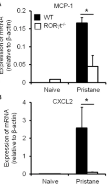
RORct Alters the Expression of MCP-1 in the Mesentery
It has been demonstrated that chemokines are responsible for myeloid cell migration. We reasoned that chemokine production may be altered in the absence of RORct during pristane-induced inflammation. We observed a significant reduction in the mRNA expression level of monocyte chemoattractant protein-1 (MCP-1) by mesenteric cells in RORct2/2 mice compared with the WT controls (Figure 3A). This finding may support the observation that RORct promotes macrophage recruitment to the mesentery Figure 3. RORct deficiency alters the expression of chemokines
by mesenteric cells during pristane-induced inflammation. Levels of mRNA of indicated chemokines in mesenteric cells from naı¨ve or pristane-injected WT and RORct2/2mice were measured by
quantitative PCR. Data show the average 6 SD of three samples, representing three independent experiments. *p,0.05, compared with
the control group.
doi:10.1371/journal.pone.0079497.g003
Figure 4. RORct deficiency reduces splenic macrophage expansion during pristane-induced inflammation.Total cells (A), macrophages (B) and neutrophils (C) in the spleens of naı¨ve or pristane-treated WT or RORct2/2mice were numerated. Data show the average
6SD of three samples, representing three independent experiments. *p,0.05, compared with the control group.
after pristane treatment. However, the expression of CXCL2, which mediates the migration of neutrophils, followed a similar pattern in the absence of RORct (Figure 3B), although RORct had no impact on the recruitment of neutrophils during pristane-induced inflammation (Figure 1I). This discrepancy suggests that the recruitment of neutrophils, or even other inflammatory leukocytes, may be also dependent on factors other than chemokine expression.
Effect of RORct on the Reservoir of Leukocytes in the Spleen during Pristane-induced Inflammation
The spleen is a site known for the storage and rapid deployment of leukocytes during inflammation [23]. Therefore, we asked whether the effects of RORct deficiency on myeloid cell recruitment to the mesentery after pristane treatment are also determined by the size of the reservoir of myeloid cells in the spleen. In concordance with previous reports [24], we found an increase in splenic cellularity in the absence of RORct at steady-state (Figure 4A). However, RORct deficiency had no impact on the number of macrophages or neutrophils (Figure 4B–C) at steady-state. Three weeks after pristane treatment, a further increase was observed in the total cellularity in the spleens of RORct2/2 mice (Figure 4A). However, macrophages was expanded to a less extend in the spleens of RORct2/2 mice compared with the WT controls (Figure 4B), which indicates that fewer splenic macrophages were available to mobilize towards the inflamed mesenteric tissue. Neutrophils, unlike macrophages, exhibited comparable expansion in the spleen when responding to pristane treatment in the WT controls and RORct2/2 mice (Figure 4C). These data correlated with the accumulation of macrophages and neutrophils in the mesenteric tissue during pristane-induced inflammation (Figure 1H–I).
Discussion
Hydrocarbon oils are commonly incorporated as adjuvants during the development of vaccines to augment the response to immunization [25,26]. However, exposure to hydrocarbon oils is also associated with the development of chronic inflammation and a number of pathologies including granulomas, plasmacytomas and autoimmune manifestations [1,2,3,4].
The granulomas act as a physical barrier that walls off substances. The disruption of granuloma structures can lead to the spread of the isolated substances, which results in further reactivation of diseases associated with the inducing stimuli [5,27]. Thus, it is crucial to understand the mechanisms that underlie hydrocarbon oil-induced chronic inflammation and granuloma formation.
Among the most potent hydrocarbon oils that induce chronic inflammation is the medium-length alkane pristane. After intra-peritoneal injection of pristane, a variety of leukocytes are recruited into the peritoneal cavity and later migrate to the mesentery [14]. Those inflammatory leukocytes include macro-phages, neutrophils, dendritic cells, B lymphocytes and T lymphocytes [15,28]. Small volumes of pristane are phagocytosed by macrophages, while larger volumes of pristane become surrounded by neutrophils and other inflammatory leukocytes to form oil-cell aggregates that adhere to peritoneal surfaces, which result in the formation of oil granulomas [5]. Therefore, macrophages and neutrophils play a critical role in controlling the development of inflammation because of their ability to entrap pristane. Defects in the recruitment of macrophages and neutrophils will result in inefficient development of oil granulomas and thereby prolong inflammation in the peritoneal cavity. This
phenomenon is supported by observations that when responding to pristane, fewer macrophages and neutrophils are recruited to the mesentery in B cell deficient mice or TNFa knockout mice, which results in the defective formation of oil granulomas [15]. Depending on the genetic background of mice, oil granulomas may represent a form of ectopic lymphoid tissue [27].
RORct is a transcription factor that regulates the development of lymph nodes and Peyer’s patches, the development of Th17 cells, and the progression of airway neutrophilic inflammation [17,20,29]. In this study, we reported that RORct promotes the formation of oil granulomas during pristane-induced inflammation by accelerating the recruitment of macrophages to the mesentery. However, RORct deficiency has no impact on the recruitment of neutrophils to the MG. The recruitment of macrophages to the inflammatory site is dependent on both the expression of corresponding chemokines and the reservoir of macrophages in lymphoid organs such as the spleen. This finding was supported by our observation that RORct2/2mice displayed less expansion in splenic macrophages and less MCP-1 expression in the mesentery compared with the wild type controls after pristane treatment. Macrophages migrate into the peritoneal cavity via circulation during inflammation. Therefore, we predicted that there are fewer circulating macrophages in RORct2/2mice compared with the wild type controls during pristane-induced inflammation; this finding should be examined further.
RORct induces the expression of Th17 cytokines including IL17, IL21 and IL22, which promotes tissue inflammation by the induction of other proinflammatory mediators and by the recruitment of leukocytes to the sites of inflammation. For example, IL17 is able to recruit macrophages via the expression of MCP-1 in rheumatoid arthritis synovial fibroblasts and macrophages [22]. IL17 induction of MCP-1 was mediated by phosphoinositide 3-kinase (PI3K) extracellular signal-regulated kinase (ERK) pathways in macrophages [22]. Based on the published data and our current findings, we hypothesized that RORct modulates pristane-induced inflammation through IL17 production, which recruits macrophages to the inflamed mesen-tery via MCP-1 expression.
T cells were shown to express RORct [30,31], but mice that lacked T cells were still able to develop oil granulomas competently [15]. This finding suggests that the RORct signal on T cells is dispensable for the development of mesenteric oil granulomas during pristane-induced inflammation. In addition to T cells, macrophages were also shown to express RORct [32]. The present study was unable to uncover the mechanisms by which pristane activates RORct in macrophages and how the RORct signal increases the number of macrophages in the spleen. We also could not determine if the RORct signal is what attributes to the formation of oil granulomas instead of local or circulating macrophages. These questions should be addressed in the future.
Supporting Information
Figure S1 Identification of cell infiltrate in mesentery after pristane treatment by flow cytometry. Single cell suspensions prepared from the spleens of pristane injected C57BL/6J mice (300ml pristane, 3 weeks) were stained and analyzed by flow cytometry. (A–B) Live cells were positively selected for analysis of CD11c expression, which results in identification of CD11c+
DCs and CD11c2 fraction. (C) Analyzing the expression of B220 of CD11c2 cells revealed B220+
fraction (conventional B cells). (D) The
the expression of CD11b and Gr-1 into CD11b+
Gr-1low (macrophages) and CD11b+
Gr-1hi(neutrophils). (TIF)
Figure S2 Influx of inflammatory leucocytes into the peritoneal cavity following pristane injection. Peritoneal cells were harvested from naı¨ve or pristane injected C57BL/6J mice at 3 weeks. Numbers of total cells (A), B cells (B), macrophages (C) and neutrophils (D) in peritoneal cavity were analyzed by flow cytometry.
(TIF)
Author Contributions
Conceived and designed the experiments: XS RC HC. Performed the experiments: QW XS LX. Analyzed the data: XL RC HC. Contributed reagents/materials/analysis tools: QW JF XS. Wrote the paper: QW XS JF HC.
References
1. Reeves WH, Lee PY, Weinstein JS, Satoh M, Lu L (2009) Induction of autoimmunity by pristane and other naturally occurring hydrocarbons. Trends Immunol 30: 455–464.
2. Spickard A 3rd, Hirschmann JV (1994) Exogenous lipoid pneumonia. Arch Intern Med 154: 686–692.
3. Satoh M, Reeves WH (1994) Induction of lupus-associated autoantibodies in BALB/c mice by intraperitoneal injection of pristane. J Exp Med 180: 2341– 2346.
4. Anderson PN, Potter M (1969) Induction of plasma cell tumours in BALB-c mice with 2,6,10,14-tetramethylpentadecane (pristane). Nature 222: 994–995. 5. Potter M, Maccardle RC (1964) Histology of Developing Plasma Cell Neoplasia
Induced by Mineral Oil in Balb/C Mice. J Natl Cancer Inst 33: 497–515. 6. Potter M, Walters JL (1973) Effect of intraperitoneal pristane on established
immunity to the adj-PC-5 plasmacytoma. J Natl Cancer Inst 51: 875–881. 7. Avcu F, Ural AU, Yilmaz MI, Ozcan A, Ide T, et al. (2005) The bisphosphonate
zoledronic acid inhibits the development of plasmacytoma induced in BALB/c mice by intraperitoneal injection of pristane. Eur J Haematol 74: 496–500. 8. Patten C, Bush K, Rioja I, Morgan R, Wooley P, et al. (2004) Characterization
of pristane-induced arthritis, a murine model of chronic disease: response to antirheumatic agents, expression of joint cytokines, and immunopathology. Arthritis Rheum 50: 3334–3345.
9. Hopkins SJ, Freemont AJ, Jayson MI (1984) Pristane-induced arthritis in Balb/c mice. I. Clinical and histological features of the arthropathy. Rheumatol Int 5: 21–28.
10. Satoh M, Kumar A, Kanwar YS, Reeves WH (1995) Anti-nuclear antibody production and immune-complex glomerulonephritis in BALB/c mice treated with pristane. Proc Natl Acad Sci U S A 92: 10934–10938.
11. Satoh M, Hamilton KJ, Ajmani AK, Dong X, Wang J, et al. (1996) Autoantibodies to ribosomal P antigens with immune complex glomerulone-phritis in SJL mice treated with pristane. J Immunol 157: 3200–3206. 12. Chowdhary VR, Grande JP, Luthra HS, David CS (2007) Characterization of
haemorrhagic pulmonary capillaritis: another manifestation of Pristane-induced lupus. Rheumatology (Oxford) 46: 1405–1410.
13. Lee PY, Li Y, Kumagai Y, Xu Y, Weinstein JS, et al. (2009) Type I interferon modulates monocyte recruitment and maturation in chronic inflammation. Am J Pathol 175: 2023–2033.
14. Chen H, Liao D, Cain D, McLeod I, Ueda Y, et al. (2010) Distinct granuloma responses in C57BL/6J and BALB/cByJ mice in response to pristane. Int J Exp Pathol 91: 460–471.
15. Chen H, Liao D, Holl TM, Snowden P, Ueda Y, et al. (2010) Genetic regulation of pristane-induced oil granuloma responses. Int J Exp Pathol 91: 472–483. 16. Lee PY, Kumagai Y, Xu Y, Li Y, Barker T, et al. (2011) IL-1alpha modulates
neutrophil recruitment in chronic inflammation induced by hydrocarbon oil. J Immunol 186: 1747–1754.
17. Ivanov, II, McKenzie BS, Zhou L, Tadokoro CE, Lepelley A, et al. (2006) The orphan nuclear receptor RORgammat directs the differentiation program of proinflammatory IL-17+T helper cells. Cell 126: 1121–1133.
18. Sun Z, Unutmaz D, Zou YR, Sunshine MJ, Pierani A, et al. (2000) Requirement for RORgamma in thymocyte survival and lymphoid organ development. Science 288: 2369–2373.
19. Kurebayashi S, Ueda E, Sakaue M, Patel DD, Medvedev A, et al. (2000) Retinoid-related orphan receptor gamma (RORgamma) is essential for lymphoid organogenesis and controls apoptosis during thymopoiesis. Proc Natl Acad Sci U S A 97: 10132–10137.
20. Ano S, Morishima Y, Ishii Y, Yoh K, Yageta Y, et al. (2013) Transcription factors GATA-3 and RORgammat are important for determining the phenotype of allergic airway inflammation in a murine model of asthma. J Immunol 190: 1056–1065.
21. Summers SA, Hoi A, Steinmetz OM, O9Sullivan KM, Ooi JD, et al. (2010) TLR9 and TLR4 are required for the development of autoimmunity and lupus nephritis in pristane nephropathy. J Autoimmun 35: 291–298.
22. Shahrara S, Pickens SR, Mandelin AM, 2nd, Karpus WJ, Huang Q, et al. (2010) IL-17-mediated monocyte migration occurs partially through CC chemokine ligand 2/monocyte chemoattractant protein-1 induction. J Immunol 184: 4479– 4487.
23. Swirski FK, Nahrendorf M, Etzrodt M, Wildgruber M, Cortez-Retamozo V, et al. (2009) Identification of splenic reservoir monocytes and their deployment to inflammatory sites. Science 325: 612–616.
24. Zhang N, Guo J, He YW (2003) Lymphocyte accumulation in the spleen of retinoic acid receptor-related orphan receptor gamma-deficient mice. J Immunol 171: 1667–1675.
25. Wilner BI, Evers MA, Troutman HD, Trader FW, McLean IW (1963) Vaccine Potentiation by Emulsification with Pure Hydrocarbon Compounds. J Immunol 91: 210–229.
26. Ehrich WE, Halbert SP, Mertens E, Mudd S (1945) Mechanism of the Augmenting Action of Mineral Oil on Antibody Production : Tissue Reactions and Antibody Response to Dysentery Vaccine in Saline, and in Saline-Lanolin-Mineral Oil Emulsion. J Exp Med 82: 343–360.
27. Nacionales DC, Kelly KM, Lee PY, Zhuang H, Li Y, et al. (2006) Type I interferon production by tertiary lymphoid tissue developing in response to 2,6,10,14-tetramethyl-pentadecane (pristane). Am J Pathol 168: 1227–1240. 28. Cancro M, Potter M (1976) The requirement of an adherent cell substratum for
the growth of developing plasmacytoma cells in vivo. J Exp Med 144: 1554– 1567.
29. Eberl G, Littman DR (2003) The role of the nuclear hormone receptor RORgammat in the development of lymph nodes and Peyer’s patches. Immunol Rev 195: 81–90.
30. Eberl G, Littman DR (2004) Thymic origin of intestinal alphabeta T cells revealed by fate mapping of RORgammat+cells. Science 305: 248–251. 31. Bezbradica JS, Hill T, Stanic AK, Van Kaer L, Joyce S (2005) Commitment
toward the natural T (iNKT) cell lineage occurs at the CD4+8+stage of thymic ontogeny. Proc Natl Acad Sci U S A 102: 5114–5119.