DM
September | 2014
Dina Maria Sousa Maciel
MASTER IN APPLIED BIOCHEMISTRYCell-Responsive Nanogels
for Anticancer Drug Delivery
SUPERVISOR Yulin Li
Dina Maria Sousa Maciel
MASTER IN APPLIED BIOCHEMISTRY
Cell-Responsive Nanogels
for Anticancer Drug Delivery
MASTER DISSERTATION
CELL-RESPONSIVE NANOGELS FOR ANTICANCER
DRUG DELIVERY
Tese apresentada à Universidade da Madeira com vista à
obtenção do grau de Mestre em Bioquímica Aplicada
Dina Maria Sousa Maciel
Sob a orientação de:
Doutor Yulin Li
Professora Doutora Helena Maria Pires Gaspar Tomás
Centro de Competências de Ciências Exatas e da Engenharia,
Centro de Química da Madeira
Funchal – Portugal
iii
Acknowledgments
The accomplishment of this master thesis was possible due to several people and I am
grateful to everyone that helped and contributed in any way to the execution of my project.
I want to acknowledge my supervisor Dr. Yulin Li for the support and for all the help
in clarifying and understanding during this project work.
To my supervisor Prof. Dr. Helena Tomás, for all the goodwill and motivation
throughout the year. And to whom I also show my appreciation for the readiness and
generosity.
To Prof. Dr. João Rodrigues and CQM for the support and for providing the facilities
to carry out this project.
I also would like to acknowledge Prof. Xiangyang Shi (CQM/UMa, Portugal and
Donghua University, Shanghai) for his collaboration, especially for the Scanning Electron
Microscopy analyses.
I am also grateful for the help and the readiness of the laboratory technicians Paula
Andrade and Paula Vieira, for providing the lab materials and reagents and in all that I
needed.
To my colleagues of the Molecular Materials Research Group (MMRG) for all the
support and enthusiasm. In particular, to my friends and colleagues Mara Gonçalves, Carla
Alves, Débora Capelo, Rita Castro, Cláudia Camacho, Nilsa Oliveira and Manuel Jardim for
their friendship, support and motivation. A special thanks to Mara, Rita, Débora and Carla
for the precious help in this project work.
To my dearest friends and Master colleagues Marisa Faria, Igor Fernandes, João
Micael Leça, and Marisela Santos for their friendship, support, enthusiasm and motivation
iv
I want to demonstrate my true and special thanks to my parents for their endless
support, concern and love. A special thanks to my brother Francisco, for always supporting
and encouraging me all the time.
This master project was financially supported by Fundaç̧ão para a Ciência e a
Tecnologia (FCT) through the Projects NAN/112428/2009 and
PTDC/CTM-NAN/116788/2010, the NMR Portuguese Network (PTNMR-2014) and the CQM strategic
project (Ref. PEst-OE/QUI/UI0674/2014). The work of Dr. Yulin Li (my supervisor) at
CQM was possible through the FCT Science 2008 Programme. For these reasons, I am deeply
grateful to FCT.
A great and sincere THANK YOU to all whom, directly or indirectly, contributed in
v This Master thesis was performed at Centro de Química da Madeira (CQM),
University of Madeira, consisting in the preparation and characterization of nanogels for drug
delivery.
For the student, it was an opportunity to learn/gain more experience on techniques
used in the preparation of nanogels, more specifically in emulsion methods, as well as in
bioconjugation synthetical procedures. The student also worked on animal cell culture,
UV-Vis, Fourier transformed infrared and fluorescence spectroscopies, dynamic light scattering
vii
Abstract
One of the main goals in Nanomedicine is to create innovative drug delivery systems
(DDS) capable of delivering drugs into a specific location with high efficiency. In the
development of DDS, some essential properties are desired, such as biocompatibility and
biodegradability. Furthermore, an ideal DDS should be able to deliver a drug in a controlled
manner and minimize its side effects. These two objectives are still a challenge for researchers
all around the world.
Nanogels are an excellent vehicle to use in drug delivery and several other applications
due to their biocompatibility. They are polymer-based networks, chemically or physically
crosslinked, with at least 80-90% water in their composition. Their properties can be tuned,
like the nanogel size, multifunctionality and degradability. Nanogels are capable of carrying
in their interior bioactive molecules and deliver them into cells.
The main objective of this project was to produce nanogels for the delivery of
anticancer drugs with the ability of responding to existent stimuli inside cells
(cell-responsiveness nanogels) and/or of controlled drug delivery. The nanogels were mainly based
on alginate (AG), a natural biopolymer, and prepared using emulsion approaches. After their
synthesis, they were used to encapsulate doxorubicin (Dox) which was chosen as a model
drug. In the first part of the experimental work, disulfide-linked AG nanogels were prepared
and, as expected, were redox-sensitive to a reducing environment like the intracellular
medium. In the second part, AG nanogels crosslinked with both calcium ions and cationic
poly(amidoamine) dendrimers were developed with improved sustained drug delivery. The
prepared nanogels were characterized in terms of size, chemical composition, morphology,
and drug delivery behavior (under redox/pH stimuli). The in vitro cytotoxicity of the nanogels
was also tested against CAL-72 cells (an osteosarcoma cell line).
ix
Resumo
Um dos principais objetivos da Nanomedicina é criar um sistema inovador de entrega
de fármacos, capaz de entregar com elevada eficácia os fármacos em locais específicos. No
desenvolvimento destes, são desejáveis propriedades como a biocompatibilidade e a
biodegradabilidade. Um sistema de entrega de fármacos ideal é capaz de entregar o fármaco
e minimizar os efeitos secundários a ele associados. Estes dois objetivos continuam a
representar um desafio para os investigadores de todo o mundo.
Os nanogéis são constituídos por redes à base de polímeros reticulados química ou
fisicamente, com pelo menos 80-90% de água na sua composição. São um excelente veículo
para uso na entrega de fármacos e em várias outras aplicações por apresentarem excelente
biocompatibilidade. Devido à sua estrutura, os nanogéis podem transportar no seu interior
moléculas ativas e entregá-las nas células. As suas propriedades podem ser controladas, tais
como o tamanho, a multifuncionalidade e a degradabilidade.
O principal objetivo deste projeto foi criar nanogéis para a entrega de fármacos
anticancerígenos com a capacidade de responder a estímulos presentes no interior das células
e/ou de libertar o fármaco de forma controlada. Os nanogéis foram constituídos à base de
alginato (AG), um biopolímero natural, e sintetizados utilizando métodos de emulsão. Após
a sua síntese, os nanogéis foram usados no encapsulamento de doxorrubicina (Dox),
escolhida como fármaco modelo. Na primeira parte do trabalho experimental, foram
preparados nanogéis de AG reticulados através de pontes dissulfureto e capazes de responder
a ambientes redutores como aqueles existentes no interior das células. Na segunda parte,
desenvolveram-se nanogéis de AG reticulados por ligações estabelecidas por iões cálcio e
dendrímeros catiónicos de poli(amidoamina) com uma capacidade melhorada de entregar o
fármaco de forma controlada. Estes nanogéis foram caracterizados em termos de dimensão,
composição química, morfologia e comportamento de libertação do fármaco (sob estímulos
do tipo redox/pH). A citotoxicidade dos nanogéis foi também testada usando células
CAL-72 (uma linha de células de osteossarcoma).
Palavras-chave: entrega de fármacos, nanogéis, responsividade celular, alginato,
xi
Table of Contents
CELL-RESPONSIVE NANOGELS FOR ANTICANCER DRUG DELIVERY ... i
Acknowledgments ... iii
Abstract ... vii
Resumo ... ix
List of Figures ... xiii
List of Tables ... xvii
List of Acronyms... xix
CHAPTERI.GENERAL INTRODUCTION ... 1
1. GENERAL INTRODUCTION ... 3
1.1. Drug Delivery Systems ... 3
1.2. Drug delivery systems based on polymers ... 4
1.3. Nanogels ... 5
1.4. Nanogels based on Alginate ... 7
1.5. Stimuli-responsive nanogels ... 9
1.6. General objectives of the thesis ... 12
References ... 14
CHAPTER II. REDOX-RESPONSIVE ALGINATE NANOGELS WITH ENHANCED ANTICANCER CYTOTOXICITY ... 19
Abstract ... 21
Introduction ... 22
Materials and Methods ... 25
Results and Discussion ... 29
Conclusions ... 38
References ... 39
CHAPTER III.DENDRIMER-ASSISTED FORMATION OF FLUORESCENT NANOGELS FOR DRUG DELIVERY AND INTRACELLULAR IMAGING ... 45
Abstract ... 47
xii
Materials and Methods ... 50
Results and Discussion ... 54
Conclusions ... 66
References ... 67
xiii
List of Figures
Figure 1. Types of nanocarriers used for transporting drugs, nucleic acids or proteins (adapted
from reference 2). ... 5
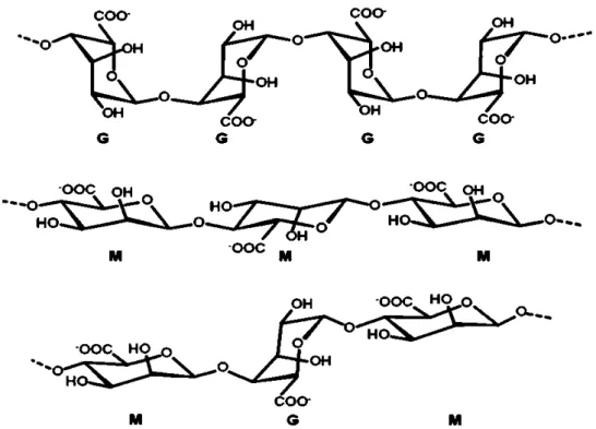
Figure 2. Structure of the β–D-mannuronic acid (M block) and α–L-guluronic acid (G block)
residues, and the alternating blocks in alginate (adapted from reference 34). ... 8
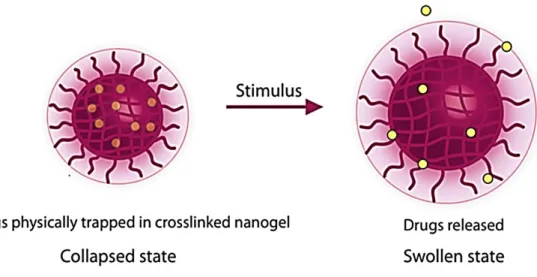
Figure 3. Behavior of nanogels with temperature, pH or other stimuli. e.g. the nanogel tend
to swallow at lower temperature and shrink/collapse at higher temperature (adapted
from reference 26). ... 9
Figure 4. Schematic illustration of the formation and drug release of Dox-loaded
(AG/Cys-Dox) nanogels. ... 26
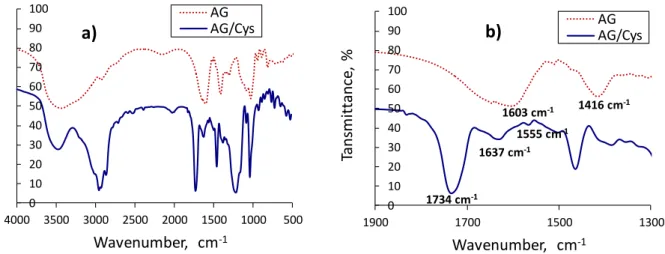
Figure 5. FTIR spectra of pure AG and AG/Cys nanogels (a); (b) is an enlarged view of the
spectra in the range of 1300 to 1900 cm-1. ... 29
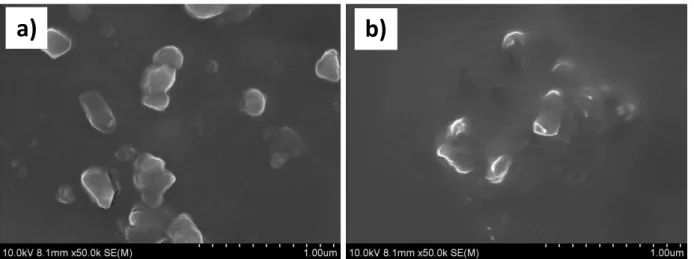
Figure 6. Scanning electron microscope (SEM) images of the AG/Cys (a) and AG/Cys-Dox
(b) nanogels. ... 31
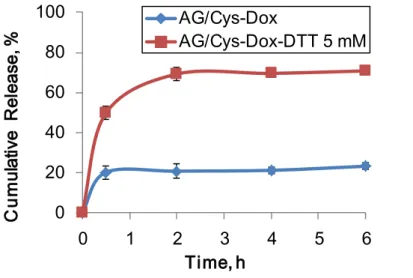
Figure 7.In vitro cumulative release of Dox from AG/Cys-Dox nanogels in the presence and
absence of DTT (5 mM) in PBS buffer (pH 7.4) at 37ºC. The results are expressed as the
mean + standard deviation (n = 3). ... 32
Figure 8. Cytotoxicity of free Dox, AG/Cys-Dox nanogels (with equivalent Dox
concentration), and AG/Cys nanogels (with equivalent weight concentration of the
corresponding AG/Cys-Dox nanogels) was analyzed after 48 h of cell culture using
CAL-72 cells. Results are reported as the mean + standard deviation (n = 4). One-way ANOVA
with Tukey’s Post Hoc test was used to assess the statistical difference between the group
means (**p < 0.01, ***p < 0.001). ... 34
Figure 9. Cell morphology (optical microscopy) of CAL-72 cells after 48 h in culture with (a)
control, (b) AG/Cys, (c) free Dox (0.5 µM), and (d) AG/Cys-Dox nanogels with an
equivalent amount of Dox (0.5 µM). ... 35
Figure 10. Bright field and fluorescence microscope images of CAL-72 cells after 2 and 4 h
culture with free Dox (0.5 µM) and AG/Cys-Dox with an equivalent amount of Dox (0.5
µM). The cell nucleus (blue) is stained with DAPI; Dox emits a red fluorescence signal.
... 36
Figure 11. Bright field and fluorescence microscope images of CAL-72 cells after 48 h culture
xiv
concentration (0.5 and 1.5 µM). The cell nucleus (blue) is stained with DAPI; Dox emits
a red fluorescence signal (The scale bar represents 100 µm). ... 37
Figure 12. Schematicoverview of the nanogels conjugated with FI, with Dox encapsulation
and the dual-crosslink. ... 49
Figure 13. Schematic illustration of the formation of nanogels through a double emulsion
method. Usually, an aqueous solution of hydrophilic polymers (precursor) is emulsified
in a surfactant organic solvent to form a water-in-oil (W/O) system. The mixture is then
re-emulsified in an aqueous solution of a second surfactant to obtain a
water-in-oil-in-water (W/O/W) system. The double-emulsified drops undergo physical and/or
chemical crosslinking, followed by organic solvent removal and purification (e.g.,
centrifugation) to obtain nanogels. ... 54
Figure 14. Scanning Electron Microscope (SEM) images of the AG-Dox (a) and
AG/G5-Dox (b) nanogels. ... 55
Figure 15. Sizes of AG, AG-Dox, AG/G5 and AG/G5-Dox nanogels in PBS as a function
of time at the pH values of 7.4 and 5.5. The results are expressed as the mean ± standard
deviation (n = 3). ... 56
Figure 16. The cumulative release profile of Dox from AG-Dox and AG/G5-Dox nanogels
in PBS buffer at the pH values of 7.4 and 5.5. An enlarged graph of the first 8 h (a), and
during 12 days (b). The results are expressed as the mean ± standard deviation (n = 3).
... 58
Figure 17. Cytotoxicity of AG/G5-Dox nanogels after 48 h using CAL-72 cells (a) and NIH
3T3 cells (b). AG-Dox, AG/G5-Dox and free Dox had equivalent Dox concentrations.
G5, AG/G5 and AG/G5-Dox nanogels had equivalent weight concentrations. Results
are reported as the mean ± standard deviation (n = 4). One-way ANOVA with Tukey’s
Post Hoc test was used to assess the statistical difference between the group means (*p <
0.05, ***p < 0.001). ... 59
Figure 18. Cell morphology of CAL-72 cells after 48 h in culture with (a) control, (b) AG/G5,
(c) AG, and (d) free Dox (2.78 μM), and (e) AG/G5-Dox nanogels and (f) AG-Dox with
an equivalent amount of Dox (2.78 μM). ... 60
Figure 19. Optical and fluorescence microscope images of CAL-72 cells after 4 h culture with
free Dox (0.50 μM), AG/G5-Dox and AG-Dox nanogels with an equivalent amount of
xv Figure 20. Enlarged optical and fluorescence microscope images of CAL-72 cells after 4 h
culture with AG/G5-Dox nanogels with an amount of Dox (0.50 μM). The cell nucleus
(blue) is stained with DAPI; Dox emits a red fluorescent signal (300x magnification). 62
Figure 21. Optical and fluorescence microscopy images of CAL-72 cells after 48 h culture
with (a-d) AG/G5-FI nanogels (50 μg/mL), (e-h) G5-FI (50 μg/mL). ... 63
Figure 22. Enlarged optical and fluorescence microscopy images of CAL-72 cells after 48 h
culture with AG/G5-FI nanogels (50 μg/mL). The cell nucleus (blue) is stained with
DAPI; FI emits a green fluorescent signal (300x magnification). ... 63
Figure 23.1H NMR spectrum of G5-FI in D2O. ... 64
Figure 24. Cytotoxicity of G5, G5-FI and AG/G5-FI nanogels after 48 h incubation with
CAL-72 cells. G5, G5-FI, and AG/G5-FI nanogels had equivalent weight
concentrations. Results are reported as the mean ± standard deviation (n = 3). One-way
ANOVA with Tukey’s Post Hoc test was used to assess the statistical difference between
xvii
List of Tables
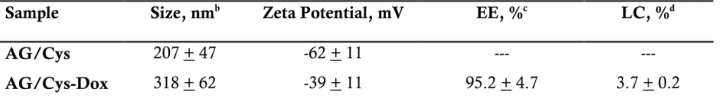
Table 1. Characterization of Dox-loaded AG/Cys Nanogels ... 30
xix
List of Acronyms
AA Antibiotic-antimycotic
Abs Absorbance
AG Alginate
AOT Dioctyl sodium sulfosuccinate
Cr Cumulative release
CST Critical solution temperature
Cys Cystamine
DAPI 4’,6-diamidino-2-phenyindole dilactate
DCM Dichloromethane
DDS Drug delivery system
DLS Dynamic light scattering
D-MEM Dulbecco’s modified eagle medium
DMSO Dimethyl sulfoxide
DNA Deoxyribonucleic acid
Dox Doxorubicin
DTT D,L-dithiothreitol
EDC 1-ethyl-3-(3-dimethylamino propyl)carbodiiamide hydrochloride
EPR Enhanced permeation and retention
FBS Fetal bovine serum
FDA Food and Drug Administration
FI Fluorescein isothiocyanate
FTIR Fourier transformed infrared spectroscopy
G5 Generation 5
GILT γ-interferon-inducible lysosomal thiol reductase
GSH Glutathione
hMSC Human mesenchymal stem cells
IC50 Half maximal inhibitory concentration
ITS Insulin-transferrin-selenium
LCST Lower critical solution temperature
MWCO Molecular weight cut off
NMR Nuclear magnetic resonance
xx
PBS Phosphate buffer saline
PEG Polyethylene glycol
PGA Polyglycolic acid
pKa Acid dissociation constant
PLGA Poly(lactic-co-glycolic acid)
PNIPAM Poly(N-isopropylacrylamide)
PVA Polyvinyl alcohol
RES Reticuloendothelial system
RNA Ribonucleic acid
rpm Revolutions per minute
SEM Scanning electron microscopy
UCST Upper critical solution temperature
UP Ultrapure
CHAPTER I. General Introduction
3
1.
GENERAL INTRODUCTION
1.1.
Drug Delivery Systems
Nowadays, nanoscience and nanotechnology are getting a huge attention due to the
advances that they are bringing to different scientific areas. The biomedical field, for example,
is one with increasing research developments and with real applications in diagnosis,
prevention and treatment of several diseases(1, 2). Other applications include tissue
engineering, biomedical implants and bionanotechnology(3).
Nanomedicine strongly relies on drug delivery systems (DDS) at the nanoscale which
shows unique physical, chemical and biological properties(1). DDS are being developed to
target and treat specific areas in the body taking advantage of complex formulations and drug
delivery controlled release(4). The DDS can also be designed having in mind the type of
administration route which can be oral, intravenous, arterial, transdermal, suppository, nasal,
subcutaneous, sublingual, amongst others(5). Issues related to low aqueous solubility of some
drugs, drug degradation by the biological system leading to lower drug efficiency and
undesirable drug accumulation in organs or tissues are unwanted effects that nanoscience
need to control(5). In anticancer therapy, for example, the DDS are often administrated
directly intravenously and it is important that the drug diffuses from the bloodstream to the
exact location, more specific/precisely to the tumor cells(6).
To accomplish the nanomedicine objectives, one should consider some factors when
designing DDS, like the chemical and physical properties of the drug, the route of
administration, the nature of the delivery vehicle, the drug release mechanism, the potential
for cell/tissue targeting and, above all, the biodegradability and biocompatibility of the
system(4, 7, 8). All these factors have a huge impact when designing an ideal DDS, but are not
easy to be taken in mind in a single system.
The main goal to develop DDS is to improve the bioavailability and pharmacokinetics
of the therapeutic agents, with systems capable of passing through the biological barriers and
deliver the drugs into a specific location(1, 9, 10). However, DDS still present some problems
that need to be overcome, like poor intracellular delivery, lack of control over the release
behavior and difficulty in targeting the diseased cells/tissues which can lead to important side
effects (the DDS, themselves, can show some toxicity)(11). In summary, DDS should: present
a high loading capacity; maintain an optimal therapeutic drug concentration in the blood;
show a sustained drug delivery with predictable and reproducible release rates with no early
CHAPTER I. General Introduction
4
tissues; conduct to reduced side effects by being made of biocompatible/biodegradable
materials; and allow patient compliance and optimized therapy(4, 5, 12). One important fact is
that these systems can even modulate and change the biodistribution profile of the drugs
depending on the different methods of administration(1). Furthermore, with the use of an ideal
DDS, just a minor amount of drug will be required to obtain a therapeutic effect and a
decrease on the side effects(13).
1.2.
Drug delivery systems based on polymers
The DDS used in nanomedicine include viral vectors, polymer-based vehicles (e.g.
dendrimers, peptides and polymersomes), lipid based nanosystems (e.g. micelles and
liposomes), carbon nanotubes, and inorganic nanomaterials (e.g. nanodiamonds, gold and
mesoporous silica nanoparticles) (Figure 1)(2, 14).
DDS based on polymer are from utmost importance. For their preparation, the
polymers can be from natural or synthetic origin and degradable or nondegradable. Natural
polymers are polysaccharides, such as alginate(15, 16), chitosan(7), gelatin(15), cellulose(7),
hyaluronic acid(17), amongst others(18). These natural polymers are abundant in nature, from
renewable sources, with low cost, nontoxic, and present a high content of functional groups
(such as hydroxyl, amino and carboxylic acid groups) that can be used in reactions for further
bioconjugation (for example, with the purpose of cell targeting)(3, 18, 19). Synthetic polymers are
those that are produced by polymerization, such as dendrimers(9), polyglycolic acid (PGA)(7),
polyethylene glycol (PEG)(9, 13, 17), and poly(lactic-co-glycolic acid) (PLGA)(7, 20). Compared to
natural polymers, synthetic ones have better controllable physicochemical properties.
Degradable polymers contain labile bonds such as ester-, amide- and anhydride-bonds that
are susceptible to hydrolysis or enzymatic degradation (surface degradation or bulk
degradations)(21). These DDS based on polymers can physically encapsulate bioactive
molecules within the polymeric network, including small drugs, proteins, and DNA/RNA.
Alternatively, they can immobilize the bioactive molecules through chemical linkages. The
incorporation of inorganic materials (such as quantum dots, and magnetic and gold
nanoparticles) inside the polymer structure can also be done to obtain multifunctional systems
(theranostic systems) that may have a therapeutic action beyond drug delivery(22).
Actually, these polymer-based DDS can be found in a vast diversity of
designs/architectures such as linear or branched polymers, dendrimers, polymersomes, and
CHAPTER I. General Introduction
5 The design of a polymer-based nanocarrier is important for its chemical, interfacial,
mechanical and biological functions(4). For example, the nanocarrier surface properties, such
as hydrophilicity, lubricity, smoothness and surface energy, have a strong influence on its
interaction with tissues and blood. Other important properties of a nanocarrier is its
durability, permeability and degradability(4).
Figure 1. Types of nanocarriers used for transporting drugs, nucleic acids or proteins (adapted from
reference 2).
1.3.
Nanogels
Nanogels are a special group of DDS based on hydrophilic polymers (it retains water
in their structure). They are crosslinked hydrogel particles(3), within a size range between 10-9
CHAPTER I. General Introduction
6
nanomaterials that have excellent interior structures for drug encapsulation(7), and present all
the properties of the bulk hydrogels, but at the nanoscale dimensions. So, nanogels are
crosslinked polymeric particles with a high water content.
The crosslinking in nanogels can be established by non-covalent physical associations,
covalent chemical linkages, or combinations between them(23). The crosslinking methods that
can be used to prepare nanogels include the use of ions, radiation, molecules with special
functional groups, self-assembly, crystallization and crosslinking polymerization(24). Nanogels
formed by physical crosslinking occur via non-covalent attractive forces, namely
hydrophilic-hydrophilic interactions, hydrophobic-hydrophobic interactions, ionic interactions and/or
hydrogen bonding(23, 24). The properties of these nanogels are dependent on
polymer/crosslinking agent composition and concentration, temperature and ionic strength
of the medium. Nanogels formed by chemical crosslinking imply the reaction between
crosslinking points along the backbone of the polymer chain(24) and usually makes use of
crosslinking molecules. The properties of these nanogels (such as porosity and swelling) are
strongly determined by the type of crosslinker and the extent of crosslinking reactions. The
chemically crosslinked nanogels are, in principle, more stable than the physically
crosslinked(23) but the use of crosslinkers may raise concerns related with toxicity(13).
Because water is in their composition in large percentage, nanogels are usually
biocompatible, and have suitable mechanical properties for DDS formulation(18, 22, 24). The
physical properties that are mutual between the nanogels and the living tissues, are the
consistency (soft and rubbery) and the low interfacial tension with water or biological fluids,
which reduces the chances of a negative immune reaction (because protein adsorption and
cell adhesion is minimized)(23). Furthermore, they show a huge loading capacity of
water-soluble compounds(13, 25). They are an excellent reservoir for drugs, oligonucleotides and
imaging agents, which is due to the porosity inside the crosslinked network that also protects
their cargo from possible environmental degradation(24, 25). Nanogels can have multifunctional
properties that are dependent on their crosslinking density, chemical functional groups, and
surface-active and stimuli-responsive constituents(24). In fact, among other applications,
nanogels have been used for drug delivery, but can also be applied in other fields such as
sensing, diagnostics and bioengineering(26).
These nanocarriers present several advantages such as a three-dimensional (3D)
tunable size and physical structure, a large surface area for multivalent bioconjugation, a
network for the incorporation of biomolecules and biodegradability for a sustained drug
CHAPTER I. General Introduction
7 circulation time and the option of being actively or passively targeted for a specific location,
like tumor sites(27).
Some important issues in nanogel development must be considered. For example, their
stability in biological fluids which is essential to avoid aggregation(13, 24). It has been reported
that nanogels with sizes around 100-200 nm have a higher cellular internalization efficiency,
whereas small nanogels may result in a lower drug encapsulation and a fast drug release(13).
On the other hand, negatively charged nanogels are better in terms of resistance to protein
interaction (they have a longer blood circulation half-life), while positively charged nanogels
are more susceptible to interact with the serum components (which may cause aggregation
and minimize the blood circulation half-life)(28). Nevertheless, negatively charged nanogels
may suffer repulsion by the negatively charged cell membrane, whereas the positively charged
nanogels are more easily internalized by cells(28).
The combination of specific properties like targetability and stimuli-responsiveness can
be used to create the perfect nanogel(29). Indeed, nanogels have a characteristic which make
them very interesting materials. They can be designed to be responsive to environmental
stimuli like pH, temperature, ion strength or reduction agents (D,L-dithiothreitol (DTT),
etc)(30), thus being an exceptional platform for biomedical applications.
1.4.
Nanogels based on Alginate
Polysaccharides are known for their excellent physicochemical properties,
biocompatibility and biodegradability(31). Alginate is a linear polysaccharide composed of β–
D-mannuronic acid and α–L-guluronic acid residues (Figure 2)(18, 27, 32-34). It is a biocompatible
and biodegradable natural polymer, extracted from marine brown algae or produced by
bacteria(16, 33-35). This polymer is considered safe by the U.S. Food and Drug Administration
(FDA) for use in biomedical applications and is easily crosslinked in the presence of
multivalent cations (calcium, barium, strontium, iron and aluminum) to form hydrogels
which can be prepared at the nanoscale(32, 35-37). Ionic crosslinking with calcium ions is the
most used method to prepare alginate hydrogels and consists in the combination of an
aqueous alginate solution with a solution of divalent calcium cations(34). It is believed that the
divalent cations bind to the guluronic blocks of the alginate chains, and form junctions in the
CHAPTER I. General Introduction
8
Figure 2. Structure of the β–D-mannuronic acid (M block) and α–L-guluronic acid (G block) residues,
and the alternating blocks in alginate (adapted from reference 34).
As a consequence of the negative charge of its residues, alginate has a high degree of
aqueous solubility, and a tendency for gelation under suitable conditions giving rise to
nontoxic gels with a high porosity, low immunogenicity, and flexible to random geometries
(27, 37). Alginate nanogels have many biomedical applications due to their exceptional
properties, being able to deliver bioactive peptides and proteins, genes, and small drugs.
Alginate macro or microgels can even encapsulate living cells, such as fibroblasts, human
mesenchymal stem cells (hMSC), or others (16, 32). Beyond these applications, alginate can also
be used in scaffolds for protein immobilization, and for neural, bone and cartilage tissue
engineering(34, 37). However, ionic crosslinked alginate nanogels usually exhibit poor
mechanical properties, uncontrolled degradation under the physiological conditions and a
burst drug release because of cation exchange in the biological medium (e.g., exchange of
calcium ions for sodium ions)(36). The use of an anionic surfactant, like dioctyl sodium
sulfosuccinate (AOT), can overcome this limitation, through the formation of a bilayer
around alginate nanogels, producing a sustained release of the drug(36). Another method to
improve alginate based nanogel properties is through covalent crosslinking (e.g., by the
reaction of the hydroxyl and carboxylic acid functional groups) with reactive groups present
CHAPTER I. General Introduction
9
1.5.
Stimuli-responsive nanogels
Stimuli-responsive nanogels are known to be sensitive/responsive when exposed to
external signals, such as physical or chemical changes of the environment, are capable of
changing their behavior. They are also named as “environment sensitive”, “smart” or
“intelligent” polymers(27, 29, 31). These nanogels are able of controlled drug release in vivo when
a specific stimuli is triggered in the target site(21). The response of nanogels to changes in the
environment may be physical (e.g., variations in solubility, macromolecular structure, surface
properties, swelling, and disassembly) or even a chemical reaction(29, 38). The external signals
can be also classified in physical signals (such as changes in temperature, electric or magnetic
fields, and mechanical stresses (ultrasound)), and chemical signals (such as changes in pH,
ionic strength, and concentration of specific molecules like enzymes or reducing agents)
(demonstrated in Figure 3)(10, 29, 31, 39). The release profile of the nanogels can be regulated by
the stimuli-responsive units that are incorporated in the nanogel network(24).
Dual responsive nanogels can also be prepared and, in fact, nanogels sensitive to pH
and temperature variations have been widely studied(30, 39, 40). Nanogels that are simultaneously
sensitive to pH and redox potential have been also reported(41). Indeed, although many
different stimuli can be used to control the behavior of nanogels (in particular their drug
release behavior), the pH, the temperature and the redox potential can be considered the most
important.
Figure 3. Behavior of nanogels with temperature, pH or other stimuli. e.g. the nanogel tend to swallow
CHAPTER I. General Introduction
10
pH-responsive nanogels
It is well known that the extracellular environment of solid tumors may exhibit an
acidic pH value (around 6.5)(3), whereas normal tissues present a pH around 7.4(31, 41).
Furthermore, even more acidic pH values can be found in the human body, like in some
intracellular compartments (endosomes and lysosomes presents a pH between 5.0 to 5.5)(3, 29).
For this reason, efforts have been done to develop pH-responsive nanogels that are stable at
physiological pH but reacts to pH changes towards lower values for a more efficient delivery
of small drugs, nucleic acids or proteins in target sites(29, 31). pH-responsive polymers can be
classified into two categories: polymers with ionizable groups, and polymers with degradable
linkages(24, 31). In the first category, polymers having weak acid or base groups, such as
carboxylic acids, phosphoric acids and amines, can present a change in the ionization state
by varying the pH(31). The nanogels made from these polymers show an accelerated drug
release that can be controlled through disassembly or dissolution(29, 31). These polymers also
have an important characteristic which is the ability to dissociate and associate with protons
in the aqueous environment, and therefore can be used as proton donors or acceptors(29, 40).
The second category includes polymers that contain acid-degradable linkages(31). The
nanogels made from these polymers may suffer induced cleavage in acidic conditions that
results in an increase of the porosity and, possibly, further polymer dissolution(31).
In response to changes in pH, nanogels based on polymers with ionizable groups go
through a volume deformation(40). When the pH of the solution is higher than the pK
a of the
nanogel, e.g. poly(acrylic acid), the carboxylic groups are deprotonated carrying a negative
charge, and resulting in the swelling of the nanogel. If these nanogels are loaded with
hydrophilic drugs, then the swallow will result in a controlled drug release into the external
medium. However, when the pH is lower than the pKa of the nanogel, the carboxylic groups
are protonated shrinking the nanogel(40).
The applications of pH-responsive nanogels in drug delivery is very important and, in
particular, in the delivery of anticancer therapeutics.
Thermoresponsive nanogels
Thermoresponsive (or temperature-responsive nanogels) are prepared from
thermosensitive polymers. These polymers show a temperature-dependent phase transition in
CHAPTER I. General Introduction
11 (29). For biomedical applications the transition temperature of the polymers should be between
10-40ºC to be effective(17). If a polymer is water soluble below a defined temperature value
and exists as a separated phase above that value, then it shows a lower critical solution
temperature (LCST). If a polymer reveals the opposite behavior, then it shows an upper
critical solution temperature (UCST)(29).
The most studied thermoresponsive polymer is poly(N-isopropylacrylamide),
PNIPAM. Since it was first reported in 1968, PNIPAM is extensively used in biomaterials,
bioconjugates, actuators, and sensors(17). This polymer present a LCST about 32ºC because it
goes through a reversible phase transition in water, from a swollen state (below 32ºC) to a
collapsed state (above 32ºC)(29, 39, 40, 42, 43). By showing this behavior, it can be used to
incorporate hydrophilic and hydrophobic molecules(17, 29, 30, 39, 40, 42, 44) that are released in
response to temperature changes (PNIPAM hydrogels present a reversible
swelling-deswelling behavior)(13). At 32ºC, PNIPAM becomes insoluble in water, shrinking, due to a
disruption of the hydrogen bonds formed between NIPAM units and water molecules(17, 30, 39).
With these features, thermoresponsive nanogels with PNIPAM may have very interesting
and promising applications in the biomedical field, like the treatment of certain cancers
through hyperthermia(45). They can be loaded with an anticancer drug and, at the target
location, by moderately increasing the temperature above the LCST, the nanogel can change
of volume and the drug release can be accelerated(13, 29).
Redox-sensitive nanogels
The intracellular and the extracellular environment have a huge difference in terms of
reduction potential, and that is being explored for triggering the intracellular delivery of
drugs(41). For example, a reductive environment, such as the presence of an excess of
glutathione (GSH), could be a powerful stimulus for drug delivery in the case of nanogels
containing reducible bonds, such as disulfide bonds(28). Actually, it was reported that the
cytosol of cancer cells has a concentration of GSH around 2-10 mM, which corresponds to
1000 times more than that existent in the extracellular fluids, that presents a concentration
between 2-20 µM(3, 11, 41, 46, 47). So, the presence of GSH in the cytosol can promote the cleavage
of disulfide bonds existent in nanogels and help the release of the encapsulated drugs when
using DDS(11, 28). On the other side, because of the low GSH concentration, the disulfide
linkages should be stable in the circulation in normal physiological conditions(41). It was
-CHAPTER I. General Introduction
12
interferon-inducible lysosomal thiol reductase (GILT) and also of cysteine, both possibly
contributing for a strong reducible environment(48).
1.6.
General objectives of the thesis
The main goal of this thesis was to develop alginate (AG)-based nanogels for
anticancer drug delivery with cell-responsiveness and/or sustained drug delivery behavior.
The anticancer drug used in this study was Doxorubicin (Dox), a model drug extensively used
to treat several types of cancers.
In more detail:
a) The first objective (Chapter II) was to prepare biocompatible redox-responsive
nanogels based on disulfide-linked AG for intracellular delivery of Dox. The
reducible nanogels were obtained through in situ crosslinking of AG by cystamine
(Cys) via a miniemulsion method. The nanogels were characterized by Fourier
transformed infrared spectroscopy (FTIR), dynamic light scattering (DLS), zeta
potential measurements and scanning electron microscopy (SEM). Dox was
loaded into the nanogels by simply mixing in aqueous solution, and the in vitro
drug release was studied under normal and reductive conditions by UV-Vis
spectroscopy. The antitumor activity was quantitatively and qualitatively studied
against CAL-72 cells (an osteosarcoma cell line) using a cell metabolic activity
assay and fluorescence microscopy, respectively.
b) The second goal (Chapter III) was to develop AG nanogels with dual crosslinking
for improved sustained drug delivery. The nanogels were prepared through an
emulsion method using calcium ions and cationic poly(amidoamine) (PAMAM)
dendrimers of generation 5 (G5) as crosslinkers. Furthermore PAMAM
dendrimers were conjugated with fluorescein isothiocyanate (FI), a fluorescent
marker, for following the path of nanogels once inside cells. The characterization
techniques involved nuclear magnetic resonance (NMR), dynamic light scattering
CHAPTER I. General Introduction
13 The in vitro drug release was studied by UV-Vis spectroscopy and the biological
assays (antitumor activity of the Dox loaded nanogels and their tracking inside
cells) were performed using CAL-72 cells (an osteosarcoma cell line) and NIH 3T3
fibroblasts (a non-carcinogenic cell line, used as a model of normal cells). Also in
this case, quantitatively and qualitatively results were obtained through a cell
CHAPTER I. General Introduction
14
References
1. Kumar A, Chen F, Mozhi A, Zhang X, Zhao Y, Xue X, et al. Innovative
pharmaceutical development based on unique properties of nanoscale delivery formulation.
Nanoscale. 2013;5:8307-8325.
2. Berindan-Neagoe I, Braicu C, Craciun L, Irimie A, Takahashi Y. Nanopharmacology
in translational hematology and oncology. Int J Nanomed. 2014;9:3465-3479.
3. Wen Y, Oh JK. Dual-stimuli reduction and acidic pH-responsive bionanogels:
intracellular delivery nanocarriers with enhanced release. RSC Adv. 2014;4:229-237.
4. del Valle EMM, Galán MA, Carbonell RG. Drug Delivery Technologies: The Way
Forward in the New Decade. Ind Eng Chem Res. 2009;48: 2475–2486.
5. Verma G, Hassan PA. Self assembled materials: design strategies and drug delivery
perspectives. Phys Chem Chem Phys. 2013;15:17016-17028.
6. Baguley BC. Multiple drug resistance mechanisms in cancer. Mol Biotechnol.
2010;46:308-316.
7. Vashist A, Vashist A, Guptac YK, Ahmad S. Recent advances in hydrogel based drug
delivery systems for the human body. J Mater Chem B. 2014;2:147-166.
8. Park K, Mrsny RJ. Controlled Drug Delivery: Present and Future. Park K, Mrsny RJ,
editors. In: Controlled Drug Delivery. 752. United States of America: American Chemical
Society; 2000. p. 2-12.
9. Khandare J, Calderon M, Dagia NM, Haag R. Multifunctional dendritic polymers in
nanomedicine: opportunities and challenges. Chem Soc Rev. 2012;41:2824-2848.
10. Gao W, Chan JM, Farokhzad OC. pH-Responsive Nanoparticles for Drug Delivery.
Mol Pharmaceutics. 2010;7:1913-1920.
11. Liu J, Detrembleur C, Hurtgen M, Debuigne A, De Pauw-Gillet M-C, Mornet S, et al.
Reversibly crosslinked thermo- and redox-responsive nanogels for controlled drug release.
Polym Chem. 2014;5:77-88.
12. Zhang L, Li Y, Yu JC. Chemical modification of inorganic nanostructures for targeted
and controlled drug delivery in cancer treatment. J Mater Chem B. 2014;2:452-470.
13. Qian Z-Y, Fu S-Z, Feng S-S. Nanohydrogels as a prospective member of the
nanomedicine family. Nanomedicine. 2013;8:161-164.
14. Ang CY, Tan SY, Zhao Y. Recent advances in biocompatible nanocarriers for delivery
CHAPTER I. General Introduction
15 15. Sarker B, Papageorgiou DG, Silva R, Zehnder T, Gul-E-Noor F, Bertmer M, et al.
Fabrication of alginate–gelatin crosslinked hydrogel microcapsules and evaluation of the
microstructure and physico-chemical properties. J Mater Chem B. 2014;2:1470-1482.
16. Higham AK, Bonino CA, Raghavan SR, Khan SA. Photo-activated ionic gelation of
alginate hydrogel: real-time rheological monitoring of the two-step crosslinking mechanism.
Soft Matter. 2014;10:4990-5002.
17. Moon HJ, Ko du Y, Park MH, Joo MK, Jeong B. Temperature-responsive compounds
as in situ gelling biomedical materials. Chem Soc Rev. 2012;41:4860-4883.
18. Oh JK, Lee DI, Park JM. Biopolymer-based microgels/nanogels for drug delivery
applications. Prog Polym Sci. 2009;34:1261-1282.
19. Nicolas J, Mura S, Brambilla D, Mackiewicz N, Couvreur P. Design,
functionalization strategies and biomedical applications of targeted
biodegradable/biocompatible polymer-based nanocarriers for drug delivery. Chem Soc Rev.
2013;42:1147-1235.
20. Weinberg BD, Patel RB, Exner AA, Saidel GM, Gao J. Modeling doxorubicin
transport to improve intratumoral drug delivery to RF ablated tumors. J Control Release.
2007;124:11-19.
21. Fu Y, Kao WJ. Drug release kinetics and transport mechanisms of non-degradable
and degradable polymeric delivery systems. Expert Opin Drug Deliv. 2010;7:429-444.
22. Oh JK, Drumright R, Siegwart DJ, Matyjaszewski K. The development of
microgels/nanogels for drug delivery applications. Prog Polym Sci. 2008;33:448-477.
23. Wu W, Zhou S. Hybrid micro-/nanogels for optical sensing and intracellular imaging.
Nano Reviews. 2010;1:5730.
24. Yallapu MM, Jaggi M, Chauhan SC. Design and engineering of nanogels for cancer
treatment. Drug Discov Today. 2011;16:457-463.
25. Moya-Ortega MD, Alvarez-Lorenzo C, Concheiro A, Loftsson T. Cyclodextrin-based
nanogels for pharmaceutical and biomedical applications. Int J Pharm. 2012;428:152-163.
26. Chacko RT, Ventura J, Zhuang J, Thayumanavan S. Polymer nanogels: A versatile
nanoscopic drug delivery platform. Adv Drug Deliver Rev. 2012;64:836-851.
27. Hamidi M, Azadi A, Rafiei P. Hydrogel nanoparticles in drug delivery. Adv Drug
Deliv Rev. 2008;60:1638-1649.
28. Guo X, Shi C, Yang G, Wang J, Cai Z, Zhou S. Dual-Responsive Polymer Micelles
CHAPTER I. General Introduction
16
29. Li Y, Dong H, Wang K, Shi D, Zhang X, Zhuo R. Stimulus-responsive polymeric
nanoparticles for biomedical applications. Sci China Chem. 2010;53:447-457.
30. Peng J, Qi T, Liao J, Fan M, Luo F, Li H, et al. Synthesis and characterization of novel
dual-responsive nanogels and their application as drug delivery systems. Nanoscale.
2012;4:2694-2704.
31. Binauld S, Stenzel MH. Acid-degradable polymers for drug delivery: a decade of
innovation. Chem Commun. 2013;49:2082-2102.
32. Li Y, Rodrigues J, Tomás H. Injectable and biodegradable hydrogels: gelation,
biodegradation and biomedical applications. Chem Soc Rev. 2012;41:2193-2221.
33. Coviello T, Matricardi P, Marianecci C, Alhaique F. Polysaccharide hydrogels for
modified release formulations. J Control Release. 2007;119:5-24.
34. Lee KY, Mooney DJ. Alginate: properties and biomedical applications. Prog Polym
Sci. 2012;37:106-126.
35. Jejurikar A, Seow XT, Lawrie G, Martin D, Jayakrishnan A, Grøndahl L. Degradable
alginate hydrogels crosslinked by the macromolecular crosslinker alginate dialdehyde. J
Mater Chem. 2012;22:9751-9758.
36. Vrignaud S, Benoit JP, Saulnier P. Strategies for the nanoencapsulation of hydrophilic
molecules in polymer-based nanoparticles. Biomaterials. 2011;32:8593-8604.
37. Zhao W, Jin X, Cong Y, Liu Y, Fu J. Degradable natural polymer hydrogels for
articular cartilage tissue engineering. J Chem Technol Biot. 2013;88:327-339.
38. Cao Z, Ziener U. Synthesis of nanostructured materials in inverse miniemulsions and
their applications. Nanoscale. 2013;5:10093-10107.
39. Pasparakis G, Vamvakaki M. Multiresponsive polymers: nano-sized assemblies,
stimuli-sensitive gels and smart surfaces. Polym Chem. 2011;2:1234-1248.
40. Lim HL, Hwang Y, Kar M, Varghese S. Smart hydrogels as functional biomimetic
systems. Biomater Sci. 2014;2:603-618.
41. Chen J, Qiu X, Ouyang J, Kong J, Zhong W, Xing MM. pH and reduction
dual-sensitive copolymeric micelles for intracellular doxorubicin delivery. Biomacromolecules.
2011;12:3601-3611.
42. Doring A, Birnbaum W, Kuckling D. Responsive hydrogels - structurally and
dimensionally optimized smart frameworks for applications in catalysis, micro-system
technology and material science. Chem Soc Rev. 2013;42:7391-7420.
43. Schmaljohann D. Thermo- and pH-responsive polymers in drug delivery. Adv Drug
CHAPTER I. General Introduction
17 44. Chiang WH, Ho VT, Huang WC, Huang YF, Chern CS, Chiu HC. Dual
stimuli-responsive polymeric hollow nanogels designed as carriers for intracellular triggered drug
release. Langmuir. 2012;28:15056-15064.
45. Quan CY, Sun YX, Cheng H, Cheng SX, Zhang XZ, Zhuo RX. Thermosensitive
P(NIPAAm-co-PAAc-co-HEMA) nanogels conjugated with transferrin for tumor cell
targeting delivery. Nanotechnology. 2008;19:275102.
46. Zhu J, Shi X. Dendrimer-based nanodevices for targeted drug delivery applications. J
Mater Chem B. 2013;1:4199-4211.
47. Wang X, Cai X, Hu J, Shao N, Wang F, Zhang Q, et al. Glutathione-triggered
"off-on" release of anticancer drugs from dendrimer-encapsulated gold nanoparticles. J Am Chem
Soc. 2013;135:9805-9810.
48. Cheng R, Feng F, Meng F, Deng C, Feijen J, Zhong Z. Glutathione-responsive
nano-vehicles as a promising platform for targeted intracellular drug and gene delivery. J Control
CHAPTER II. Redox-Responsive Alginate Nanogels with
Enhanced Anticancer Cytotoxicity
*This Chapter is based on the following publication:
Maciel D, Figueira P, Xiao S, Hu D, Shi X, Rodrigues J, Tomás H, Li Y. Redox-Responsive
Alginate Nanogels with Enhanced Anticancer Cytotoxicity. Biomacromolecules.
CHAPTER II. Redox-Responsive Alginate Nanogels with Enhanced Anticancer Cytotoxicity
21
Abstract
Although doxorubicin (Dox) has been widely used in the treatment of different types
of cancer, its insufficient cellular uptake and intracellular release is still a limitation. Herein,
we report an easy process for the preparation of redox-sensitive nanogels which were shown
to be highly efficient in the intracellular delivery of Dox. The nanogels (AG/Cys) were
obtained through in situ crosslinking of alginate (AG) using cystamine (Cys) as a crosslinker
via a miniemulsion method. Dox was loaded into the AG/Cys nanogels by simply mixing it
in aqueous solution with the nanogels, that is, by the establishment of electrostatic
interactions between the anionic AG and the cationic Dox. The results demonstrated that the
AG/Cys nanogels are cytocompatible, have a high drug encapsulation efficiency
(95.2±4.7%), show an in vitro accelerated release of Dox in conditions that mimic the
intracellular reductive conditions, and can quickly be taken up by CAL-72 cells (an
osteosarcoma cell line), resulting in higher Dox intracellular accumulation, and a remarkable
cell death extension when compared with free Dox. The developed nanogels can be used as
a tool to overcome the problem of Dox resistance in anticancer treatments, and possibly be
used for the delivery of other cationic drugs in applications beyond cancer.
CHAPTER II. Redox-Responsive Alginate Nanogels with Enhanced Anticancer Cytotoxicity
22
Introduction
Cancer is one of the most serious diseases around the world. Doxorubicin (Dox), one
of the smallest anticancer drugs, has been widely used for chemotherapy of several kinds of
cancers of different organs, including bone(1), liver(2, 3) or breast(4). Dox is a member of the
anthracycline family of anticancer drugs, and its use for cancer treatment can lead to a
sequence of complications, such as tumor resistance, cellular toxicity and particularly
cardiotoxicity(5). In the other hand, Dox presents antitumor activity since it intercalates in the
DNA double helix and, as a consequence, inhibits DNA replication and the biosynthesis of
macromolecules(5, 6).
However, Dox is a weak base with a pKa of 8.30 and tends to undergo ion trapping in
acidic conditions of the extracellular microenvironment of solid tumors (pH of 6.5 to 6.9) and
in the internal milieu of endolysosomes (pH of 5.0 to 5.5)(6, 7). The ion trapping phenomenon
is caused by the acidic regions of solid tumors creating a physiological barrier for the cellular
uptake of weak bases, that are seized by acidic compartments, leading to drug resistance(7, 8).
This occurs when there is a big difference between the permeabilization of ionized and
nonionized species of a drug(7).
Weak base drugs, such as Dox, ionize in solution, and an equilibrium is established
between the protonated species with the uncharged, unprotonated form of the drug(8). While
uncharged Dox can freely permeate membranes, the protonated Dox has a lower membrane
permeability becoming trapped inside acidic compartments. Furthermore, Dox has been
reported to have multidrug resistance(9, 10), possibly because of the p-glycoprotein, also known
as the multidrug resistance protein, that is responsible for pumping unfamiliar molecules out
of the cell. Therefore, free Dox can likely be pumped out of the cells by p-glycoprotein which
shows enhanced activity in acid environments(9). Both these situations limit the therapeutic
bioactivity of free Dox. So, to keep its desirable therapeutic efficacy, a large dosage or an
increased number of injections may be needed which can lead to adverse side effects in normal
tissues, especially in the heart and kidneys, causing heart failure and cardiomyopathy among
others malignancies and thus limiting its clinical applications(10-13). Due to these adverse
effects, it is extremely important to develop biocompatible platforms for effective Dox
delivery into the cytoplasm and/or the cell nucleus.
Encapsulating anticancer drugs into nanocarriers can be the answer. The
encapsulation of drugs may reduce or avoid the toxicity associated to the free drug, sustain
CHAPTER II. Redox-Responsive Alginate Nanogels with Enhanced Anticancer Cytotoxicity
23 site via the Enhanced Permeation and Retention (EPR) effect(14-17). Furthermore, the
encapsulation of Dox inside nanocarriers can also protect it from recognition by the
p-glycoprotein, resulting in an improved intracellular accumulation and Dox resistance
reduction(18). Cell-responsive nanocarriers, which are sensitive to intracellular
microenvironmental stimuli, such as temperature(19), pH(20) and reduction potential(21), can be
used as mean of controlling the drug release. After arrival in tumor tissues, these smart
nanocarriers can be endocytosed by cells and release the loaded drug triggered by intracellular
stimuli, consequently exerting maximal antitumor activity and minimal side effects to the
body(22).
It is reported that the glutathione (GSH) concentration in the cytoplasm (about 2-10
mM) is about 1000 times higher than that in the extracellular environment (about 2-20 μM)(23).
Additionally, the GSH concentration in tumor cells is several times higher than in normal
cells(24). Protection and detoxification are some of the functions of GSH, which can be one of
the reasons that explains the decrease in cytotoxicity of many chemotherapeutic agents(24). It
was also reported that endolysosomes contain a high content of a specific reducing enzyme γ-interferon-inducible lysosomal thiol reductase (GILT) and also of cysteine(23). As such, disulfide bonds present in the nanocarriers will be easily degraded in this reducing
environment, while remain more stable in the extracellular space with lower -SH
concentration. The development of reducible nanosystems (containing -SH-cleavable
disulfide bonds) for efficient delivery of antitumor drugs is a challenge for researchers(23-25).
Compared to other nanocarriers, such as liposomes(26, 27), micelles(28), dendrimers(29, 30)
hydroxyapatite nanoparticles(31), and nanotubes(32, 33), nanogels show good biocompatibility,
high aqueous dispersability and stability, well-defined structure and multifunctional
possibilities(34, 35). As a natural and nontoxic biodegradable polymer, alginate (AG) has been
widely investigated for therapeutic applications(36). AG is an anionic polymer that can form
gels and encapsulate cationic molecules very effectively due to their high binding ability
(through electrostatic interactions) and thus increased drug loading capacity(37). Recently,
calcium-crosslinked AG nanogels (Ca2+-AG) have been fabricated and used for delivery of
Dox with improved antitumor activity(38, 39). However, pure Ca2+-crosslinked AG nanogels
have uncontrollable stability and often give a burst drug release, probably caused by the rapid
exchange of Ca2+ with other cations present in phosphate buffered saline (PBS) solution(38, 40).
Additionally, only limited amount of Dox is released from Ca2+-AG nanogels, which hampers
the Dox antitumor efficacy(39). Chang et al. synthesized oxidized sodium alginate and then
CHAPTER II. Redox-Responsive Alginate Nanogels with Enhanced Anticancer Cytotoxicity
24
nanoparticles in water by oxidation of AG-SH in air (41). However, the synthesis process was
very complicated, and the AG-SH was difficultly stored due to its sensitivity to oxygen in air.
Also, aggregation was a problem during the disulfide crosslinking process.
In this work, a simple approach was employed to develop biocompatible
reduction-responsive nanogels based on disulfide-linked alginate for efficient intracellular delivery of
Dox. The reducible nanogels with controllable size were synthesized through in situ
crosslinking of alginate by cystamine via a miniemulsion method. The nanogels were shown
to have excellent biocompatibility. Dox was loaded into the nanogels by simply mixing in
aqueous solution, and the in vitro drug release was accelerated in intracellular reductive
conditions. These Dox-loaded nanogels showed improved antitumor activity towards
CAL-72 cells (an osteosarcoma cell line), compared to free Dox. This study is expected to be helpful
for the design of more effective and safer nanogel-based drug carrier systems which may find
CHAPTER II. Redox-Responsive Alginate Nanogels with Enhanced Anticancer Cytotoxicity
25
Materials and Methods
Materials
Alginate acid sodium salt (from brown Algae, Mw from 12 to 58 kD, cell culture
tested) (AG) was purchased from Sigma, USA. Cystamine dihydrochloride (Cys) was bought
from Fluka. 1-Ethyl-3-(3-Dimethylamino propyl) carbodiiamide hydrochloride (EDC) was
bought from J&K Chemical Ltd. Dioctyl sodium sulfosuccinate (AOT) was obtained from
Sigma-Aldrich. Dichloromethane (DCM) HPLC grade was purchased from Fisher. Polyvinyl
Alcohol (PVA, Mw 72000 Da) was bought from Merck, Germany. Doxorubicin
hydrochloride (Dox) was obtained from Aldrich and used as received. D,L-Dithiothreitol
(DTT) was purchased from Sigma Aldrich. CAL-72 cells were purchased DSMZ, Germany.
4’, 6-Diamidino-2-phenyindole dilactate (DAPI) was bought from Sigma, USA.
Glutaraldehyde was obtained from Merck, Germany. Dulbecco’s phosphate buffer saline
(PBS) (without Ca2+ and Mg2+) was bought from Invitrogen Corporation, USA. All the other
reagents were purchased from Sigma, unless otherwise stated.
Preparation and Characterization of the AG/Cys and AG/Cys-Dox Nanogels
AG nanoparticles were prepared by adapting a published double emulsion method(38,
39). A total of 2 g of 1 wt% AG aqueous solution was dropped into 0.25 mL of ultrapure (UP)
water with 5.5 mg EDC, followed by stirring at 400 rpm for 3 h at room temperature. The
mixture was dropped into 4 mL of 2.5 wt% AOT solution in DCM under stirring at 1000
rpm. The mixture was stirred under 400 rpm for 5 min, and then was dropped into 30 g of 2
wt% PVA aqueous solution, followed by stirring at 400 rpm for 10 min. 50 mg of Cys in 1
mL UP water was dropped into the above solution, and then stayed overnight under stirring
at 400 rpm for DCM evaporation. The obtained mixture was centrifuged (15000 rpm for 5
min) and washed with distilled water (25 mL x 3 times). The precipitate was freeze-dried for
3 days to get AG/Cys nanogels.
The AG/Cys-Dox nanogels were prepared according to Figure 4 by dropping 2 mg
Dox in 1 mL water into 5 mL UP water containing 50 mg of AG/Cys. The mixture was kept
overnight under magnetic stirring, followed by centrifuge to remove free Dox. The
supernatant was determined spectrophotometrically at 490 nm using an ultraviolet-visible
CHAPTER II. Redox-Responsive Alginate Nanogels with Enhanced Anticancer Cytotoxicity
26
encapsulation efficiency (EE). The analysis was performed based on a Dox calibration curve.
The least-squares approach was used to fit the data (the regression equation and the
correlation coefficient at 490 nm were y = 12611x + 0.0299 and 0.9979, respectively). The
experimental molar absorption coefficient for Dox was 12611 M-1. The precipitate was
lyophilized and kept at 4 °C for further study.
The Fourier transform infrared (FTIR) spectra of AG and AG/Cys nanogels recorded
on a Spectrometer (Spectrum Two, Perkin-Elmer) in a transmission mode ranging from 650
to 4000 cm-1 under ambient conditions.
The particle size and the zeta potential of the AG/Cys and AG/Cys-Dox nanogels
were measured using a Zetasizer Nano ZS (Malvern Instruments) equipment. The nanogels
were dispersed in PBS and sonicated for 15 min before measurements.
The morphology of the AG/Cys and AG/Cys-Dox nanogels were examined by
scanning electron microscopy (SEM, JSM-5600LV, JEOL Ltd., Japan) with an operating
voltage of 15 kV. Before measurement, the samples were dispersed in UP water under
sonication (SK1200H, 50 W) for 10 min. The aqueous suspensions of the samples were
dropped onto an aluminum foil, air-dried, and Au-sputtered coated before analysis.
Figure 4. Schematic illustration of the formation and drug release of Dox-loaded (AG/Cys-Dox)
nanogels. Alginate (AG) Anionic Doxorubicin (Dox) Cationic D o x lo a d in g 3) Cystamine 2) Miniemulsion 1) EDC activation
CHAPTER II. Redox-Responsive Alginate Nanogels with Enhanced Anticancer Cytotoxicity
27 In vitro drug release studies
In triplicate, 1 mg of AG/Cys-Dox nanogels was dispersed in 2 mL of PBS at 37 ºC
under a pH value of 7.4, in the absence and presence of 5 mM DTT. At different time
intervals, the solutions were centrifuged at 12000 rpm for 5 min. The supernatants were
analyzed spectrophotometrically at 490 nm using an UV-Vis spectrometer for the Dox release
analysis. The cumulative release (Cr) of Dox against time was obtained according to the
equation:
𝐶𝑟 = 100 ∗ 𝐴𝑏𝑠t
𝐴𝑏𝑠tot (1)
Where Abst and Abstot are the cumulative amount of drug released at time t and total
drug contained in the nanogels used for drug release, respectively.
Cell Biological Evaluation
CAL-72 cells (an osteosarcoma cell line) were cultured in Dulbecco’s Modified Eagle
Medium (D-MEM) containing 10% (v/v) fetal bovine serum (FBS, Gibco) and 1% (v/v) of
an antibiotic-antimycotic 100x solution (AA, Gibco, with penicillin, streptomycin, and
amphotericin B). The medium was supplemented with 1% (v/v) of L-glutamine 100x solution
(Gibco) and 1% (v/v) of insulin-transferin-selenium 100x solution (ITS, Gibco). The cells
were grown at 37ºC, in a humidified atmosphere, in an atmosphere of 5% carbon dioxide.
Afterwards, the cells were harvested at 70-80% confluence, using trypsin-EDTA solution for
the enzymatic detachment of the cells from the plastic substrate.
For the cytotoxicity experiments, CAL-72 cells were first plated in 24-well plate for
24 h at a seeding density of 16 x 103 cells per well. After one day, free Dox and AG/Cys-Dox
nanogels solutions (with equivalent Dox concentrations), prepared in PBS buffer, were added
to the cell culture media and then incubated for 48 h at 37ºC before the resazurin reduction
assay. Solutions of PBS and AG/Cys nanogels in PBS (containing equivalent mass
concentrations to those used in AG/Cys-Dox nanogel solutions) were used as controls.
The cell viability was quantified by the measurement of the metabolic activity of the
cells in the culture through the resazurin reduction assay. Briefly, after the 48 h incubation
time, the cell culture medium was replaced with fresh medium containing resazurin at a
concentration of 0.1 mg/mL and kept at 37ºC for 3 h. Afterward, aliquots of the cell
CHAPTER II. Redox-Responsive Alginate Nanogels with Enhanced Anticancer Cytotoxicity
28
nm, λem=590 nm) was measured using a microplate reader (model Victor3 1420,
Perkin-Elmer). Statistical analysis was performed using the IBM SPSS Statistics 20 software (IBM
Inc., Armonk). One-way ANOVA with Tukey Post Hoc test was used to assess the statistical
difference between group means.
For the cell uptake study, cells were plated 24 h before the incubation, to allow the
attachment. In these experiments, solutions of free Dox and AG/Cys-Dox nanogels were
used at the same Dox concentration (the final concentrations in the wells were 0.5 µM). Cells
were then incubated with the test solutions at 37ºC for 2 and 4 h. Subsequently, the cultures
were washed with sterilized PBS buffer, fixed with 3.7% glutaraldehyde, stained with DAPI