Vol. 51, Special Number: pp. 31-37, December 2008
ISSN 1516-8913 Printed in Brazil BRAZILIAN ARCHIVES OF
BIOLOGY AND TECHNOLOGY
A N I N T E R N A T I O N A L J O U R N A L
Imaging and Therapy with Radionuclide Labeled Magnetic
Nanoparticles
Perihan Ünak*
Department of Nuclear Applications; Institute of Nuclear Sciences; Ege University, 35100;
[email protected]; Bornova Izmir - Turkey
ABSTRACT
Magnetic nanoparticles offer exciting new opportunities including the improvement of the quality of magnetic resonance imaging (MRI), hyperthermic treatment for malignant cells, site-specific drug delivery and also the recent research interest of manipulating cell membranes. The biological applications of these nanomaterials require these nanoparticles to have high magnetization values, size smaller than 20 nm, narrow particle size distribution and a special surface coating for both avoiding toxicity and allowing the coupling of biomolecules. In this review, we focus on the feasibility of radionuclide labeled magnetic nanoparticles, as drug carriers, and summarize recent advances in this field.
Key words: Magnetite Nanoparticles, radionuclide, imaging, therapy, targeted drug delivery, iron oxide
* Author for correspondence
INTRODUCTION
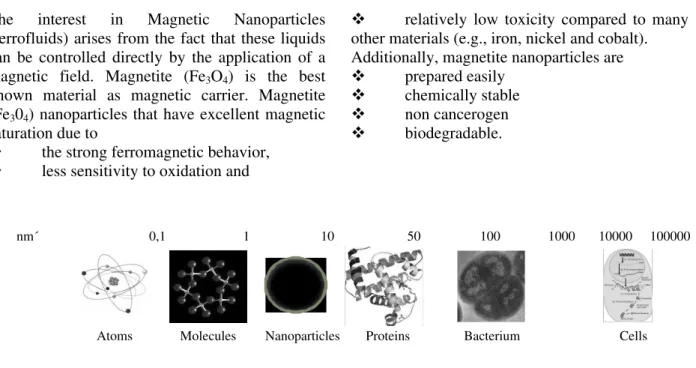
Nanoparticles are colloidal vesicular systems that vary in size from 10 to 1000 nm, and the drug and/or imaging probe of interest is either entrapped therein or attached thereon. The application of methodologies to produce nanoparticles with bioresponsive properties has opened the way for producing useful tools for molecular diagnostics, therapeutics, and biotechnology (Bjerner et al., 2004; Campo, et al., 2005; Jung, 1995; Pintaske et al., 2006; Vassallo et al., 1994; Daldrup et al., 1999). Metal, semiconductor, and magnetic colloidal nanoparticles are presently under intensive study for potential applications (Fuente et al., 2006). Nano-sized structures range from 1-100 nm - for comparative purposes, a single turn of the DNA helix is 3.4 nm in length, and a eukaryotic cell has dimensions in the range 10-100 mm (Fig. 1).
Ünak, P. 32
The interest in Magnetic Nanoparticles (ferrofluids) arises from the fact that these liquids can be controlled directly by the application of a magnetic field. Magnetite (Fe3O4) is the best known material as magnetic carrier. Magnetite (Fe304) nanoparticles that have excellent magnetic saturation due to
the strong ferromagnetic behavior,
less sensitivity to oxidation and
relatively low toxicity compared to many other materials (e.g., iron, nickel and cobalt). Additionally, magnetite nanoparticles are
prepared easily
chemically stable
non cancerogen
biodegradable.
Figure 1 - The figure depicts the sizes of nanoparticles in relation to other biological objects.
Magnetically targeted drug delivery by magnetite nanoparticles is an efficient method of delivering drugs to localized disease sites, such as tumors. Thus high concentrations of carrier molecules such as therapeutic radiopharmaceuticals can be achieved near the target site without any toxic effects to normal surrounding tissue. The interest in magnetite nanoparticles (ironfluids) arises from the fact that these liquids can be controlled directly by the application of a magnetic field.
The ability of Magnetic nanoparticles to enhance proton relaxation of specific tissues and serve as MR imaging contrast agents is one of the most promising applications of nanomedicine. MNPs in the form of superparamagnetic iron oxides (SPIO) have been actively investigated as MR imaging contrast agents for over two decades (Sun et al., 2008). Different targets may be obtained with varying the particles size. For example; large iron oxide particles were used for bowel contrast [AMI-121 (i.e. Lumirem and Gastromark) and OMP (i.e Abdoscan), mean diameter no less than 300 nm] (Jung, 1995) and liver/spleen imaging [AMI-25 [i.e. Endorem and Feridex IV, diameter 80-150 nm); SHU 555A (i.e. Resovist, mean diameter 60 nm)] (Pintaske et al., 2006). Smaller iron oxide particles also may be applied for lymph node imaging [AMI-227 (i.e. Sinerem and Combidex, diameter 20-40 nm)] (Vassallo et al., 1994), bone marrow imaging (AMI-227) (Daldrup et al., 1999), perfusion imaging [NC100150 (i.e.
Clariscan, mean diameter 20 nm)] and Magnetic resonance (MR) angiography (NC100150) (Bjerner et al., 2004). Even smaller monocrystalline iron oxide nanoparticles may be used for receptor imaging and magnetically labeled cell probe imaging (Stevens et al., 2008).
Synthesis and surface modification of magnetite nanoparticles
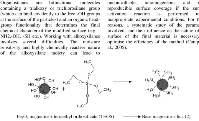
Surface-modification of the magnetite nanoparticle core with a suitable and functional organic core represents an important issue in nanoparticle research for several reasons such as stability, prevent agglemeration or increase affinity to living systems. Mostly relying on ligand exchange reactions has been reported, enabling the modification with polymeric (Thunemann et al., 2006), organic (Sonvico et al., 2005), and enzyme (Johnson et al., 2008). In the most of the works, modification of Fe2O3-nanoparticle-surfaces was performed by use of the amino silane coupling agent, N-[3-(trimethyoxysilyl) propyl]-ethylenediamine (SG-Si900) followed by the ligand or pro-drug was covalently linked to the amine group using glutaraldehyde as cross-linker (Ünak et al., 2008). (Figs. 2, 3 and 4). Thiols, disulfides, amines, nitriles, carboxylic acids and phosphines are the other groups that may be used for modification of metal nanoparticles (Neouze and Schubert, 2008).
nm´ 0,1 1 10 50 100 1000 10000 100000
Organosilanes are bifunctional molecules containing a trialkoxy or trichlorosilane group (which can bind covalently to the free -OH groups at the surface of the particles) and an organic head-group functionality that determines the final chemical character of the modified surface (e.g. -NH2,-OH, -SH etc.). Working with alkoxysilanes involves several difficulties. The moisture sensitivity and highly chemically reactive nature of the alkoxysilane moiety can lead to
uncontrollable, inhomogeneous and non-reproducible surface coverage if the surface activation reaction is performed under inappropriate experimental conditions. For these reasons, a systematic study of the parameters involved, and their influence on the nature of the surface of the final material is necessary to optimise the efficiency of the method (Campo et al., 2005).
O Si H3
SiH3 O
SiH3 SiH3 O Si H3 SiH
3
O Si H3
Si H3 O
O H
OH
OH OH
O OH OH O+ O H
O H
O H
OH O H
+
Si O O
O O
C H3
CH3
C H3 C H3
Fe3O4 magnetite + tetraethyl orthosilicate (TEOS) Base magnetite-silica (2)
Figure 2 - Silica-coated magnetite nanoparticles.
Figure 3 - Procedure for covalently linking Amino silan (SG-Si900) onto magnetic nanoparticles.
Si
C H3
O C H3
O CH3 N
H2
NH
O Si H3 SiH3
O SiH3
SiH3
O Si H3 SiH3
O Si H3
Si H3
O SiH2
NH SiH2
NH2
N H
NH2 O
SiH2
NH
NH2
NH NH2
O SiH2
SiH3
NH N
H2
O Si H3
SiH2 NH
N H2
NH
CH3
N H2
NH
C H3
Ünak, P. 34
A B
Figure 4 - A) Tetraethyl orthosilicate conjugated magnetite nanoparticles, B) Amino silan conjugated magnetite nanoparticles.
Surface modification of silica-coated magnetite nanoparticles with amino silane (SG-Si900)
The preparation procedure for silica-coated magnetite nanoparticles is shown in Equations (1) and (2). In step 1, nanomagnetite is formed, which is then coated in step 2 with a silica layer:
6Fe3+ + SO32- + 18NH3.H2O
→ 2Fe3O4 +
SO42- + 18NH4+ + 9H2O (1)
Normally surface modification involves development of core-shell structures to create a new types of material in which unique magnetic properties of iron oxide-core are combined with biological functionalities of shell. Each of the coating materials has its own importance in biomedical point of view. Polymers like polyethylene glycol, polyvinylpyrrolidone and dextran are used in order to improve the biocompatibility, blood circulation time and to stabilize the colloidal solution (Gupta and Gupta, 2005).
Gelatin is thermally and hydrolytically denatured product of collagen, the most abundant protein in animals, and further, it has been utilized extensively for industrial, pharmaceutical, and medical applications. Properties like biodegradability, biocompatibility and presence of multifunctional groups make gelatin a promising candidate for the surface modification (Maria, et al., 2002). Ideally, gelatin can be used to encapsulate the magnetic nanoparticles and pharmaceutical drugs could be attached on its
surface or in their bulk and could be guided to the target organs (Muniyandy et al., 2004). Development of magnetic microspheres and nanoparticles, using gelatin as a carrier system, is a new area and only few research works have been carried out (Maria et al., 2002; Muniyandy et al., 2004; Saravanan et al., 2003; Gaihre et al., 2008).
Potential radionuclides for labeling of nanoparticles for therapy and imaging
MRI system was used for imaging with magnetic particles, however in the case of radionuclide labeling magnetic particles, hybride imaging such as SPECT-MRI or PET-MRI may be efficiently depend on the decay properties of radionuclide or therapeutic agents or both (imaging and therapy) may be obtained. Several radionuclides labeled magnetic particles such as Re-188 (Jeong and Chung, 2003), In-111 (Heckl, 2007),Y-90 (Shabat et al., 2002) and I-125/131 Lankester et al., 2007; Dağdeviren et al., 2007), Tc-99m (Ünak and Medine, 2007; Bekiş et al., 2008) have been reported in the literature.
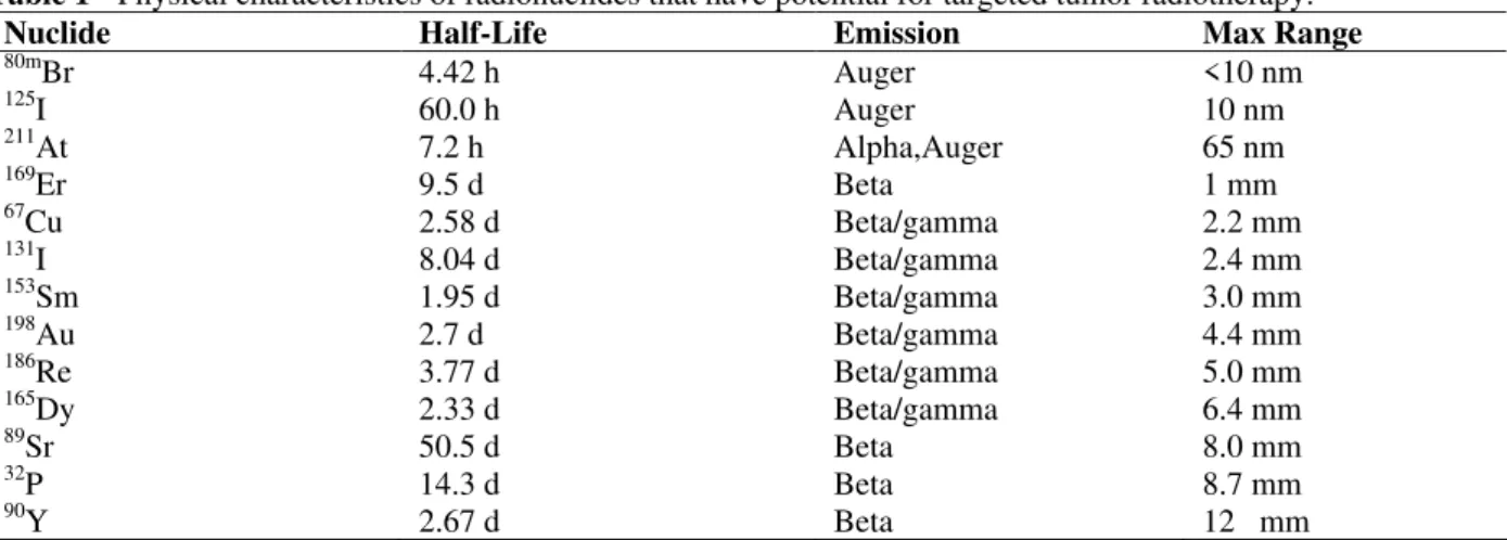
Table 1 - Physical characteristics of radionuclides that have potential for targeted tumor radiotherapy.
Nuclide Half-Life Emission Max Range
80mBr
125I 211
At
169
Er
67Cu
131I 153
Sm
198
Au
186Re
165Dy
89
Sr
32
P
90Y
4.42 h 60.0 h 7.2 h 9.5 d 2.58 d 8.04 d 1.95 d 2.7 d 3.77 d 2.33 d 50.5 d 14.3 d 2.67 d
Auger Auger Alpha,Auger Beta
Beta/gamma Beta/gamma Beta/gamma Beta/gamma Beta/gamma Beta/gamma Beta Beta Beta
<10 nm 10 nm 65 nm 1 mm 2.2 mm 2.4 mm 3.0 mm 4.4 mm 5.0 mm 6.4 mm 8.0 mm 8.7 mm 12 mm
Table 1 shows some radionuclides, which have different decay characteristics, and emit particles with different LET and different range. Some of these radionuclides emit photons in addition to particle emission which make them suitable for monitoring the therapy with imaging, and for continuous follow-up of the absorbed dose distribution. Also, preadministration of the therapeutic ligands is possible in estimating the absorbed dose per unit administered activity. Where there is no emission of photons, bremstrahlung imaging might be a solution. Another solution is to label ligands with a gamma-emitting radionuclide with similar chemical properties, preferably an isotope of the same element as the therapeutic radionuclide or an isotope, which have similar chemical properties. Thus, gamma emitting radionuclide can be used for imaging and mechanisms of follow up, other one can be used for therapy. Tc-99m and Re-186, Y-88 and Y-90, I-123 and I-131, 111 and In-114m are this type of couples.
CONCLUSIONS AND PERSPECTIVES
Colloidal magnetite nanoparticules are alternative in bioimaging and diagnostic applications due to ease of preparation and bioconjugation as well as non-cytotoxic properties. It could be possible to increase this specificity by targeting of the nanoparticles. Because of their size range, 10-100 nm, nanoparticles are very suitable for manipulations at the molecular level, for example cell-receptor binding for site-selective imaging and targeting, localization of encapsulated therapeutics
for delivery, and decoration of expression systems for substrate-based nanosensing.
Targeted drug delivery by nanoparticles is a promising method of delivering drugs to localized disease sites, such as tumors. Magnetite, conjugated to radionuclide labeled therapeutic agent may be used for this aim. Thus, high concentrations of carrier molecules such as therapeutic radiopharmaceuticals can be achieved near the target site without any toxic effects to normal surrounding tissue.
We expect that a variety of applications of radionuclide labeled magnetic nanoparticles for hybrid imaging systems such as PET-MRI or SPECT-MRI together with thepeutic radionuclide labeled magnetic nanoparticles for targeted therapy will be developed in the near future.
RESUMO
Nanopartículas magnéticas oferecem novas oportunidades interessantes, incuindo a melhora da qualidade da imagem de ressonância magnética (MRI), no tratamento hipertérmico para células malignas, na administração de medicamentos sítio-específicos e também no recente interesse da manipulação de membranas celulares. As aplicações biológicas desses nanomateriais requer que essas nanopartículas tenham valores altos de magnetização, tamanho menor que 20 nm, partículas de dimensão de distribuição restrita e um revestimento especial de superfície para evitar a toxicidade e permitir o acoplamento de biomoléculas. Nessa revisão, focalizamos na viabilidade de nanopartículas magnéticas
Ünak, P. 36
transportadoras de medicamentos, e resumimos os recentes avanços nesse campo.
REFERENCES
Ahlberg, A.; Mikulowski, P.; Odelberg-Johnson, O. (1969), Intra-articular injection of radioactive gold in treatment of chronic synovial effusion in the knee. Acta Rheumatol Scand.,15, 81-89.
Bekiş, R.; Medine, I.; Dağdeviren, K.; Ünak, P. (2008), A new method for sentinel lenf detection. Turkish J Nucl Med.,17, 49.
Bjerner, T.; Wikstrom, G.; Johansson, L.; Ahlström H. (2004), High in-Plane Resolution T2-Weighted Magnetic Resonance Imaging of Acute Myocardial Ischemia in Pigs Using The Intravascular Contrast Agent NC100150 Injection. Invest Radiol., 39, 470-478.
Campo, A. D.; Sena T.; Lellouche, J. P.; Bruce, I. J. (2005), Multifunctional magnetite and silica-magnetite nanoparticles: Synthesis,surface activation and applications in life sciences. J Magn Magn Mater., 293, 33-40.
Dağdeviren, K.; Ünak, P.; Bekiş, R.; Biber, Z.; Akdurak, S.; Ulker, O.; Ergur, B.; Ertay, T.; Durak, H. (2007), Radioiodinated magnetic targeted carriers (I-131-MTC). J Radioanal Nucl Chem., 273, 635-639.
Daldrup, H. E.; Link, T.M.; Blasius, S; Kšnemann, S.; JŸrgens, H.; Rummeny, E. J. (1999), Monitoring Radiation-Induced Changes in Bone Marrow Histopathology with Ultra-Small Superparamagnetic Iron Oxide (USPIO)-Enhanced MRI. J Magn Resonance, 9, 643-652.
Dobson, J. (2006), Magnetic micro- and nano-particle-based targeting for drug and gene delivery. Nanomedicine,1, 31-37.
Fortina, P.; Kricka, L. J.; Graves, D. J.; Park, J.; Hyslop, T.; Tam, F.; Halas, N.; Surrey, S.; Waldman, S. A. (2007), Applications of nanoparticles to diagnostics and therapeutics in colorectal cancer. Opin Trends Biotechnol., 25, 145-153.
Fuente, J. M.; Alcantara, D.; Eaton, P.; Crespo, P.; Rojas, T. C.; Fernandez, A.; Hernando, A.; Penades, S.; (2006), Gold and Gold-Iron Oxide Magnetic Glyconanoparticles: Synthesis, Characterization and Magnetic Properties, J Phys Chem. B, 110, 13021-13028.
Gaihre, B.; Aryal, S.; Khil, M. S.; Kim, H. Y. (2008), Encapsulation of Fe3O4 in gelatin nanoparticles: Effect of different parameters on size and stability of the colloidal dispersion. J Microencapsul., 25, 21-30. Gupta, A. K.; Gupta, M. (2005), Synthesis and surface
engineering of iron oxide nanoparticles for biomedical applications. Biomaterials, 26, 3995-4021.
Heckl, S. (2007), Future contrast agents for molecular imaging in stroke. Curr Med Chem.,14, 1713-1728. Jeong, J. M.; Chung, K. K. (2003), Therapy with
Re-188-labeled radiopharmaceuticals: an overview of promising results from initial clinical trials. Cancer Biother Radiopharm., 18, 707-717.
Johnson, A. K.; Zawadzka, A. M.; Deobald, L. A.; Crawford, R. L., Paszczynski, A. J. (2008), Novel method for immobilization of enzymes to magnetic nanoparticles. J Nanopart Res., 10, 1009-1025. Jung, C. (1995), Surface properties of
superparamagnetic iron oxide MR contrast agents: Ferumoxides, ferumoxtran, ferumoxsil. Magn Reson Imaging, 13, 675 - 691.
Kreuter, J. (2007), Nanoparticles a historical perspective. Inter J Pharm., 331, 1-10.
Lankester, K. J., Maxwell, R. J., Pedley, R. B., Dearling, J. L.; Qureshi, U. A.; El-Emir, E.; Hill, S. A.; Tozer, G. M. (2007), Combretastatin A-4-phosphate effectively increases tumor retention of the therapeutic antibody, I-131-A5B7, even at doses that are sub-optimal for vascular shut-down. Int J Oncol.,
30, 453-460.
Maria, G. C.; Claudia, C.; Zhouhai, Z. (2002), Gelatin nanoparticles produced by a simple W/O emulsion as delivery system for methotreaxate. J Mater Sci Mater Med., 13, 523-526.
Moses, C.; Kent, E.; Boatman, J. B.; Cole, R. D.; Sunder, J. H.; George, R. S.; Russ, C.; Ford, W. B.; Kutz, E. R. (1955), Experimental and clinical studies with radioactive colloidal gold in the therapy of serous effusions arising from cancer. Cancer, 8, 417-423.
Muniyandy, S., Bhaskar, K., Maharajan, G., Pillar, K. S. (2004), Ultrasonically controlled release and targeted delivery of diclofenac sodium via gelatin magnetic microspheres. Int J Pharm.,283, 71-82. Neouze, M. A.; Schubert, U.; (2008), Surface
modification and functionalization of metal and metal oxide nanoparticles by organic ligands, Monatshefte Fur Chemie, 139, 183-195.
Neves, M.; Reis, F.; Waerenborgh, F.; Martinho, E.; Patricio, L. (1987a), 166Holmium: a potential lanthanide element in radiotherapy. Inorg Chim Acta,
140, 359-360.
Neves, M.; Waerenborgh, F.; Patricio, L. (1987b), 109Palladium and 166Holmium potential radionuclides for synoviotherapy-radiation absorbed dose calculations. Appl Radiat Isot., 38, 745-749. Pintaske, J.; Bantleon, R.; Kehlbach, R.; Claussen, C.
Saravanan, M.; Bhaskar, K.; Narayanan, V. S. N.; Maharajan, G.; Pillai, K. S. (2003), Diclofenace sodium loaded gelatin magnetic microspheres for intra-arterial administration: Formulation, characterization and in vitro release studies. Boll Chim Farm.,142, 347-351.
Shabat, S.; Kollender, Y.; Merimsky, O.; Isakov, J.; Flusser, G.; Nyska, M.; Meller, I. (2002), The use of surgery and Yttrium 90 in the management of extensive and diffuse pigmented villonodular synovitis of large. Rheumatology,41, 1113-1118. Sonvico, F.; Mornet, S.; Vasseur, S.; Dubernet, C.;
Jaillard, D.; Degrouard, J.; Hoebeke, J.; Duguet, E.; Colombo, P.; Couvreur, P. (2005) Folate-conjugated iron oxide nanoparticles for solid tumor targeting as potential specific magnetic hyperthermia mediators: synthesis, physicochemical characterization, and in vitro experiments. Bioconjugate Chem., 16, 1181-1187.
Stevens, N.; O'Connor, N.; Vishwasrao, H.; Samaroo, D.; Kandel, E. R.; Akins, D. L.; Drain, C. M.;Turro, N. J. (2008), Two Color RNA Intercalating Probe For Cell Imaging Applications. J Am Chem Soc., 130, 71-82.
Sun, C.; Lee, J. S. H.; Zhang, M. (2008), Magnetic nanoparticles in MR imaging and drug delivery. Adv Drug Deliv Rev., 60, 1252-1265.
Sun, H.; Yua, J.; Gonga,, P.; Xua, D.; Zhanga, C.; Yao, S. (2005), Novel core-shell magnetic nanogels synthesized in an emulsion-free aqueous system under UV irradiation for targeted
radiopharmaceutical applications. J Magn Magn Mater., 294, 273-280.
Thunemann, A. F.; Schutt, D.; Pison, U.; Mohwald, H. (2006), Maghemite Nanoparticles Protectively Coated with Poly(ethylene imine) and Poly(ethylene oxide)-block-poly(glutamic acid), Langmuir, 22, 2351-2357.
Ünak, P.; Medine, E. Đ. (2007), Tc-99m Labeled Magnetite Nanoparticles As Drug Carriers, EANM'07 - Annual Congress of the European Association of Nuclear Medicine. Eur J Nucl Med Mol Imaging, 34, 133.
Ünak, P.; Medine, E. I.; Sakarya, S.; Yürekli, Y. (2008),
Synthesis of radionuclide labeled magnetic
nanoparticules and investigation of their therapeutic potentials in cells. TUBITAK research project report no SBAG 3293 (105S486), 135 p.
Vassallo, P.; Matei, C.; Heston, W. D.; McLachlan, S. J.; Koutcher, J. A.; Castellino, R. A. (1994), AMI-221-enhanced MR lymphography -usefulness for differentiating reactive from tumor-bearing lymph-nodes. Radiology,193, 501-506.
Volkert, W. A.; Hoffman, T. J. (1999), Therapeutic radiopharmaceuticals. Chem. Rev.,9, 2269-2292. Wessels, B. W.; Mears, C. F. (2000), Physical and
chemical properties of radionuclide therapy. Semin Radiat Oncol., 10, 115-122.