Mastoid Obliteration with Autologous Bone in
Mastoidectomy Canal Wall Down Surgery: a
Literature Overview
Ricardo Dourado Alves
1Francisco Cabral Junior
1Anna Carolina de Oliveira Fonseca
1Ricardo Ferreira Bento
11Department of Otorhinolaryngology, Universidade de São Paulo, São Paulo, SP, Brazil
Int Arch Otorhinolaryngol 2016;20:76–83.
Address for correspondenceRicardo Dourado Alves, MD, Department of Otolaryngology, University of Sao Paulo, Av. Dr. Enéas de Carvalho Aguiar, 255 6° andar - sala 6167, São Paulo, SP 05403-000, Brazil (e-mail: [email protected]).
Introduction
Usually, for a successful surgical eradication of medium ear diseases, the otologic surgeon must remove diseased ana-tomic structures and, sometimes, even normal structures. Canal wall down mastoidectomy (CWD) is one of those common surgical techniques with variations of long-term outcomes. Although the majority of patients experience little to no long-term problems postoperatively, there is a small but expressive number of patients with
chronic complaints associated with the persistent mastoid
bowl.1
Recurrent drainage and infection are the most common cause of discontent and medical return for patients with mastoid bowls. Other frequent complaints may include water intolerance, leading to infection, the need for frequent oto-microscopic cleaning, calorically induced vertigo from either water or air exposure, barometrically induced vertigo, and, in those with compromising hearing loss, being unable to wear
traditional hearing aids.2
Keywords
►
cholesteatoma
►
middle ear
►
mastoid obliteration
►
mastoidectomy
►
otitis media
►
suppurative
►
bone and bones
►
tympanomastoi-dectomy
Abstract
Introduction
The objectives of mastoidectomy in cholesteatoma are a disease-free
and dry ear, the prevention of recurrent disease, and the maintenance of hearing or the
possibility to reconstruct an affected hearing mechanism. Canal wall down
mastoidec-tomy has been traditionally used to achieve those goals with greater or lesser degrees of
success. However, canal wall down is an aggressive approach, as it involves creating an
open cavity and changing the anatomy and physiology of the middle ear and mastoid. A
canal wall up technique eliminates the need to destroy the middle ear and mastoid, but
is associated with a higher rate of residual cholesteatoma. The obliteration technics arise
as an effort to avoid the disadvantages of both techniques.
Objectives
Evaluate the effectiveness of the mastoid obliteration with autologous
bone in mastoidectomy surgery with canal wall down for chronic otitis, with or without
cholesteatoma.
Data Synthesis
We analyzed nine studies of case series comprehending similar surgery
techniques on 1017 total cases of operated ears in both adults and children, with at least
12 months follow-up.
Conclusion
Mastoid Obliteration with autologous bone has been utilized for many
years to present date, and it seems to be safe, lowcost, with low recurrence rates
-similar to traditional canal wall down procedures and with greater water resistance and
quality of life improvements.
received May 11, 2015 accepted June 2, 2015 published online August 24, 2015
DOI http://dx.doi.org/ 10.1055/s-0035-1563382. ISSN 1809-9777.
Copyright © 2016 by Thieme Publicações Ltda, Rio de Janeiro, Brazil
Systematic Review
Mosher, in 1911, started the idea of mastoid obliteration to promote healing of a mastoidectomy defect. Mosher de-scribed an obliteration technique using a superiorly based
postauricular soft tissueflap.3The researcher noticed that the
muscle atrophied over time, causing a progressive enlarge-ment in cavity size. This observation is supported by
histo-logical data from the temporal bone study of Linthicum,4
which demonstrates the replacement of muscle withfi
bro-connective tissue and fat. These findings encouraged
sur-geons to associate other filler materials inside the bowl.
Palva5modified and popularized the technique, further
add-ing to it the use of bone chips and bone pate in combination
with an anteriorly based musculoperiosteal flap.5Over the
course of the last decades, there have been a large number of reports detailing a multiplicity of techniques for obliterating the mastoid cavity. The most frequent and popular techniques
consist of either localflaps (muscle, periosteum, or fascia) or
free autologous grafts (bone, cartilage, fat, fascia), or even alloplastic grafts (hydroxyapatite, silicon, synthetics bones,
among others).1
The decision whether to perform a intact canal wall mastoidectomy (ICW) or CWD operation in patients with chronic ear disease is usually based on several factors, such as the extent of disease, an assessment of middle ear ventilation, the hearing in the ear in question, the state of the opposite ear, any preoperative complications, the condition of the
patient, the possibility for follow-up, and the surgeon’s
preference.6
The benefits and drawbacks of ICW and CWD for
choles-teatoma are well established. The greatest problem with ICW techniques are the recidivism rates, reported as high as 40 to 60% in children and 20% in adults. This high rate of recurrence
is associated with the relatively deficient exposure during
surgery, the persistence of Eustachian tube dysfunction, and the persistence of mucosa in the mastoid that keeps resorbing gas and creates a negative pressure environment for
resurg-ing of retraction pockets.7
Although the CWD technique is known to have lower residual and recurrent cholesteatoma rates, as mentioned previously, it is often accompanied by the problems associat-ed by the mastoid cavity such as crust accumulation, water intolerance and intermittent discharge. The principle behind the mastoid obliteration is that it combines the advantages of
both techniques (CWD and ICW).3
Objectives
The purpose of this review is to evaluate the effectiveness of mastoid obliteration with autologous bone in mastoidectomy surgery with canal wall down for chronic otitis, with or without cholesteatoma, mainly for infection control and drainage, recurrence of cholesteatoma and water tolerance.
Search Methods
In January 2015, we searched online databases for the
fol-lowing keywords:“mastoid obliteration bone chronic otitis
canal wall down.”We searched MEDLINE, LILACS and EBSCO
databases. We were only able to find journal articles at
MEDLINE, where 26 articles were found. We included other different and interesting articles that have met our selection criteria, which we obtained through the references in the articles initially searched.
Selection Criteria
First, we tried to locate articles with randomized case-control studies, with no success. We selected articles with case series
that included the technique used in the surgery–specifically
those that use autologous bone to obliterate the mastoid
cavity associated with a canal wall down procedure–with or
without posterior reconstruction of the wall, and at least one year of mean follow-up of patients. We have included articles that used cartilage, fascia, skin grafts, or musculoperiosteal
flaps to cover the obliterated bowl.
We excluded any article that: was not specific to surgery in
chronic otitis; was on other associated mastoidfiller
materi-als–like silicon, ceramics or other alloplastic materials; was
on musculoperiosteal and/or cartilage without using autolo-gous bone parts; was on total tympanomastoid obliteration; was not written in English, Spanish, or Portuguese.
Review of Literature
After applying the selection criteria to the initial 26 articles,
we selected six of them. They all had Level 4 evidence:five
were retrospective and one was a prospective case series, according to Oxford Centre for Evidence-based Medicine (►Table 1). One article, by Walker et al7 was an updated
version of a previous one,8so we discarded the less current
version.
When reviewing the references the articles mentioned above, as well as others pertinent to mastoid obliteration, we found four new studies that met the selection criteria. These were all retrospective case series, without a control group or
randomization, and with Level 4 evidence.►Table 2
summa-rizes the articles found.
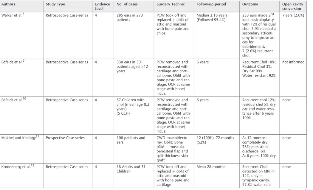
Walker et al7showed a retrospective case series of
conse-cutive patients treated from 1997 to 2011 with a Canal Wall Reconstruction (CWR) tympanomastoidectomy with mastoid obliteration using bone pate. The sample consisted of 285 ears with cholesteatoma in 273 patients, with a mean age of 35 years. There were 25 children under 10 years of age that had undergone surgery (average 6.9 years old). Thirteen patients (4.6%) were lost to follow-up right after the surgery. Thirty percent of the patients had previous surgery, with 20% having undergone ICW mastoidectomy. The authors collected bone pate from healthy cortical bone with a sheehy pate collector and performed a simple mastoidectomy and attic-otomy. They removed the incus and the malleus head, cut the posterior bone canal (PBC) superiorly and inferiorly with a saw and removed it. After removing the PBC, they cleaned the entire middle ear of cholesteatoma, put the PBC back, placing a large single block of bone harvested from mastoid tip blocking the attic to avoid retraction of the pocket into the attic space. Smaller bone chips are placed to block the facial recess and prevent bone pate from entering the middle ear.
The authorsfilled the bowl with bone pate and the original
meatus skin would cover the PBC. Typically, a second-look
International Archives of Otorhinolaryngology Vol. 20 No. 1/2016
tympanoplasty with ossiculoplasty is performed 6 months after the initial tympanomastoidectomy. In this case series, 253 (89%) of 285 ears underwent the second-look operation, of which 30 (12%) had residual cholesteatoma only in the middle ear, which was successfully removed. Of the returning patients, 38 (14%) developed retraction pockets toward the attic; however, only 16 (5.9% of total) required an endaural atticotomy to improve access for debridement. These ears are now dray and self-cleaning. After the attic blocking technique was replaced with a single large bone block in 2005, only 7.1% of the patients developed retraction pockets. Prior to the change, this number was 21%. Seven patients (2.6%) devel-oped a recurrence of cholesteatoma, having to convert to an open cavity. Average time until recurrence was 3.68 years. When making the canal wall cuts, 14 ears (4.9%) had
intra-operative cerebral spinal fluid leakage, all of which were
immediately detected and repaired. Only one patient required a secondary operation.
Edfeldt et al9published an article with a series of 330
operated ears in adults (over 12 years in age) with choles-teatoma in 301 patients. They underwent an operation performed by three senior surgeons between 1982 and 2004 using an identical technique. From this sample, 156 ears (47%) had undergone previous surgeries, while 61% had undergone one previous operation. The surgeons performed a CWD mastoidectomy, meatoplasty, and used cartilage from the tragus or meatus to rebuild the wall. The mastoid and epitympanic spaces were obliterated with cartilage and bone pate. When necessary, they reconstructed the ossicular chain at the same stage with autologous cortical bone or shaped incus. A large temporal fascia was used for myringoplasty and to cover the reconstructed ear canal. They followed the patients for at least 6 years. They did not use Computer Tomography (CT) nor Magnetic Resonance Imaging (MRI). Nine cases (3%) had residual disease and 33 cases (10%) had recurrent disease during the study period. These patients underwent miscellaneous revision surgery. The study did not inform whether there was a need to convert to an open cavity
in any of the cases. They did report, however, that only one case had recurrent ear discharge, after 6 years of follow-up.
Edfeldt et al10published another retrospective case series
that included only children under 12 years of age, with a mean age of 8.2 years. The group consisted of 57 children with
cholesteatoma,five of which presented congenital
cholestea-toma. They all underwent operations by three senior sur-geons between 1983 and 2004, who used the exact surgical technique described in the previous paragraph. Four patients (7%) had undergone previous surgeries. All of the patients had a follow-up period of at least 6 years. No imaging was
performed. The authors confirmed and checked three
resid-ual (5%) and seven recurrent cholesteatoma (12%). Three (42%) of the recurrent cholesteatoma were located in the reconstructed ear canal. None of them underwent conversion to open cavity. After six years, all cases were dry and water tolerant. The authors compared these results to their data-base from operated adults, and found that they were similar. They did not observe any extrusion of autologous material.
Mokbel and Khafagy11 showed a prospective case series
with 100 adults operated between 2003 and 2010. The inclusion criteria were patients with unilateral chronic sup-purative otitis media, with no history of mastoidectomy or systemic debilitating condition. The minimum follow-up period was 12 months, extending up to 72 months (52% of the patients). There were 64% of patients with cholesteatoma. The authors performed a CWD mastoidectomy, large meato-plasty, anterior and inferior canalomeato-plasty, obliterated with cortical bone pate, and covered it with musculoperiosteal
flap. Theflap and exposed bone was covered with temporal
fascia and split-thickness skin grafts. No ossicular chain reconstruction was performed. All cases completed the 12-month follow-up and 78% had complete dry cavity, 16% had intermittent otorrhea and 6% with persistent discharge. Throughout the follow-up period, 10 patients presented persistent discharge, all of them caused by the presence of granulation tissues. These patients were treated with a revi-sion surgery (6%) and cauterization (4%), which resulted in
Table 1 Levels of Evidence
Level of Evidence Grading Criteria Grade of
Recommendation
1a Systematic review of Randomized Controlled Trials (RCT), including meta-analysis A
1b Randomized Controlled Trial with narrow confidence interval A
1c All or none studies B
2a Systematic Review of cohort studies B
2b Cohort study and low quality RCT (e.g.,<80% follow-up) B
2c Outcomes research studies; ecological studies C
3a Systematic review of case-control studies C
3b Case-control study C
4 Case-series, poor quality cohort and case control studies C
5 Expert opinion D
Notes: Last updated on March 2009.
Source: Oxford Centre for Evidence-based Medicine (http://www.cebm.net/oxford-centre-evidence-based-medicine-levels-evidence-march-2009/).
International Archives of Otorhinolaryngology Vol. 20 No. 1/2016
Mastoid Obliteration with Autologous Bone in Mastoidectomy Canal Wall Down Surgery Alves et al.
Table 2 A summary of articles on mastoid obliteration with autologous bone in mastoidectomy with canal wall down for chronic otitis
Authors Study Type Evidence
Level
No. of cases Surgery Technic Follow-up period Outcome Open cavity
conversion
Walker et al.7 Retrospective Case-series 4 285 ears in 273 patients
PCW took off and replacedþoblit of attic and mastoid with bone pate and chips
Median 3.16 years (Followed 95.4%)
253 ears made 2nd look ossiculoplasty with 12% of residual chol. 5.9% needed a secondary atticot-omy to improve ac-ces for
debridement. 7 (2.6%) recurrent chol.
7 ears (2.6%)
Edfeldt et al.9 Retrospective Case-series 4 330 ears in 301 patients aged>12 years
PCW removed and reconstructed with cartilage and corti-cal bone. Oblit with bone paste and car-tilage. OCR at same stage with bone/ incus.
6 years Recurrent Chol 10%; Residual Chol 3%; Dry Ear 99% Water resistant 92%
not informed
Edfeldt et al.10 Retrospective Case-series 4 57 Children with chol (mean age 8.2 years)
(5 CCH)
PCW removed and reconstructed with cartilage and corti-cal bone. Oblit with bone paste and car-tilage. OCR at same stage with bone/ incus.
6 years Recurrent chol 12%; residual chol 5%; dry ear and water resis-tance after 6 years 100%
none
Mokbel and Khafagy11 Prospective Case-series 4 100 patients and ears
CWD mastoidecto-my. Oblit: Bone pâtéþ musculo-periostealflap and split-thickness skin graft
12 (100%) -72 months (52%)
At 12 months: completely dry: 78%; persistent discharge: 6% At 6 years: 100% dry
none
Kronenberg et al.12 Retrospective Case-series 4 18 Adults and 31 Children
PCW took off and replacedþoblit of attic and mastoid with bone pate and cartilage
Mean 28 months Recurrent Chol detected on MRI in 12%, only in tympanic cavity. 77.8% water-safe
none
(Continued)
In
te
rn
at
io
nal
A
rc
hi
ve
s
o
f
O
to
rh
in
ol
ar
yn
g
o
lo
g
y
V
o
l.
2
0
N
o
.
1
/2
01
6
Ma
st
oid
O
bliteration
w
ith
A
ut
ologous
Bone
in
Ma
st
oidec
tom
y
C
anal
W
a
ll
Down
Surger
y
Al
ve
s
e
t
a
l.
Table 2 (Continued)
Authors Study Type Evidence
Level
No. of cases Surgery Technic Follow-up period Outcome Open cavity
conversion
Sun et al13 Retrospective Case-series 4 48 ears in 45 chil-dren (5–12 years)
CWD mastoidecto-my with oblit with cartilage and bone pasteþtemporalis fascia
2–5 years (mean 3,1) Residual chol: 0%; Recurrent chol: 4.2%; Dry ear 95.8%
none
Beutner et al14 Retrospective Case-series 4 26 patients with previous CWD mastoidectomy
Bone pateþ con-chal cartilage plates and fascia
6 years Mean follow-up 100% epithelialized and dry.
Before surgery 54% had vertigo on ca-loric stimuli; none had it after. 0% chol
none
Ramsey et al15 Retrospective Case-series 4 60 ears (59 patients)
Bone pateþ inferi-orly pedicled peri-ostealflapþ split-thickness skin grafting
>12 months (mean: 31 months)
No cholesteatomas 82% dry; 8% inter-mittent discharge; 6 ears(10%) fre-quent discharge within 4 had meatal stenosis
none
Roberson et al2 Retrospective Case-series 4 62 ears (56 patients)
Bone pateþfascia graft
Average 18,5 months (0.2–54.8 months)
6% residual Chol, none after second stage surgery. 87% dry cavity; 5% had pate resorption after early infection.
none
Abbreviations: CCH, congenital Chol; Chol, Cholesteatoma; No., number; Oblit, obliteration; OCR, Ossicular Chain Reconstruction; PCW, Posterior Canal Wall; yrs, years.
In
ter
n
a
tio
na
l
A
rc
hi
ve
s
o
f
O
to
rh
in
ola
ry
n
g
o
lo
g
y
V
o
l.
2
0
N
o
.
1
/2
01
6
Ma
st
oid
O
bliteration
w
ith
A
ut
ologous
Bone
in
Ma
st
oidec
tom
y
C
anal
W
a
ll
Down
Surger
y
Al
ve
s
e
t
a
l.
8 patients becoming dry. None of the follow-up cases pre-sented residual or recurrent cholesteatoma.
Kronenberg et al12came forth with a retrospective case
series that included 49 consecutive patients (31 children and 18 adults) that had undergone surgery between 2008 and 2011. They all had cholesteatoma and their mean follow-up period was 28 months (the authors did not mention the minimum or the maximum follow-up time). Thirty patients
were undergoing theirfirst ear surgery. The authors used a
technique similar to that used by Walker et al7whereby they
collected bone pate from cortical bone, made a simple mastoidectomy and atticotomy, removed the incus and the malleus head, cut the posterior bone canal (PBC) superiorly and inferiorly, and removed it. After removing the PBC, they fully cleaned the middle ear of cholesteatoma, examined the sinus tympani with a 30° endoscope, restored the PBC, placed
cartilage blocking the attic, and filled the bowl with bone
pate. They covered the PBC with tragus perichondrium and the tympanic membrane with temporalis fascia. In the sec-ondary surgery group, if there was damage to the PBC, the patients underwent a reconstruction using cartilage. The authors found recurrent cholesteatoma in six patients
(12%); three from the group undergoing first surgery and
three in the other group. The authors identified all the
cholesteatomas using non-EPI Diffusion-weighted (DW) MRI, and observed that they were small and located only in
the tympanic cavity. Thirty-five patients (77.8%) were water
safe during the follow-up period. In primary surgery, howev-er, the surgeons achieved 85.7% water-safe and 90% with dry ear, contrasting with the other group, which had 64% and 73%, respectively.
Sun et al13 presented a retrospective case series that
consisted of 45 children aged between 5 and 12 years (mean age was 10 years), with a total of 48 ears that had undergone procedures in the period between 1999 and 2006. Only primary surgery cases were included. They were
fol-lowed-up for two tofive years (16 patients– 35%), with a
mean follow-up after 3.1 years. All the children had choles-teatoma. The surgeons performed a CWD mastoidectomy, removed the incus and the head of the malleus, performed a meatoplasty and harvested cartilage from concha. They used bone pate to seal the epitympanum and bone pate, cartilage,
and musculoperiosteal flap to cover the mastoid. Then, a
temporalis fascia graft was placed to reconstruct the tym-panic membrane and cover the obliteration. The study found recurrent cholesteatoma in two patients (4.16%) (at 16 months and 33 months) and all were located in the tympanic cavity. Epithelization of the mastoid bowl was
completed within 8–10 weeks, and all ears were dry within
the same 8–10 weeks.
Beutner et al14demonstrated a case series of patients that
had already been submitted to CWD mastoidectomy and were undergoing a revision surgery with CWD mastoidecto-my with obliteration using autologous bone pate, covered with cartilage plates. The surgeon performed a meatoplasty and reconstructed the tympanic membrane with thinned slices of cartilage. The entire surgery was performed by the same surgeon. The authors selected 26 patients, but only 18 of
them agreed to a complete follow-up, including vestibular testing. The median follow-up period was 6 years and mean age was 46 years. None of the selected patients had residual or recurrent cholesteatoma. In analyzing preoperative data, 14 patients of the 26 patients (53.8%) had reported that caloric stimuli (such as wind, water, or suction cleaning) regularly induced vertigo. After the surgery, none of the patients reported similar symptoms in the same situations and all patients had dry ear with complete epithelization.
Ramsey et al15presented a retrospective clinical study of
60 consecutive surgeries between 1995 and 2000 for active chronic otitis media. All patients had CWD mastoidectomy with simultaneous tympanoplasty, including split-thickness skin grafting. An inferiorly pedicled periosteal-pericranial
flap was used in conjunction with autologous bone pate to
obliterate the mastoid cavity. The surgeon performed an anterior and inferior canalplasty and a large meatoplasty. The sample consisted of 60 years from 59 patients. The ages ranged from 4 to 84 years, with a mean of 39 years. Fifty-three ears (88%) had cholesteatoma, and the others presented granulation tissue without cholesteatoma. The minimum follow-up period was 12 months, with a mean of 32 months (maximum 80 months). The authors followed-up on 36 ears (60%) for over 24 months and 18 ears (30%) for over
36 months. Of all procedures performed, fifty-four (90%)
were successful in controlling patientśinfections. Six patients
(10%) had frequent discharge, of which four had meatal stenosis and underwent revision surgery. The other two cases
were attributable to granulation tissue, treated with office
management debridement with secondary split-thickness skin grafting. There were no cases of residual or recurrent cholesteatoma.
Roberson et al2presented a retrospective case series of 57
patients with 62 operated ears. The average of patients with
previous surgery before obliteration was 2.2 (ranging from 0–
7). Twenty-seven patients had cholesteatoma at the time of the obliteration; other indications were recurrent infections, water intolerance, hearing device intolerance, excessive re-current cleaning, and caloric induced vertigo or vestibular
fistula. The technique was a CWD mastoidectomy or revision
of it, associated with an adequate meatoplasty, if necessary, and the removal of incus and head of malleus. The cavity obliteration was performed with healthily cortical bone pate, covered with fascia. Another piece of fascia was used to reconstruct tympanic membrane. It is important to avoid the exposure of any pate to either the middle ear or the external auditory canal, without a fascia covering it. The mean
follow-up period was 18.5 months (ranging from 0.2–54.8
months). Thirty-six ears underwent second-stage reconstruc-tive surgery and four patients (6.4% of 62 ears) had residual cholesteatoma. Two patients presented partial reabsorption of bone pate, having early and recurrent infections. For both of these patients, the surgical indications were recurrent infec-tion. Eight patients had early canal infections, 6 of them with subsequent clearing, and the other two were the ones pre-senting reabsorption. Ninety-two percent of patients who had complete take of the bone graft did not require any cleaning (87% of all ears).
International Archives of Otorhinolaryngology Vol. 20 No. 1/2016
Discussion
Usually, otologists treat recurrent mastoid disease with tech-niques that remove tissue and further changes to the normal anatomy. CWD mastoidectomy removes the entire posterior bony wall, showing excellent exposure of the middle ear and epitympanum. This helps to complete disease elimination with lower rates of recidivism, reported herein as 2% to 17%.
The open cavity procedure is widely considered the“gold
standard” for cholesteatoma management due to the low
recurrence rates.7 It is generally accepted that the goals
that lead to a trouble-free cavity include complete remove of the disease, a smoothly contoured cavity with a low facial
ridge, and extensive meatoplasty.16
Patients may undergo multiple surgeries in an attempt to achieve such goals. In many cases, however, greater tissue removal during revision surgery yields disappointing results or may even be counterproductive. Although this strategy is successful in the majority of patients, some continue to have issues, such as: recurrent infections; the need for continued microscopic debridement; water intolerance; calorically in-duced vertigo; or the inability to wear a hearing device, because the large mastoid cavity becomes easily infected when the external auditory canal is occluded, allowing moisture and bacterial proliferation within an existing canal
wall down mastoid cavity.2Throughout many years, surgeons
have been developing different techniques to reduce mastoid cavity and epitympanic space in the hope of avoiding such complications. Isolating the attic from the middle ear and obliterating the attic and mastoid with bone pate prevents
retraction pockets and new cholesteatoma development.17
There is no evidence to date that indicates that one
particularfiller material is better than another is. Autologous
bone and cartilage, and traditional alloplastic materials such as hydroxyapatite and SerenoCem (Miamisburg, USA) have all stood the test of time. The factors that are most likely to
influence the surgeońs choice of filler materials are the
materialś user-friendliness and cost and the surgeońs
per-sonal preference, rather than scientific reasons.3The diversity
offlaps available indicates that there is no idealflap for this
purpose.18
Bone pate is an autologous material that is readily available in primary and revision cases. After reviewing all those
articles, we can safely affirm that Mastoid obliteration with
autogenous cranial bone pate is a safe and extremely effective option in the treatment of problematic canal wall down mastoid cavities, which result in a dry, trouble-free mastoid
cavity. Linthicum4reported that the bone pate became
en-circled byfibroconnective tissue without inflammation. In
the immediate surrounding area, the author identified
oste-oid deposition and osteoblasts, showing new bone deposi-tion. This may contribute to the maintenance of volume with time.
The removal of the canal wall grants improved direct visualization of the whole epitympanum. Removing the head of the malleus allows for an inspection of the total posterior epitympanic space and removal of the cog. This maneuver gives access to the anterior epitympanic space. It is
hard to inspect the anterior epitympanum and tympanum
together with the canal wall in place, and this deficiency of
exposure may partially account for higher rates of recurrence
in ICW mastoids.7
Most surgeons consider cholesteatoma in children to be
more aggressive and difficult to treat than in adults.10This
may mean that toddler cholesteatoma has a different biology, with an elevated grade of cell proliferation, which would explain the higher rate of recurrence and residual disease in children. Consequently, optimal functional outcome could be
more intricate to accomplish, requiring the “second look”
principle.19
Looking over the selected articles, we could find great
infection control rates, with four articles presenting 0% recurrence of cholesteatoma, going up to 15%. These achieve-ments are similar to those expected from CWD mastoidecto-my without obliteration, with the added advantage of
patients’having greater water resistance and needing less
clinical care in the cavity.
Vartiainen20reported on CWD results with 10 years of
follow-up. He compared the cohort to a group with less than 10 years of up and found that the group with a follow-up period of 10 years or greater had higher rates of recur-rence. For the group with 10 years or greater of follow-up, the recurrence rate was 17%, but those with less than 10 years had
only 8.8% recurrence.20This implies that even CWD
mastoid-ectomy needs to be followed-up in the long term, in clinical
offices and in cohort studies. Based on thesefindings and on
the increasing cholesteatoma rates over time found in the
Walker et al7case series, we come to the conclusion that we
need more long-term follow-up articles, closer to or greater than a 10-year minimum.
One of the most criticized aspects of mastoid obliteration is the possibility of a silent cholesteatoma within the obliter-ated cavity, resulting in a severe complication. Perhaps one of the most exciting developments in cholesteatoma research is magnetic resonance imaging (MRI). This technique allows the differentiation of cholesteatoma from granulation,
cholester-ol granuloma, and variousfiller materials within the mastoid
cavity.3Aarts et al examined the result of three
non-echo-planar (non-EPI) diffusion-weighted (DW) MRI studies and
found that the corresponding pooled sensitivity, specificity,
positive predictive value and negative predictive value in
cholesteatoma detection were 97% to all four.21
The recent development of MRI means there is now a reliable way to detect cholesteatoma within the obliterated
mastoid cavities, which mitigates concerns that
hidden cholesteatoma could be missed. In reality, many otologists had been performing mastoid obliteration surgery long before DW MRI imaging was available, and the long-term outcome was favorable. Therefore, non-availability of non-EPI DW MRI should not be an obstacle for the introduction of
mastoid obliteration into one’s otological practice.3
Final Comments
All the articles mentioned in►Table 2have a“C”grade for
recommendation level (►Table 1). For a more confident
International Archives of Otorhinolaryngology Vol. 20 No. 1/2016
Mastoid Obliteration with Autologous Bone in Mastoidectomy Canal Wall Down Surgery Alves et al.
recommendation level, the studies should have an improved design, adopting prospective long-term randomized case control studies. In analyzing the current studies, we are not able to determine the better technique: the CWD or the CWD with obliteration with autologous bone. However, compiling all the conclusions found in the articles selected for this study, we can conclude that mastoid obliteration with autologous bone has been utilized for many years now and that is has proven to be a safe low-cost technique with low recurrence rates. Although it is similar to traditional canal wall down procedures, it produces more favorable results in terms of water resistance and quality of life for patients.
The procedure could be done in primary surgery, or upon revision surgery on patients with unstable cavities, even after radical mastoidectomy. Nowadays, mastoid obliteration is the preferred treatment for discharging mastoid cavities. There
are many different surgical techniques andfiller materials for
mastoid obliteration, and so far, there has not been any evidence of a better one. The main factor in selecting a technique seems to be the surgeons previous experience
and cost.3
References
1 Mehta RP, Harris JP. Mastoid obliteration. Otolaryngol Clin North Am 2006;39(6):1129–1142
2 Roberson JB Jr, Mason TP, Stidham KR. Mastoid obliteration: autogenous cranial bone pate reconstruction. Otol Neurotol 2003;24(2):132–140
3 Yung M, Bennett A. Use of mastoid obliteration techniques in cholesteatoma. Curr Opin Otolaryngol Head Neck Surg 2013; 21(5):455–460
4 Linthicum FH Jr. The fate of mastoid obliteration tissue: a histo-pathological study. Laryngoscope 2002;112(10):1777–1781 5 Palva T. Mastoid obliteration. Acta Otolaryngol Suppl 1979;
360:152–154
6 Heo KW, Kang MK, Park JY. Alternative to canal wall-down mastoid-ectomy for sclerotic mastoid cavities: epitympanoplasty with mas-toid obliteration. Ann Otol Rhinol Laryngol 2014;123(1):47–52 7 Walker PC, Mowry SE, Hansen MR, Gantz BJ. Long-term results of
canal wall reconstruction tympanomastoidectomy. Otol Neurotol 2014;35(6):954–960
8 Gantz BJ, Wilkinson EP, Hansen MR. Canal wall reconstruction tympanomastoidectomy with mastoid obliteration. Laryngoscope 2005;115(10):1734–1740
9 Edfeldt L, Strömbäck K, Kinnefors A, Rask-Andersen H. Surgical treatment of adult cholesteatoma: long-term follow-up using total reconstruction procedure without staging. Acta Otolaryngol 2013; 133(1):28–34
10 Edfeldt L, Kinnefors A, Strömbäck K, Köbler S, Rask-Andersen H. Surgical treatment of paediatric cholesteatoma: long-term follow up in comparison with adults. Int J Pediatr Otorhinolaryngol 2012; 76(8):1091–1097
11 Mokbel KM, Khafagy YW. Singleflap with three pedicles, bone pate and split-thickness skin graft for immediate mastoid obliter-ation after canal wall down mastoidectomy. Eur Arch Otorhinolar-yngol 2012;269(9):2037–2041
12 Kronenberg J, Shapira Y, Migirov L. Mastoidectomy reconstruction of the posterior wall and obliteration (MAPRO): preliminary results. Acta Otolaryngol 2012;132(4):400–403
13 Sun J, Sun J, Hu Y, et al. Canal wall-down mastoidectomy with mastoid obliteration for pediatric cholesteatoma. Acta Otolaryngol 2010;130(2):259–262
14 Beutner D, Helmstaedter V, Stumpf R, et al. Impact of partial mastoid obliteration on caloric vestibular function in canal wall down mastoidectomy. Otol Neurotol 2010;31(9):1399–1403 15 Ramsey MJ, Merchant SN, McKenna MJ. Postauricular
periosteal-pericranialflap for mastoid obliteration and canal wall down tympanomastoidectomy. Otol Neurotol 2004;25(6):873–878 16 van Hasselt CA. Toynbee Memorial Lecture 1994: mastoid
surgery and the Hong Kong Flap. J Laryngol Otol 1994; 108(10):825–833
17 Vercruysse JP, De Foer B, Somers T, Casselman JW, Offeciers E. Mastoid and epitympanic bony obliteration in pediatric choles-teatoma. Otol Neurotol 2008;29(7):953–960
18 O’Sullivan PG, Atlas MD. Use of soft tissue vascular flaps for mastoid cavity obliteration. Laryngoscope 2004;114(5):957–959 19 De Corso E, Marchese MR, Scarano E, Paludetti G. Aural acquired
cholesteatoma in children: surgicalfindings, recurrence and func-tional results. Int J Pediatr Otorhinolaryngol 2006;70(7): 1269–1273
20 Vartiainen E. Ten-year results of canal wall down mastoidectomy for acquired cholesteatoma. Auris Nasus Larynx 2000;27(3): 227–229
21 Aarts MC, Rovers MM, van der Veen EL, Schilder AG, van der Heijden GJ, Grolman W. The diagnostic value of diffusion-weight-ed magnetic resonance imaging in detecting a residual choles-teatoma. Otolaryngol Head Neck Surg 2010;143(1):12–16
International Archives of Otorhinolaryngology Vol. 20 No. 1/2016