Abstract
Objective: To comparatively evaluate P-wave dispersion (PWD) in patients with β-thalassemia major (TM) and healthy control subjects for the early prediction of arrhythmia risk.
Methods: Eighty-one children with β-TM, aged 4-19 years, and 74 healthy children (control group) underwent routine electrocardiography and transthoracic echocardiography for cardiac evaluation. PWD was calculated as the difference between the maximum and the minimum P-wave duration.
Results: There was a statistically signiicant difference between study and control groups in peak early (E) mitral inlow velocity and E/late (A) velocity ratio. Maximum P-wave duration and PWD were found to be signiicantly higher in β-TM patients than in control subjects.
Conclusions: Increased PWD in our β-TM patients might be related to depression of intra-atrial conduction due to atrial dilatation and increased sympathetic activity. These patients should be closely followed up for risk of life-threatening arrhythmias.
J Pediatr (Rio J). 2010;86(2):159-162: Thalassemia major, P-wave dispersion, ferritin, cardiac involvement.
O
RiginAlA
RtiCle 0021-7557/10/86-02/159Jornal de Pediatria
Copyright © 2010 by Sociedade Brasileira de Pediatria
159
introduction
Cardiac failure and sudden death, the latter probably due to arrhythmias, remain the major causes of death in β-thalassemia major (TM).1 Disease prognosis, however, has been modiied with regular blood transfusions and iron chelation therapy with deferoxamine.2 P-wave dispersion (PWD) is a simple electrocardiographic marker that has been reported to be associated with inhomogeneous and discontinuous propagation of sinus impulses. It has been deined as the difference between the maximum and the minimum P-wave duration.3-4 Prolonged P-wave duration and increased PWD have been shown to carry an increased risk for atrial ibrillation.4 Cardiomyopathy is associated
with a four- to six-fold increase in the risk of developing atrial ibrillation.5 The present study aimed to comparatively investigate these electrocardiographic markers in patients with β-TM and healthy control subjects for the early prediction of arrhythmia risk.
Methods
The study population consisted of two groups: Group I – 81 children with β-TM; and group II – 74 healthy children (control group) without clinically apparent cardiovascular disease by physical examination, electrocardiography
electrocardiographic markers for the early detection
of cardiac disease in patients with beta-thalassemia major
Kemal nisli,1 taner Yavuz,1 naci Oner,1 Zafer Salcioglu,2 Zeynep Karakas,3 Aygun Dindar,1 Umrah Aydogan,1 Rukiye eker,1 turkan ertugrul1
1. MD, Department of Pediatric Cardiology, Istanbul Medical Faculty, Istanbul University, Istanbul, Turkey. 2. MD, Department of Pediatric Hematology, Bakirkoy Education Hospitals, Istanbul, Turkey.
3. MD, Department of Pediatric Hematology, Istanbul Medical Faculty, Istanbul University, Istanbul, Turkey.
No conflicts of interest declared concerning the publication of this article.
Suggested citation: Nisli K, Yavuz T, Oner N, Salcioglu Z, Karakas Z, Dindar A, et al. Electrocardiographic markers for the early detection of cardiac disease in patients with beta-thalassemia major. J Pediatr (Rio J). 2010;86(2):159-162.
Manuscript submitted Nov 26 2009, accepted for publication Dec 28 2009.
160 Jornal de Pediatria - Vol. 86, No. 2, 2010
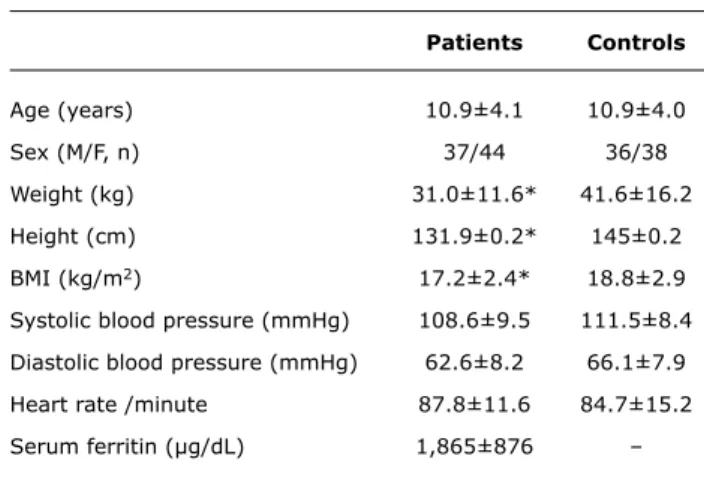
Patients Controls
Age (years) 10.9±4.1 10.9±4.0
Sex (M/F, n) 37/44 36/38
Weight (kg) 31.0±11.6* 41.6±16.2
Height (cm) 131.9±0.2* 145±0.2
BMI (kg/m2) 17.2±2.4* 18.8±2.9
Systolic blood pressure (mmHg) 108.6±9.5 111.5±8.4 Diastolic blood pressure (mmHg) 62.6±8.2 66.1±7.9
Heart rate /minute 87.8±11.6 84.7±15.2
Serum ferritin (µg/dL) 1,865±876 –
table 1 - Characteristics of β-thalassemic patients and healthy controls
BMI = body mass index; F = female; M = male. * p < 0.05 vs. healthy controls.
Results expressed as mean ± standard deviation.
Blood samples were drawn in the pretransfusion period, but physical examination was performed in the posttransfusion period.
Cardiac involvement in beta-thalassemia major - Nisli K et al.
and echocardiography. All participants were selected from among subjects attending the hematology service of two local hospitals. Patients with diabetes mellitus, valvular heart disease, ventricular preexcitation, and atrioventricular conduction abnormalities were excluded from the study. Cardiac evaluation was performed during the interval between blood transfusions, at a mean of 5 days (3-7 days) after transfusions. All patients and control subjects underwent routine transthoracic echocardiographic examination (Vivid 3, General Electric, USA), during which M-mode measurements of left ventricular (LV) end-diastolic and end-systolic diameters and LV ejection fraction (EF) were made according to the recommendations of the American Society of Echocardiography.6 To assess overall LV diastolic function, typical spectral LV illing curves were obtained with conventional pulsed Doppler from a sample volume positioned at the tips of the mitral valve lealets. Peak mitral inlow velocities in early diastole (E) and after atrial contraction (A) were expressed as cm/s. Deceleration time of peak E illing velocity was measured in milliseconds. Overall LV diastolic dysfunction was deined as the presence of any abnormality of the mitral valve.
Twelve-lead electrocardiogram (ECG) was recorded for each patient and control subject at a rate of 50 mm/s. At the time of electrocardiographic recording, all subjects were in sinus rhythm and none were taking any type of antiarrhythmic agent. P-wave duration was measured manually by two of the investigators who were blinded to the participants’ clinical status. All measurements were performed using calipers and magnifying lens to improve accuracy. The onset of P-wave was deined as the junction between the isoelectric line and the beginning of the P-wave delection, and the offset of P-wave as the junction between the end of the P-wave delection and the isoelectric line. Maximum and minimum P-wave durations were measured from the 12-lead surface ECG. Patients with measurable P-waves in nine or fewer ECG leads were excluded from the study. PWD was calculated as the difference between maximum P-wave duration (Pmax) and minimum P-wave duration (Pmin) (PWD = Pmax - Pmin).7
Statistical analysis was performed using Mann-Whitney U test and the chi-square test whenever appropriate. Pearson correlation test was used to determine the correlation between electrocardiographic and echocardiographic variables. Numerical variables were expressed as mean ± standard deviation, and categorical variables were expressed as percentage. P values of < 0.05 were considered to be statistically signiicant.
Results
The study group was composed of 81 β-TM patients (37 boys and 44 girls) aged between 4 and 19 (10.9±4.1) years, and the control group was composed of 74 healthy subjects
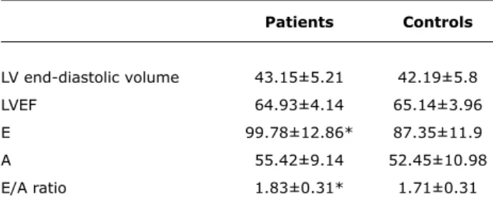
(36 boys and 38 girls) aged between 4 and 20 (10.9±4.1) years. Body mass index (BMI) values in the study and control groups were 17.2±2.4 and 18.8±2.9, respectively. Serum ferritin level in β-TM patients ranged from 375 to 4,900 (1,865±876) µg/L, indicating that compliance to chelation therapy was not uniform within this group. At the time of the study, all subjects’ hemodynamic parameters were normal, and none of them showed clinical signs of cardiovascular disease. There were no statistically signiicant differences between study and control groups regarding sex, mean age, heart rate, and systolic and diastolic blood pressure (p > 0.05). However, BMI was lower in β-TM patients than in control subjects (Table 1). Mean LV end-diastolic volume and EF were, respectively, 43.1±5.2 mm and 64.9±4.1% in the study group, and 42.2±5.8 mm and 65.1±3.9% in the control group. There was a statistically signiicant difference between study and control groups in the measurements of peak E mitral inlow velocity and E/A velocity ratio (Table 2). Maximum P-wave duration and PWD were found to be signiicantly higher in β-TM patients than in control subjects. However, there was no statistically signiicant difference between study and control groups regarding minimum P-wave duration (Table 3).
Discussion
Jornal de Pediatria - Vol. 86, No. 2, 2010 161
Patients Controls
Pmax 102.99±4.64 92.85±4.97*
Pmin 67.4±3.06 65.32±7.76
PWD 35.07±4.80 26.85±4.55*
table 3 - Electrocardiographic indings in β-thalassemic patients and healthy controls
Pmax = maximum P-wave duration; Pmin = minimum P-wave duration; PWD = P-wave dispersion.
* p < 0.05 vs. healthy controls.
Results expressed as mean ± standard deviation.
Patients Controls
LV end-diastolic volume 43.15±5.21 42.19±5.8
LVEF 64.93±4.14 65.14±3.96
E 99.78±12.86* 87.35±11.9
A 55.42±9.14 52.45±10.98
E/A ratio 1.83±0.31* 1.71±0.31
table 2 - Echocardiographic indings in β-thalassemic patients and healthy controls
A = peak mitral inflow velocity after atrial contraction; E = peak mitral inflow velocity in early diastole; LV = left ventricle; LVEF = left ventricular ejection fraction. * p < 0.05 vs. healthy controls.
Results expressed as mean ± standard deviation.
In ß-thalassemic patients, echocardiography was performed in the posttransfusion period.
is a restrictive cardiomyopathy that manifests as systolic or diastolic dysfunction and/or ventricular arrhythmias secondary to increased iron deposition in the myocardium. Chelation therapy with deferoxamine has been associated with a marked decrease in morbidity and mortality in patients with TM.9 Recent studies have suggested that deferiprone provides greater cardiac protection against iron-induced heart disease than deferoxamine.10 In our study, all patients underwent standard deferoxamine therapy (40-50 mg/kg, subcutaneously, 5 days/week).
Currently, there is a need for a reliable test for the presymptomatic identiication of cardiomyopathy, since conventional assessment of ventricular function by echocardiography and nuclear techniques has not proven adequate.11 Cardiac MRI measurement with T2* shows a weak relationship with cardiac function until a critical level is reached, followed by rapid deterioration, which explains why identiication of abnormal systolic function is a late sign of iron toxicity. Iron clears more slowly from the heart than the liver, thus contributing to the high mortality rates observed in patients with established cardiomyopathy despite intensive chelation therapy. This T2* technique enables us to identify much earlier those patients who need intensive chelation therapy prior to the onset of systolic dysfunction, which may ultimately reduce the mortality
associated with manifest heart failure.12 Tissue Doppler imaging (TDI) analysis disclosed, in these patients, an early myocardial dysfunction likely to be related speciically to this initial iron overload, as suggested by the correlation with cardiac T2* values.13 In our study we could not evaluate patients by cardiac MRI or TDI, which becomes the main limitation of this study.
Restrictive LV illing is known to be associated with iron-induced cardiomyopathy. Thus, the prognostic signiicance of diastolic echocardiographic indings has been questioned even in TM.14 The E/A ratio, however, which is one of the most widely used criteria for restrictive LV illing, was signiicantly increased in TM both in it patients and in those with evident heart disease, probably representing the effect of iron overload.15 In our study, the main difference of β-TM patients vs. controls was restrictive diastolic illing pattern, even when they were asymptomatic and had normal LV function.
Cardiac complications of TM were irst described prior to the introduction of chelation therapy. Atrial arrhythmias were observed in half of the patients, and repetitive ventricular tachycardia was present in a minority.16 However, the value of monitoring cardiac function to the long-term management of TM remains unclear, which is partly because the prognostic signiicance of diastolic abnormalities, which appear early in the disease process, is unknown.14 Additionally, once congestive heart failure was present, systolic abnormalities were evident by echocardiography. These observations have led some investigators to question the value of noninvasive monitoring of cardiac function in the management of thalassemia.17
Depression of intra-atrial conduction causes a lengthening of P-wave.18 In addition, it is now well accepted that a new and simple electrocardiographic marker, PWD, also represents inhomogeneous and discontinuous atrial conduction and has predictive value especially for paroxysmal atrial ibrillation.19-21 In healthy children, P-wave duration has been reported to range from 50 to 100 ms.22 PWD has been studied in some other cardiac conditions such as atrial enlargement, obesity, hypertension, atrial septal defect, pulmonary stenosis, and dilated cardiomyopathy.23-24 Tükek et al.25 reported that increased sympathetic activity leads to a signiicant elevation in PWD. Therefore, the increased PWD observed in our patients might be partly related to an elevation in sympathetic activity, which might, in turn, increase the risk of arrhythmias.
Cardiac involvement in beta-thalassemia major - Nisli K et al.
References
1. Bianco I. Clinical and therapeutic aspects of Mediterranean anaemia. II Progr Med. 1986;42:471-5.
162 Jornal de Pediatria - Vol. 86, No. 2, 2010
16. Cohen AR, Galanello R, Pennell DJ, Cunningham MJ, Vichinsky E. Thalassemia. Hematology Am Soc Hematol Educ Program. 2004:14-34.
17. Borow KM, Propper R, Bierman FZ, Grady S, Inati A. The left
ventricular end-systolic pressure-dimension relation in patients
with thalassemia major. A new noninvasive method for assessing contractile state.Circulation. 1982;66:980-5.
18. Kawano S, Hiraoka M, Sawanobori T. Electrocardiographic features of P waves from patients with transient atrial ibrillation. Jpn Heart J. 1988;29:57-67.
19. Dilaveris PE, Gialafos JE. P-wave dispersion: a novel predictor of paroxysmal atrial ibrillation.Ann Noninvasive Electrocardiol. 2001;6:159-65.
20. Tanigawa M, Fukutani M, Konoe A, Isomoto S, Kadena M, Hashiba K. Prolonged and fractionated right atrial electrograms during sinus rhythm in patients with paroxysmal atrial ibrillation and sick sinus node syndrome.J Am Coll Cardiol. 1991;17:403-8. 21. Papageorgiou P, Monaham K, Boyle NG, Seifert MJ, Beswick
P, Zebede J, et al. Site-dependent intra-atrial conduction
delay. Relationship to initiation of atrial ibrillation. Circulation. 1996;94:384-9.
22. Garson A. Electrocardiography. In: Garson A, Bricker JT, Fisher DJ, Neish SR, editors. The Science and Practice of Pediatric Cardiology. 2nd ed. Vol 1. Baltimore: Williams & Wilkins; 1998. p. 735-88. 23. Seyfeli E, Duru M, Kuvandiık G, Kaya H, Yalcin F. Effect of obesity
on P-wave dispersion and QT dispersion in women. Int J Obes.
2006;30:957-61.
24. Ho TF, Chia EL, Yip WC, Chan KY. Analysis of P wave and P dispersion in children with secundum atrial septal defect. Ann Noninvasive Electrocardiol 2001;6:305-9.
25. Tükek T, Akkaya V, Demirel S, Sözen AB, Kudat H, Atilgan D, et al. Effect of Valsalva maneuver on surface electrocardiographic P-wave dispersion in paroxysmal atrial ibrillation. Am J Cardiol. 2000;85:896-9, A10.
Correspondence: Kemal Nisli
Istanbul (Çapa) Tıp Fakültesi Çocuk Kardiyolojisi Bilim Dalı Şehremini/Çapa Istanbul - Turkey Tel: +90 (221) 661.5870 Fax: +90 (212) 414.2196 E-mail: [email protected] 3. Dilaveris PE, Gialafos EJ, Sideris SK, Theopistou AM, Andrikopoulos
GK, Kyriakidis M, et al. Simple electrocardiographic markers for the prediction of paroxysmal idiopathic atrial ibrillation.Am Heart J. 1998;135:733-8.
4. Dilaveris PE, Gialafos EJ, Andrikopoulos GK, Richter DJ, Papanikolaou V, Poralis K, et al. Clinical and electrocardiographic
predictors of recurrent atrial ibrillation.Pacing Clin Electrophysiol. 2000;23:352-8.
5. Senen K, Turhan H, Riza Erbay A, Basar N, Saatci Yasar A, Sahin O, et al. P-wave duration and P-wave dispersion in patients with dilated cardiomyopathy. Eur J Heart Fail. 2004;6:567-9.
6. Sahn DJ, DeMaria A, Kisslo J, Weyman A. Recommendations
regarding quantitation in M-mode echocardiography: results of a survey of echocardiographic measurements. Circulation 1978;58:1072-83.
7. Gialafos JE, Dilaveris PE, Gialafos EJ. P-wave dispersion: a valuable electrocardiographic marker for the prediction of paroxysmal lone atrial ibrillation. Ann Noninvas Electrocardiol. 1999;4:39-45. 8. Olivieri NF, Nathan DG, MacMillan JH, Wayne AS, Liu PP, McGee
A, et al. Survival in medically treated patients with homozygous beta-thalassemia. N Engl J Med 1994;331:574-8.
9. Borgna-Pignatti C, Rugolotto S, De Stefano P, Zhao H, Cappellini MD, Del Vecchio GC, et al. Survival and complications in patients
with thalassemia major treated with transfusion and deferoxamine.
Haematologica. 2004;89:1187-93.
10. Anderson LJ, Wonke B, Prescott E, Holden S, Walker JM, Pennell DJ. Comparison of effects of oral deferiprone and subcutaneous desferrioxamine on myocardial iron concentrations and ventricular function in beta-thalassaemia. Lancet. 2002;360:516-20. 11. Hoffbrand AV. A sensitive test for early myocardial iron loading.
Eur Heart J. 2003;24:26-7.
12. Anderson LJ, Holden S, Davis B, Prescott E, Charrier CC, Bunce NH, et al. Cardiovascular T2-star (T2*) magnetic resonance for the early diagnosis of myocardial iron overload. Eur Heart J. 2001;22;2171-9.
13. Magri D, Sciomer S, Fedele F, Gualdi G, Casciani E, Pugliese P, et al. Early impairment of myocardial function in young patients with beta-thalassemia major. Eur J Haematol. 2008;80:515-22. 14. Kremastinos DT, Tsiapras DP, Tsetsos GA, Rentoukas EI, Vretou
HP, Toutouzas PK. Left ventricular diastolic Doppler characteristics in beta-thalassemia major.Circulation. 1993;88:1127-35. 15. Lubien E, DeMaria A, Krishnaswamy P, Clopton P, Koon J,
Kazanegra R, et al. Utility of B-natriuretic peptide in detecting diastolic dysfunction: comparison with Doppler velocity recordings.
Circulation. 2002;105:595-601.