Jebmh.com
Original Article
J. Evid. Based Med. Healthc., pISSN- 2349-2562, eISSN- 2349-2570/ Vol. 3/Issue 52/June 30, 2016 Page 2699
DIAGNOSTIC UTILITY OF USG-GUIDED FNAC IN HEPATIC LESIONS
Sudha P. Meena1, Priyanka Patangia2, Naresh N. Rai3
1Associate Professor, Department of Pathology, Government Medical College, Kota.
2Senior Resident, Department of Pathology, Government Medical College, Kota.
3Senior Professor, Department of Pathology, Government Medical College, Kota.
ABSTRACT
INTRODUCTION
Guided fine-needle aspiration cytology (FNAC) is an easy, rapid, minimally invasive and a cost effective diagnostic method for detecting benign and malignant lesions of liver.
AIM
The main aim of the present study was to establish the incidence of various hepatic lesions and to find out adequacy and utility of the procedure.
MATERIAL AND METHOD
A total of 174 cases were included in the study from Government Medical College, Kota and associated hospitals. All cases diagnosed to have single or multiple hepatic mass lesions on USG were included in the study.
RESULTS
Most common age group affected by hepatic lesion was 51-60 years (34.0%). 91.4% cases were having adequate aspirates. 95.6% of the total diagnosed cases were malignant and among malignant cases majority were metastatic.
CONCLUSION
USG-guided FNAC is a very useful procedure in the diagnosis of hepatic lesions as the procedure is simple and safe. Thus, FNAC is a simple and effective diagnostic tool in our hand.
KEYWORDS
FNAC, USG, Hepatocellular Carcinoma, Metastasis.
HOW TO CITE THIS ARTICLE: Meena SP, Patangia P, Rai NN. Diagnostic utility of USG-guided FNAC in hepatic lesions. J.
Evid. Based Med. Healthc. 2016; 3(52), 2699-2702. DOI: 10.18410/jebmh/2016/591
INTRODUCTION: Guided fine-needle aspiration cytology
(FNAC) is an easy, rapid, minimally invasive and a cost effective diagnostic method for detecting benign and malignant lesions of liver. Ehrlich was first to use percutaneous FNAC for liver in 1893, but it was used initially as a diagnostic method in 1923.[1]
Ultrasonography (USG) or computed tomography (CT)-guided fine needle aspiration cytology (FNAC) is an accurate method for a definite diagnosis in focal liver lesions.[2] The benefit of this technique is its high diagnostic accuracy leading to obsolete use of older technique of blind percutaneous biopsy using a coarse needle.[3]
Clinical, serological and radiological findings are not reliable to differentiate between a benign and malignant lesion but are useful in making differential diagnosis of various hepatic lesions.[4] Thus, guided FNAC has gained increasing popularity as the diagnostic procedure of choice.[4,5,6,7,8,9,10,11]
During the procedure, involvement cytopathologist increases overall diagnostic accuracy.[5] It is a valuable tool for identification of malignant lesions and also work as an accessory tool in diagnosis of benign and inflammatory disease of the liver.[12]
FNAC is the diagnostic tool with minimum risk, less discomfort to the patient with an added advantage of multiple aspirations at one sitting in the outpatient department. It has proved to be superior to core-needle or open biopsy with an advantage of minimal complications and early diagnosis of the lesion.
AIM: The main aim of the present study was to establish the incidence of various hepatic lesions and to find out adequacy and utility of the procedure.
MATERIAL AND METHOD: A total of 174 cases were
included in the study from Government Medical College, Kota and associated hospitals. All cases diagnosed to have single or multiple hepatic mass lesions on USG were included in the study. USG-guided FNAC was done using 22 G lumbar puncture needle under aseptic conditions. The smears were prepared and fixed by air drying followed by Giemsa staining.
Financial or Other, Competing Interest: None. Submission 03-06-2016, Peer Review 12-06-2016, Acceptance 22-06-2016, Published 30-06-2016. Corresponding Author:
Dr.Priyanka Patingia,
Senior Resident, Department of Pathology, Government Medical College, Kota, Rajasthan, India. E-mail: [email protected]
Jebmh.com
Original Article
J. Evid. Based Med. Healthc., pISSN- 2349-2562, eISSN- 2349-2570/ Vol. 3/Issue 52/June 30, 2016 Page 2700 For Giemsa stain, smears were removed from methanol
fixative and followed to air dry. Working staining solution was prepared by mixing of Giemsa solution 1 mL into 9 mL of working buffer solution. Smears were covered for 10 minutes with stain. Stain was then poured off and washed well in running tap water. Smears were air dried and mounted in DPX.
RESULTS: Most common age group affected by hepatic
lesion was 51-60 years (34.0%) followed by 61-70 years (27.3%). Age range of affected patients was 23-90 years. In our study, 91.4% cases were having adequate aspirates with only 8.6% being inadequate. Of the total diagnosed cases, 95.6% were malignant while only 4.4% were inflammatory or benign. Among malignant cases, majority were metastatic (123 cases) followed by primary tumours (28) and a single case of Non-Hodgkin’s lymphoma.
Metastatic adenocarcinoma was most common (79.7%) among metastatic lesions followed by metastatic small cell carcinoma (10.6%), metastatic round cell tumour (5.7%), metastatic squamous cell carcinoma (1.6%). Single cases of other metastatic lesion like large cell carcinoma, adenosquamous carcinoma and malignant mesenchymal tumour were also reported. Majority of the metastatic adenocarcinomas were from GIT and metastatic small cell carcinomas were from lung. Metastatic lesions were diagnosed on the basis of characteristic features of the primary lesion depending on the type of tumour.
In our study, 28 primary lesions from liver were detected of which 27 were HCC (27) and a single case was cholangiocarcinoma. Primary hepatocellular carcinomas were diagnosed in smears showing characteristic features like trabeculae with traversing capillaries, polygonal cells, INCI’s.
Age Frequency Percentage
0-10 0 0
11-20 1 0.42
21-30 7 2.94
31-40 21 8.82
41-50 38 15.97
51-60 81 34.03
61-70 65 27.31
71-80 21 8.82
81-90 4 1.68
91-100 0 0
Total 238 100
Table 1: Age wise Distribution of Hepatobiliary Lesion
Type of TUMOUR Frequency
Inflammatory 5
Benign
Focal Nodular Hyperplasia 1
Angiomyolipoma 1
Malignant Primary
Hepatocellular Ca. 27
Cholangiocarcinoma 1
Secondary
Metastatic Adenocarcinoma 98
Metastatic Small Cell
Carcinoma 13
Metastatic Small Round
Cell Tumour 7
Metastatic Squamous
Cell Ca. 2
Metastatic Large Cell
Carcinoma 1
Metastatic Adenosquamous
Carcinoma 1
Metastatic Mesenchymal
Tumour 1
NHL 1
Total 159
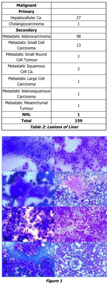
Table 2: Lesions of Liver
Figure 1
Figure 1: Giemsa-stained cytosmear showing
well-differentiated hepatocellular carcinoma showing A)
Jebmh.com
Original Article
J. Evid. Based Med. Healthc., pISSN- 2349-2562, eISSN- 2349-2570/ Vol. 3/Issue 52/June 30, 2016 Page 2701 Metastatic malignant round cell tumour (10x) G) Metastatic
adenosquamous carcinoma (20x) H) Non-Hodgkin’s
Lymphoma (20x) showing tingible body macrophage.
DISCUSSION: FNAC has proven to be an effective
diagnostic procedure for superficial swelling at various sites as well as deep seated visceral lesions under USG guidance. Hepatic lesions are commonly aspirated under USG guidance. It has an advantage of early diagnosis with minimal invasion, safety, minimal complication rate and cost effectiveness. FNAC plays an important role in diagnosis of benign as well as malignant lesions. It is primarily used in diagnosing malignant lesion and classifying them as primary or metastatic. Guided FNAC is a sensitive procedure in diagnosing hepatic lesion.
The present study had 91.4% adequate aspirates. High adequacy rate has been proven by other studies also where sensitivity for diagnosis of hepatic malignancy ranges from 75.34% to 93%.[13,14] Our study shows inadequacy rate of
8.6 % using 18 gauge spinal needle for deep‑seated lesions.
Other studies like Bell et al,[15] Talukdar[16] and Balani et al[17] reported inadequacy rate of 13%, 6.5%, and 5.7% respectively. Using 22 gauge Chiba needle, lowest inadequacy rate of 1% was reported by Guo et al.[18] In the present study, causes of inadequacy were haemorrhagic
smear and non‑representative smears.
For adequacy of smear, a representative sample must be taken as aspirate from surrounding tissue will show reactive changes while from centre may be necrotic. So under USG guidance, solid mass of tumour must be targeted. Multiple aspirations can be done to increase adequacy rate as representative material can be obtained by any one aspirate. Tao et al.[12] and Edoute et al.[19] also recommended multiple aspirations, as this may reduce diagnostic fallacies.
In our study, age range was 23 to 90 years with male predominance M:F ratio being 1.2: 1. According to Rasania et al,[8] age range from 8 to 80 years with male predominance was reported. In several studies, the youngest patient was 10 years old. The youngest patient reported with hepatic malignancy was 20 days old in a study by Das et al.[2] In our study, the youngest case was 23 years old.
In our study, majority of the cases were metastatic carcinomas (77.76%). Our results were on concordance with Tao et al[12] and Barbhuiya et al[20] with metastatic lesion being 75% and 74.9% respectively. Some studies have shown metastatic liver malignancy as high as 90%.[21] Metastatic adenocarcinoma was the most common lesion reported in our study (77.36%). Majority of them were of GI origin. This results are supported by Barbhuiya et al.[20]
In the studies performed by several authors, the complications induced by FNAC were considered. Ramdas and Chopra et al[22] and Barbhuiya et al[20] reported no complication in their observation. Similarly, in our study, no FNAC induced complications were seen. Complications like fatal bleeding and needle tract seeding were reported by
Mingoli A[23] and Patel RI et al[24] respectively. A study of 2600 cases done by Lundquist et al[25] reported only a single case of complication having intrahepatic haematoma. Thus, FNAC has proven to be a safe procedure with minimal procedure induced complication.
CONCLUSION: In the present study, it is concluded that USG-guided FNAC is a very useful procedure in the diagnosis of hepatic lesions as the procedure is simple and safe. The early diagnosis can be done by FNAC along with USG finding without any serious complications related to the procedure. FNAC also plays an important role at centres like ours where it is the only diagnostic tool for hepatic lesion where facility for liver biopsy (Tru-cut or open), other ancillary studies and surgery are not available. Thus, FNAC is a simple and effective diagnostic tool in our hand.
REFERENCES
1. Bingel A. Uber die parenchympunktion der leber. Verh Dtsch Ges Inn Med 1923;35:210-212.
2. Das DK, Tripathi RP, Chachra KL, et al. Role of guided fine needle aspiration cytology in diagnosis and
classification of liver malignancies. Trop
Gastroenterol 1997;18(3):101-106.
3. Naggada HA, Ahidjo A, Ajagi N, et al. Correlation between ultrasound findings and ultrasound guided FNAC in the diagnosis of hepatic lesions: a Nigerian tertiary hospital experience. Int J Gastroenterol 2007;5:1.
4. Swamy MC, Arathi CA, Kodandaswamy CR. Value of
ultrasonography-guided fine needle aspiration
cytology in the investigative sequence of hepatic lesions with an emphasis on hepatocellular carcinoma. J Cytol 2011;28(4):178-184.
5. Chhieng DC. Fine needle aspiration biopsy of liver-an update. World J Surg Oncol 2004;2:5.
6. Wee A. Fine needle aspiration biopsy of the liver: algorithmic approach and current issues in the diagnosis of hepatocellular carcinoma. Cyto Journal 2005;2:7.
7. Nazir RT, Sharif MA, Iqbal M, et al. Diagnostic accuracy of fine needle aspiration cytology in hepatic tumours. J Coll Physicians Surg Pak 2010;20(6):373-376.
8. Rasania A, Pandey CL, Joshi N. Evaluation of FNAC in diagnosis of hepatic lesion. J Cytol 2007;24(1):51-54.
9. Asghar F, Riaz S. Diagnostic accuracy of
percutaneous cytodiagnosis of hepatic masses, by ultrasound guided fine needle aspiration cytology. Annals 2010;16(3):184-188.
10. Bottles K, Cohen MB. An approach to fine-needle aspiration biopsy diagnosis of hepatic masses. Diagn Cytopathol 1991;7(2):204-210.
11. Ji XL. Fine-needle aspiration cytology of liver diseases. World J Gastroenterol 1999;5(2):95-97. 12. Tao LC, Donat EE, Ho CS, et al. Percutaneous
Jebmh.com
Original Article
J. Evid. Based Med. Healthc., pISSN- 2349-2562, eISSN- 2349-2570/ Vol. 3/Issue 52/June 30, 2016 Page 2702 13. Herszenyi L, Farinati F, Cecchetto A, et al. Fine-needle
biopsy in focal liver lesions: the usefulness of a screening programme and the role of cytology and microhistology. Ital J Gastroenterol 1995;27(9):473-478.
14. Shah A, Jain GM. Fine needle aspiration cytology of the liver - a study of 518 cases. J Cytol 2002;19:139-143.
15. Bell DA, Carr CP, Szyfelbein WM. Fine needle aspiration cytology of focal liver lesions. Results obtained with examination of both cytologic and
histologic preparations. Acta Cytol
1986;30(4):397‑402.
16. Talukder SI, Huq MH, Haque MA, et al. Ultrasound guided fine needle aspiration cytology for diagnosis of mass lesions of liver. Mymensingh Med J
2004;13(1):25-29.
17. Balani S, Malik R, Kapoor N, et al. Cytomorphological variables of hepatic malignancies in fine needle aspiration smears with special reference to grading of hepatocellular carcinoma. J Cytol 2013;30(2):116-120.
18. Guo Z, Kurtycz DF, Salem R, et al. Radiology guided percutaneous fine needle aspiration biopsy of the liver: retrospective study of 119 cases evaluating diagnostic effectiveness and clinical complications.
Diagn Cytopathol 2002;26(5):283-289.
19. Edoute Y, Tibon‑Fisher O, Haim SB, et al. Ultrasound
guided fine needle aspiration of liver lesions. Am J
Gastroenterol 1992;87(9):1138-1141.
20. Barbhuiya M, Bhunia S, Kakkar M, et al. Fine needle aspiration cytology of lesions of liver and gallbladder: an analysis of 400 consecutive aspirations. J Cytol 2014;31(1):20-24.
21. Orell SR, Sterrett GF, Walters MN, et al.
Retroperitoneum, liver and spleen. In: Manual and atlas of fine needle aspiration cytology. 2nd edn. Hong Kong: Churchill Livingstone 1992:217-266.
22. Ramdas A, Chopra R. Diagnostic accuracy of fine needle aspiration cytology of liver lesions. J Cytol 2003;20:121-123.
23. Mingoli A, Marzano M, Sgarzini G, et al. Fatal bleeding after fine-needle aspiration biopsy of the liver. Ital J Gastroenterol 1995;27(5):250-251.
24. Patel RI, Shapiro MJ. Biliary venous fistula: an unusual complication of fine-needle aspiration biopsy of the liver. J Vasc Interv Radiol 1995;6(6):953-956. 25. Lundquist A. Fine-needle aspiration biopsy for