ORIGIN
AL RESEAR
CH
Mailing address: Fernanda Sampaio Teles – SQS 307 Bloco K apt. 202, Asa Sul –Brasília (DF), Brazil – CEP: 70354-110 – Email: [email protected] – Funding source: Nothing to declare – Conlict of interest: Nothing to declare – Presentation: Oct. 2015 – Accepted for publication: Aug. 2016 – Approved by the Ethics and Research Committee of
1Physical Education Professor; master’s student in Physical Education – University of Brasília.
2PhD professor in Physical Education. Visiting Professor of Physical Education course – University of Brasília. 3
Master professor in Physical Education; Educational Afairs technician – Federal Police of Brazil.
4PhD professor in Physical Education; Associate Professor of Physical Education course – University of Brasília. 5PhD Professor and electrical engineer; Associate Professor of Engineering at Gama campus – University of Brasília.
ABSTRACT | The present study sought to investigate the electromyographic parameters of muscle fatigue during dynamic exercise conducted with variable resistance (elastic) and ixed resistance (pulley). Ten trained men participated voluntarily in the study. Two maximum voluntary isometric contractions (MVIC) of the elbow lexors were performed for 5 seconds and a two minutes interval rest was given between reps. Then, the volunteers performed unilateral elbow lexion repetitions until the exhaustion using ixed and elastic resistance with a pre-established rhythm of two seconds for each phase of the movement. Constant resistance was carried out at the pulley at 30% of MVIC. For the elastic resistance, load control was based on the subjective perception of the participant’s efort. The exercise order was randomized, and electromyography signal of the biceps brachii muscle was recorded during contractions. From the electromyographic signal related to every movement, linear regression lines were drawn for root mean square (RMS) variables and median power frequency (MPF). The inclination of the lines normalized by the linear coeicient of regression equations were compared using the paired t-test. No signiicant diferences between the types of resistance (elastic and pulley) were found. During elbow lexion exercise performed with elastic resistance and pulley, electromyographic parameters of fatigue did not difer. Efort perception during the exercise with an elastic implement does not inluence the electromyographic signal pattern (RMS and MPF).
Keywords | Electromyography; Muscle Fatigue
257
RESUMO | Buscou-se investigar os parâmetros eletromiográicos da fadiga muscular durante exercício dinâmico realizado com resistência variável (elástica) e resistência ixa (polia). Dez homens treinados participaram voluntariamente do estudo. Foram realizadas duas contrações voluntárias isométricas máximas (CVIM) de lexão do cotovelo com 5 segundos de duração cada e 2 minutos de intervalo entre as mesmas. Em seguida, os voluntários realizaram a lexão unilateral do cotovelo até a exaustão, utilizando resistência ixa e elástica com ritmo pré-estabelecido de 2 segundos para cada fase do movimento. A resistência constante foi realizada na polia a 30% da CVIM. Para a resistência elástica, o controle de carga era baseado na percepção subjetiva de esforço do participante. A ordem do exercício foi randomizada, e o sinal de eletromiograia do músculo bíceps braquial foi registrado durante as contrações. A partir do sinal eletromiográico referente a cada execução de movimento, foram traçadas retas de regressão linear para as variáveis RMS e frequência de potência mediana (FPM). As inclinações das retas normalizadas pelo coeiciente linear das equações de regressão foram comparadas por meio do teste t pareado. Não foram observadas diferenças signiicativas entre os tipos de resistência (elástica e polia). Os parâmetros eletromiográicos de fadiga não foram diferentes durante o exercício de lexão do cotovelo realizado com resistência elástica e polia. A percepção de esforço durante o exercício com implemento elástico não inluencia no padrão do sinal eletromiográico (RMS e FPM).
Descritores | Eletromiograia; Fadiga Muscular.
Electromyographic parameters in fatiguing exercises
performed with diferent types of resistance
Parâmetros eletromiográicos em exercícios fatigantes realizados com diferentes tipos de resistência
Estándares electromiográicos en ejercicios extenuantes empleando distintos tipos de resistencia
Fernanda Sampaio Teles1, Maria Claudia Pereira2, Valdinar de Araújo Rocha-Júnior3,RESUMEN | En esta investigación se pretendió estudiar los estándares electromiográicos de fatiga muscular durante la realización de ejercicio dinámico con resistencia variable (elástico) y resistencia ija (polea). Han participado voluntariamente diez varones entrenados. Se llevó a cabo dos contracciones voluntarias máximas (CVIM) de lexión de codo, cada cual de cinco segundos de duración y dos minutos de intervalo entre las mismas. Después los voluntarios hicieron la lexión unilateral del codo hasta sentirse cansados, y emplearon la resistencia ija y elástica con ritmo prestablecido de dos segundos para cada fase del movimiento. Se llevó a cabo la resistencia constante en la polea a 30% de la CVIM. Relativo a la resistencia elástica, el control de carga se basaba en una percepción subjetiva del esfuerzo empleado por el participante. El orden del ejercicio fue aleatorio, y
se registró la señal electromiográica del músculo bíceps durante las contracciones. Desde la señal electromiográica relativa a cada ejecución del movimiento se dibujó rectas de regresión lineal para las variables RMS y la frecuencia de potencia media (FPM). Se conirmó las inclinaciones de rectas normalizadas por el coeiciente lineal de las ecuaciones de regresión a través de la prueba t pareada. No se observaron diferencias signiicantes entre los tipos de resistencia (elástica y polea). Los estándares electromiográicas de fatiga durante la práctica de ejercicios de lexión de codo empleando resistencia elástica y polea no fueron distintos. La percepción del esfuerzo durante la práctica de ejercicio con elástico no trae consecuencias al estándar de señal electromiográico (RMS y FPM).
Palabras clave | Electromiografía; Fadiga Muscular.
INTRODUCTION
Resistance training (RT) is commonly used as a strategy to develop and improve physical capabilities. Nowadays, there are many implements that allow its practice varying according to the purpose intended1.
Exercises performed with free weights or machines have constant resistance. On the other hand, exercises practiced with variable resistance, such as elastic tubing, have load variations during the movement1.
Although resistance bands have unique features such as convenience, portability and low cost, studies that compare elastic load with constant load implements are scarce.
Besides the scarcity of studies on elastic resistance, the diiculty in obtaining a methodological procedure to compare the types of resistance, as well as physiological consequences for each intervention, makes elastic resistance use restricted and focused on articular or clinical rehabilitation programs, in which the exercises are usually carried out with submaximal intensity. According to Andersen et al.3 few studies have
analyzed physiological adaptations induced by exercises with elastic resistance compared to other contraction modalities. Moreover, previous studies found that elastic resistance used in an adequate intensity provides signiicant improvements in functional tests and changes the body composition – reduction of abdominal perimetry and body fat percentage4,5.
Surface electromyography (SEMG) allows the researcher to analyze the lower and upper limbs muscular actions during exercises. It is known that SEMG may provide relevant information about the activity of motor units and recruitment of diferent
muscle iber types6. For example, some authors
veriied an association between the amplitude and the electromyographic signal frequency and the pattern of motor units activity7,8. Moreover, a connection was
identiied between the conduction velocity obtained by SEMG and the recruitment of diferent muscle ibers. Several authors use SEMG to analyze the behavior of localized muscular fatigue9-11 and many of them
have used this tool, adding value to the discussion about neuromuscular behavior during exercises with diferent types of resistance12,13,15. However,
there is no consensus on the efectiveness using diferent resistance methods and their neuromuscular adaptations, since most of these studies assess various types of populations.
he study of Melchiorri et al.13 investigated the
neuromuscular modiications induced by exercises until fatigue with two types of resistance (elastic versus constant). However, the constant resistance used in this study was provided by a machine developed by the researchers. Also, it is not the kind of equipment commonly found in itness centers, which limits the research reproducibility.
exercises performed using pulley at a conventional weight machine to elastic resistance.
METHODOLOGY
Participants
Ten male volunteers (age 27.7±6.0; 79.8±5.5kg; 175.7±4.9cm) participated in the present study – they had at least six months of resistance training experience. Volunteers could not have any physical problems and/or osteomioarticular injuries that could be aggravated by the elbow lexion movement. Before taking part of the study, all volunteers signed a written informed consent form (ICF) that explained all research procedures. his study was carried out after approval by the Ethics and Research Committee of the Faculty of Health Sciences at the University of Brasília (UnB) –16303013.0.0000.0030.
Experimental procedures
Firstly, body weight (Líder Balanças®, model PM180, Araçatuba, SP) and height (Sanny®) were measured for the sample description. hen, individuals performed two maximum isometric voluntary contractions (MVIC) during ive seconds with a two-minute rest between them. During the MVIC execution, volunteers remained seated with their right elbow forming a 90° angle between upper arm and forearm. A load cell, model TS (AEPH do Brasil Indústria e Comércio Ltda., 50kg±10%), was ixed on the chair by an inextensible chain, and a hilt attached to it was used to perform the exercise. Verbal encouragement was given throughout the MVIC by the same investigator.
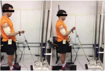
After that, the volunteers were requested to perform elbow lexion repetitions with their right limb until exhaustion. his exercise protocol was repeated in two situations: with pulley at a conventional weight machine (Gervasport itness equipment) (P) and with elastic tubing (E) as displayed in Figure 1. he order of tasks was counterbalanced with a 10-minute rest between them. he P task load corresponded to 30% of the MVIC, and the rhythm of contractions was two seconds for each phase of the movement, all controlled by a digital metronome (Seiko). he E task followed the same rhythm. However, the load adjustment of 30% of the MVIC was determined by a scale of subjective
perception efort used with elastic implements and
validated by Colado et al.14 (OMNI-RES), which
intensity (in kilos) was informed to the evaluator. he volunteers, through their efort perception, were instructed to move enough to extend the elastic resistance until the perception corresponded to 30% of the MVIC, in order to make as many repetitions as possible of elbow lexion until exhaustion or until they were unable to keep the requested cadence. he OMNI-RES scale is currently the most widely used method to determine the load for variable resistance.
All tests were conducted in the Laboratory of Biological Signal Processing and Motor Control of the Faculty of Physical Education at University of Brasília (UnB) after prior calibration of the equipment and preparation of all complementary materials for data collection: adhesive plasters, razor blades, cotton, scotch tape, and so on.
Figure 1. A volunteer exemplifying the experimental protocol with diferent resistances: pulley (P) and elastic (E), respectively
Electromyography
SEMG signals from the biceps brachii muscle were recorded by an electromyography (Bagnoli-2, Delsys, Boston, USA) with a 2,000 Hz sampling frequency. Bipolar electrodes (Ag/AgCl) pre-ampliied with a 1,000V/V gain, a 20Hz-450Hz band pass-ilter and 1-centimeter inter-electrode distance were used. he electromyography signal obtained was transferred to the computer through a 12-bit analog-to-digital board (National Instruments, PCI-6024E model, Austin,
USA). SENIAM16 recommendations for the electrode
test preparation. An electrogoniometer was positioned with its axis at the elbow lateral epicondyle and its stems aixed on the upper and forearm.
he SEMG signal was originally iltered by a
4th-order Butterworth ilter with a 20Hz-500Hz
bandwidth and correction on phase delay17. A low
pass ilter with the same characteristics and a 15 Hz cutof frequency was used to ilter the power signal18.
he electrogoniometer signal was used as a reference for the burst cutting of the electromyographic signal in each repetition. From the angular position peak, which was represented by the largest extension angle of the elbow, 500 samples were covered and a window of 2,000 samples were cut. his methodology was adopted because even with the cadence control with a metronome, there was a slight variation in the movement speed of the most acute angles of elbow lexion. With this procedure, it was possible to obtain more reliable data concerning the imposed cadence to the volunteers during the entire experiment. his procedure also provided more uniformed windowing between the bursts of the two experimental situations.
he root mean square (RMS) and median power frequency (MPF) were calculated for the bursts of each repetition. From these amplitude and frequency parameters, linear regressions were drawn to indicate the electromyographic signal behavior during the exercise. he inclinations of regression lines (angular coeicients) were normalized by their initial values (linear coeicients) to help the comparison between volunteers and experimental situations. Digital signal processing was performed using speciic routines written in Matlab 6.5 software (Mathworks software; Natick, MA, USA).
Statistical Analyses
All statistical procedures were performed using the statistical software package SPSS 20 (SPSS Inc., Chicago, IL). To verify the data normality, Shapiro-Wilk test was used. After conirming the normal distribution, comparisons between the types of resistance were made using paired t-test for each electromyographic parameter. Signiicance level adopted was 5%.
RESULTS
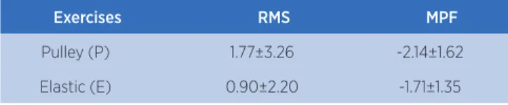
he reported data with mean values ± standard deviation related to electromyographic variables are
shown in Table 1. Considering linear regression curves for the RMS and MPF variables, there are no signiicant diferences for line inclinations between the types of resistance (P and E) (Table 2).
Table 1. Mean values and standard deviation of regression line inclinations of electromyographic variables with diferent types of resistance used (n=8)
Exercises RMS MPF
Pulley (P) 1.77±3.26 -2.14±1.62
Elastic (E) 0.90±2.20 -1.71±1.35
p<0.05; RMS: root mean square, normalized by the maximum voluntary contraction; MPF: median power frequency
Table 2. Paired t-test values for each variable with diferent types of electromyographic resistance (elastic and pulley)
Variables Paired t-test
RMS 0.38
MPF 0.45
p<0.05; RMS: root mean square, normalized by the maximum voluntary contraction; MPF: median power frequency
DISCUSSION
he study results show that there was no signiicant diference between the inclination of the regression lines of the RMS and MPF recorded during the elbow lexions performed with elastic and constant resistance for the variables studied. Despite having unique characteristics, elastic resistance has not caused greater muscle activation and fatigue, even with a varying intensity throughout all the movement extent when compared to pulley, which has a measurable load control.
In both cases, the RMS value increased and the median power frequency (MPF) decreased. his pattern is well described in the literature as an indicative of Localized Muscle Fatigue (LMF) and is justiied by an increasing recruitment due to the sum of motor units, which consequently increases the signal amplitude
over time17. We also observed an increase in the
electromyographic signal power at low frequencies, when compared to the high ones, which represents a spectral signature associated with LMF respectively19,20.
Sundstrup et al.21 investigated the recruitment strategies
of the medial deltoid, upper trapezius, splenius and infraspinatus muscles by using SEMG during shoulder lateral raise exercise, performed with maximum and submaximal loads using elastic implement. he results found showed that elastic overload use in submaximal intensity exercise conducted until muscle exhaustion promotes quite satisfactory muscle activation levels of the muscle motor units exercised. his reinforces the usability and portability of elastic implement and corroborates the RMS value increase during the task carried out with elastic in the study. In addition, regardless of the method of load determination, the use of elastic implement until exhaustion with submaximal loads is enough to increase muscle activation.
Calatayud et al.15 aimed to compare strength levels
with diferent resistances and found similar gains of strength in push-ups performed with elastic resistance and bench press, using the Smith equipment, a bar and barbells. heir result contributes to the use of elastic implements in biomechanically similar movements, i.e., individuals – who are trained and familiar with the bench press and push-ups – can use both resistance modalities for strength gains. In addition, push-ups with elastic resistance are versatile and can be carried out in multiple locations with diferent intensity settings. Adding strength to electromyographic signals can be an important inferential tool to evaluate physiological aspects of muscular system and its implications on strenght improvements12,13. hus, future studies should
include the analysis of muscle strength in results. In the case of lower limbs, Jakobsen et al.22 analyzed
the electromyographic signal of the primary motor muscles and the stabilizers muscles of hamstring knee performed with dumbbells and elastic. he authors determined 33%, 66% and 100% intensities for 10 repetition maximum. hey concluded that the greatest recruitment of the hip and knee muscles occurred with elastic resistance with mean intensity (66% of 10RM) when compared with low (33% of 10RM) and maximal intensities (100% of 10RM). Such inding suggests that when lunges are performed with the elastic band using a medium load, it can be a good alternative to inducing high levels of neuromuscular activation during muscle contractions.
However, our study has not found a pattern or a speciic behavior for the elbow lexion exercise performed with elastic implement and pulley. Our indings are in accordance with the results of the Melchiorri et
al.13 study which investigated neuromuscular changes
induced by fatiguing exercise performed with elastic resistance compared to constant load. he constant load equipment used was built by the authors themselves to perform the elbow lexion exercise. Despite being a common movement of resistance training, all results are based in an equipment that is not available in the itness market.
he present study innovates by making the comparison between an exercise performed with variable resistance and the same exercise performed in a pulley machine. In addition, the study of Melchiorri et al.13 presented an exhaustion protocol with isometric
contraction of elbow lexion. Moreover, when comparing electromyographic variables before and after exhaustion, they have found no signiicant diferences as well.
Our experimental protocol consisted in dynamic elbow lexion contractions. Despite dynamic muscle contractions present more vulnerability to movement artifacts from the electrode on the skin and muscles during a SEMG acquisition, our results of fatigue and muscle activation for both tasks (E and P) were consistent and are in accordance with those described in the literature9,19,20 – decrease of the MPF and increase
of the muscle recruitment.
herefore, considering that the studied tasks have the same indicative of muscle fatigue with distinct load curves – constant and elastic resistance –, the results suggest that the adoption of intercalated exercises between these two modalities, during the process of physical training, can become an alternative to avoid a training plateau23. An exercise practiced with a single
modality of resistance tends to achieve this plateau1 and
to minimize the results23. Based on the results, the
non-signiicance between tasks suggests that, intercalating practices that cause muscle fatigue in the same muscle group, with diferent load curves, can avoid training plateau by achieving a more efective result.
CONCLUSION
We suggest additional studies that associate these parameters of fatigue for both resistances and the force level generated during execution. he variable resistance, besides depending on the individual’s perception of intensity, also lacks quantitative load control, unlike the pulley, which has a constant and known load.
REFERENCES
1. Fleck SJ, Kraemer WJ. Fundamentos do treinamento de força muscular. 3. ed. Porto Alegre: Artmed; 2006.
2. Andersen LL, Andersen CH, Mortensen OS, Poulsen OM, Bjornlund IBT, Zebis MK. Muscle activation and perceived loading during rehabilitation exercises: comparison of dumbbells and elastic resistance. Phys Ther. 2010; 90(4):538-49.
3. Colado JC, Garcia-Masso X, Pellicer M, Alakhdar Y, Benavent J, Cabeza-Ruiz R. A comparison of elastic tubing and isotonic resistance exercises. Int J Sports Med. 2010;31(11):810-7. 4. Colado JC, Triplett NT. Efects of a short-term resistance
program using elastic bands versus weight machines for sedentary middle-aged women. J Strength Cond Res. 2008;22(5):1441-8.
5. Colado JC, Triplett NT, Tella V, Saucedo P, Abellán J. Efects of aquatic resistance training on health and itness in postmenopausal women. Eur J Appl Physiol. 2009;106(1):113-22.
6. Moritani T, Muro M. Motor unit activity and surface electromyogram power spectrum during increasing force of contraction. Eur J Appl Physiol Occup Physiol. 1987;56(3):260-5.
7. Houtman CJ, Stegeman DF, Van Dijk JP, Zwarts MJ. Changes in muscle iber conduction velocity indicate recruitment of distinct motor unit populations. J Appl Physiol. 2003;95(3):1045-54.
8. Farina D, Ferguson RA, Macaluso A, De Vito G. Correlation of average muscle iber conduction velocity measured during cycling exercise with myosin heavy chain composition, lactate threshold, and VO2max. J Electromyogr Kinesiol. 2007;17(4):393-400.
9. Gerleman DG, Cook TM. Instrumentation in manual of surface electromyography for use in the occupational setting. Washington: DHHS Publication; 1989. p. 81-126.
10. Bonato P, Gagliati G, Knalitz M. Analysis of myoelectric signals recorded during dynamic contractions. IEEE Eng Med Biol Mag. 1996;15(6):102-11.
11. Farina D, Merletti R. Methods for estimating muscle ibre conduction velocity from surface electromyographic signals. Med Biol Eng Comput. 2004;42(4):432-45.
12. Cannon J, Kay D, Tarpenning KM, Marino FE. Comparative efects of resistance training on peak isometric torque, muscle hypertrophy, voluntary activation and surface EMG between young and elderly women. Clin Physiol Funct Imaging. 2007;27(2):91-100.
13. Melchiorri G, Rainoldi A. Muscle fatigue induced by two diferent resistances: elastic tubing versus weight machines. J Electromyogr Kinesiol. 2011;21(6):954-9.
14. Calatayud J, Borreani S, Colado JC, Martin F, Tella V, Andersen LL. Bench press and push-up at comparable levels of muscle activity results in similar strength gains. J Strength Cond Res. 2015; 29(1):246-53.
15. Colado JC, Garcia-Masso X, Triplett TN, Flandez J, Borreani S, Tella V. Concurrent validation of the OMNI-resistance exercise scale of perceived exertion with Thera-band resistance bands. J Strength Cond Res. 2012;26(11):3018-24.
16. Hermens HJ, Freriks B, Disselhorst-Klug C, Rau G. Development of recommendations for SEMG sensors and sensor placement procedures. J Electromyogr Kinesiol. 2000;10(5):361-74.
17. De Luca, CJ. The use of surface electromyography in biomechanics. J Appl Biom. 1997;13:135-63.
18. Aagaard P, Simonsen EB, Andersen JL, Magnusson P, Dyhre-Poulsen P. Increased rate of force development and neural drive of human skeletal muscle following resistance training. J Appl Physiol. 2002;93(4):1318-26.
19. Lindstrom L, Petersen I. Power spectrum analysis of EMG signals and its application. In: Desmedt JE, editor. Computer-aided eletromyography: progress in clinical neurophysiology. Basel: Karger; 1970. v.10.
20. De Luca CJ. Physiology and mathematics of myoelectric signal. IEEE Trans Biomed Eng. 1979;26(6):313-25.
21. Sundstrup E, Jakobsen MD, Andersen CH, Zebis MK, Mortensen OS, Andersen LL. Muscle activation strategies during strength training with heavy loading vs repetitions to failure. J Strength Cond Res. 2012;26(7):1897-903.
22. Jakobsen MD, Sundstrup E, Andersen CH, Aagaard P, Andersen LL. Muscle activity during leg strengthening exercise using free weights and elastic resistance: efects of ballistic vs controlled contractions. Hum Mov Sci. 2013;32(1):65-78.