SERUM IgM AS AN INDICATOR
OF INTRAUTERINE
INFECTION
IN COLOMBIAN
NEWBORNS1
David N. McMurray,
2 Ana Carlina de Aly,3 and Humberto Rey*
Highb elevated levels of serum IgM (over 50 mg per cent) and aktectable leveis
of serum IgA (over 1 mg per cent) were correlated with overt infection in less than I per cent of 2,029 Colombian newboms. Toxoplasmosis and cytomegalovirus infection were the most~equent spec;fic infections encountered. Apparent4 noninfectious antigenic stimulation in utero, as evidenced by moderately elevated serum IgM (20-50 mg per cent), is quite comnwn in thk population.
Introduction
Infections in the perinatal period are a significant cause of morbidity and mortality in newborns throughout the world. The clinical manifestations of perinatal infections are dramatic. They include fetal growth retarda- tion, embryopathy, central nervous system in- volvement, perceptual and cognitive disabili- ties, and immune dysfunction (I, 2). Estimates of the incidence of intrauterine in- fection vary from 0.2 to 4 per cent of all newborns, depending upon the study popula- tion (3, 4). Determination of this incidence is complicated by the fact that many children in- fected prenatally are asymptomatic at birth (5, 6). Sequellae of infections may appear only months or years later, when the association with a perinatal event is difficult to establish
IThe work was supported by the Tulane University- Universidad del Valle International Center for Medical Research (ICMR) and by a grant (#AI-10050) from the National Institute of Allergy and Infectious Disease, Na- tional Institutes of Health, United States Public Health Service.
2Assistant Professor, Department of Medical Micro- biology and Immunology, College of Medicine, Texas A&M University, College Station, Texas 77843, U.S.A.
STechnician, Immunology Laboratory, Tulane University-Universidad de1 Valle International Center for Medical Research, Cali, Colombia.
4Associate Professor, Department of Pediatrics, Universidad de1 Valle, Cali, Colombia.
The diagnosis of “silent” intrauterine in- fections is a prerequisite for the proper treat- ment and follow-up of affected children, but
this diagnosis is complicated by lack of a reliable indicator of infection in the absence of symptoms. To help deal with this problem, several investigators have demonstrated the usefulness of measuring immunoglobulin M (IgM) in cord or newborn serum (8-10). Con- centrations of IgM in normal newborns tend to fall in the range O-20 mg per cent, while in- fected neonates usually have IgM levels ex- ceeding 20 mg per cent (II). Unfortunately, some infected fetuses do not produce elevated amounts of IgM (12); and, conversely, some children born with IgM concentrations greater than 20 mg per cent never show any signs of disease (13). Hence the clinical evaluation of both groups requires follow-up for at least several months to detect the delayed appearance or continued absence of symptoms.
A growing list of infectious agents have been associated with intrauterine infections. These include the so-called TORCH agents
(Toxoplasma gondii, rubella virus, cytomegalo- virus, herpesvirus), Treponema pallidurn, and others (3). Differentiation between these disease entities is often impossible on purely clinical grounds, necessitating identification of the causative agent by cultural or serologic means (II). The latter, employing immuno- fluorescent tests for specific IgM antibodies to
McMurray et al. l SERUM IgM AND INTRAUTERINE INFECTION 377
each of the common pathogens (1.5), has proved the most feasible identification method.
A few previous studies suggest that an- tigenic stimulation and in utero infection are more common among economically disadvan- taged populations in both industrialized (16) and developing societies (17,18). Mata et al. (19) have reported that up to 15 per cent of the newborns in a rural Guatemalan village had elevated levels of serum IgM at birth, although many of these children were not ob- viously infected (19). Similar data for Colom- bian populations are not available in the literature.
The present study was designed to ac- complish three objectives: (1) to evaluate the usefulness of measuring serum IgM as an in- dicator of intrauterine infection in Colombian newborns; (2) to determine the incidence of infection in a poor urban population; and (3) to identify some of the specific microbial pathogens responsible for intrauterine infec- tions in this population.
Patients and Methods
A total of 2,029 infants born in the Hospital Univenitario del Valle in Cali, Colombia, be- tween November 1976 and April 1978 were screened for elevated serum immunoglobulin M (IgM). Informed consent was obtained from all mothers of participating newborns. A small blood sample was taken by heel punc- ture with a lancet during the first 24 hours of life. The blood sample was allowed to clot at room temperature, and the serum was separated by centrifugation and tested within a few hours.
The IgM level in each newborn’s serum was determined by two techniques, radial im- munodiffusion (RID) and counterimmuno- electrophoresis (CIE). The RID test is com- monly used in clinical immunology laborato- ries to quantify immunoglobulins and other serum proteins (20). The test was performed by incorporating commercial monospecific anti-human IgM antiserum (Hyland-Trave-
nol) into 1 per cent Noble agar at a concentra- tion of 1:60 on a microscope slide. Holes were then cut into the agar and the infants’ sera were used to fill the wells. Forty-eight hours later, the diameter of the precipitin ring around each well was measured using an ocular micrometer. A set of four standard dilutions of human IgM were tested with each batch of infant sera, and the results from the standards were used to construct a graph on semi-logarithmic paper from which the unknown serum IgM concentrations were calculated. The results were expressed as mg of IgM per 100 ml of serum (mg per cent). The minimum detectable level of IgM using this procedure was found to be about 1 .O mg per cent.
The CIE test was designed as a rapid, semi- quantitative assay to detect elevated levels of IgM within 2 hours rather than 2 days. The test was performed using a Gelman deluxe electrophoresis chamber and employing a 1 per cent Agarose overlay on a tray of micro- scope slides. Pairs of adjacent wells were cut into the agar. A 1: 10 dilution of commercial monospecific anti-human IgM antiserum (Hyland-Travenol) was placed in each well on the anode side, and the infant’s serum was placed in the corresponding well on the cathode side. The electrophoresis was carried out using high-resolution buffer (Tris-barbi- tal - sodium barbital; pH 8.8; /J = 0.05) at a constant voltage of 250 v and an effective current flow of approximately 2-3 milliam- peres per tray of six slides. Six infant sera were tested simultaneously on each slide, making a total of 36 per tray.
for an additional 24 hours to detect the delayed appearance of a precipitate.
Infants who were identified as having elevated serum IgM based upon the CIE test were given a complete physical examination within 24 hours of birth. This examination in- cluded X rays of the long bones, chest, and cranium; a fundoscopic examination for retinopathy, a complete blood count, and palpation for organomegaly. The gestational age of the infant was determined using the physical and neurological criteria of Dubowitz (21). In addition, a venous blood sample was taken for further immunologic studies. Seven- ty-five healthy newborns with normal IgM levels (less than 20 mg per cent) were selected at random throughout the study to provide control values.
The immunologic tests included quantita- tion of serum IgG, IgA, IgD, and C3 comple- ment using a RID test similar to that de- scribed for IgM and employing monospecific commercial antisera (Hyland-Travenol). The test for IgA was modified to detect the very low levels of IgA expected in newborn sera. The minimum detectable level for this test as performed was 1.0 mg per cent. The lym- phocyte blastogenesis assay was performed in vitro on peripheral blood lymphocytes from these infants, using a whole-blood culture technique adapted from the procedure described by Han and Pauly (22).
Each patient’s cells were cultured in the presence or absence of two plant lectins- phytohemagglutinin (PHA) and pokeweed mitogen (PWM)-that stimulate blast transformation and mitosis in either thymus- derived (T) lymphocytes alone (PHA) or both T and B (bone marrow - derived) lym- phocytes (PWM). The amount of blastogenic activity was measured by incorporating tritiated thymidine (3H-TdR; specific activity 6.7 mCi/ mM; New England Nuclear) into the cell cultures after they had been kept 4 days at 37% and counting the radioactivity in a liquid scintillation counter after an addi- tional 24 hours. The results were expressed as a lymphocyte blastogenesis index, which is the
ratio of the mean counts per minute (cpm) of 3H-TdR in the stimulated culture to the mean cpm in the unstimulated (control) culture. Duplicate cultures were tested in all cases. All counts were corrected for background and quenching and were adjusted to represent the activity of 10s lymphocytes.
In addition to these tests of general immu- nologic competence, serologic tests for specific IgM antibodies to some of the common pathogens that cause intrauterine infection were performed. The indirect immunofluores- cence test was used to detect specific IgM anti- bodies against Toxoplasma gondii, herpesvirus (types 1 and 2), and cytomegalovirus. Com- mercial kits (containing an antigen prepara- tion fuced to a microscope slide) and a fluores- cein-conjugated anti-human IgM antiserum were used for each test (Microbiological Re- search Corporation). Each infant serum was tested over a series of two-fold dilutions and an end-point was determined. Each test in- cluded a negative control serum as well as one strong and one weak positive control serum. The minimum dilutions accepted as positive were 1: 100 for cytomegalovirus; 1: 100 for herpesviruses 1 and 2; and 1:250 for Toxoplas- ma gondii.
Newborns who were obviously ill upon examination were retained in the newborn unit for treatment and observation. Infants with no detectable disease were sent home with their mothers. Both symptomatic and asymptomatic children were subsequently visited in their homes at 2, 3, and 6 months of age and were examined for the delayed ap- pearance of disease.
Results
McMurray et al. l SERUM IgM AND INTRAUTERINE INFECTION 379
Table 1. Serum IgM concentrations in newborns tested at the Hospikd Univcrsitario de1 Vail.
Grouping (by Naof
observed IgM children Observed IgM concentrations(mg%) concentrationl (IXlpej (mean * SEMY
< 20 mg% 1,714 o-19.5 6.00* 0.15 20-50 mg% 286 20.0-50.0 26.60 * 0.41 > 50 mg% 29 65-520 222.67 f 35.64 A0 set-a 2,029 O-520 12.02 f 0.78
5EM = Standard error of the mean.
averages that most of the children in the 20-50 mg per cent group had IgM levels near 20 mg per cent, while most of those in the over 50 mg per cent group had IgM levels much greater than 50 mg per cent. Overall, the mean serum IgM concentration for the 2,029 children was about 12 mg per cent.
Nearly 39 per cent of the children had IgM concentrations below the minimum detectable level (1 .O mg per cent). On the other hand, nearly 1.5 per cent of the newborn sera con- tained over 50 mg per cent IgM. The highest IgM concentration measured was 520 mg per cent.
Figure 1 graphs the distribution of serum IgM levels in the newborns studied (light bars) together with the relative ability of the CIE test to detect elevated IgM concentra- tions (dark bars). Table 2 compares the results obtained with the RID and CIE tests for serum IgM. Both tests were performed on all the sera tested. The CIE results are presented as the presence (positive result) or the absence (negative result) of a precipitin band under the test conditions described. At the two ex- tremes of IgM concentration (under 20 mg per cent and over 50 mg per cent) there was complete agreement between the two tests. All children with markedly elevated (over 50 mg per cent) IgM at birth yielded positive CIE results, as did 35 of the 39 with over 40 mg per cent IgM. On the other hand, only 23 of 85 (27 per cent) of the newborns with IgM con- centrations in the range of 21-40 mg per cent yielded positive CIE results.
Of the 58 newborns identified by the CIE test as having elevated serum IgM levels, a total of 14 (24 per cent) were found to have signs and symptoms of overt disease upon ex- haustive physical examination. The most
Figure 1. Distribution of serum IgM concentrations (light bars) and positive counterimmunoelectrophoresis for IgM (dark bars) in sera from
Colombian newborns.
I< t-10 II-20 21-30 31-40 41-50 >50
Table 2. Comparison of results from radial immuno- diffusion (RID) and counterimmunoelectrophoresis
(CIE) tests used to detect elevated serum IgM in newborns at the Hospital Universitario dei Valk.
CIE results for sera with IgM detected by different IgM values radial immuno-
diffusion Sera positive No. of Total No.
(in mg%) negative of sera
NO. % sera
O-20 mg% 0 0% 1,882 1,882
21-30 mg% 16 23% 55 71
31-40 mg% 7 19% 30 37
41-50 mg% 6 60% 4 10
> 50 mg% 29 100% 0 29
Total 58 2.8% 1,971 2,029
common physical findings included hepato- magaly (10 of 14), splenomegaly (7 of 14), icterus (6 of 14), retinopathy (7 of 14), and calcifications of the cranium and/or long bones (3 of 14). The other 44 children with elevated IgM levels were completely asympto- matic.
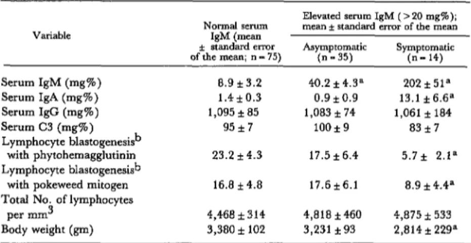
Newborns with overt diseases weighed significantly less at birth than either the con- trol cohort or the asymptomatic cohort, although there was no significant difference in their mean gestational ages. Concentrations of serum IgG and the third component of com- plement were about the same in all groups. Except for a significant elevation in serum IgM, the asymptomatic neonates appeared similar to the normal newborns with respect to the other immunoglobulins and in vitro lym- phocyte function.
Table 3 compares immunologic data for the Serologic tests for specific IgM antibodies to symptomatic and asymptomatic newborns Toxoplasma gondii, herpesvirus types 1 and 2, with elevated IgM (as detected by the CIE and cytomegalovirus were performed on 14 test) with the group of 75 newborns showing symptomatic and ‘23 asymptomatic newborns
normal serum IgM levels at birth. In addition to the striking differences in mean serum IgM concentrations, symptomatic neonates had significantly more IgA in their sera at birth. Lymphocyte blastogenesis (upon stimulation with phytohemagglutinin and pokeweed mitogen) was significantly decreased in the symptomatic infants, although the average number of circulating lymphocytes observed did not differ markedly from that found in the normal newborns.
Table 3. Comparison of selected immunologic parameters in newborns with normal and elevated serum IgM concentrations.
Variable Normal serum
Elevated serum IgM (5 20 mg%); mean f standard error of the mean IgM (mean
zt stmdardemr
of the mean; ” - 75) Asymptomatic (n-35) Symptomatic (n- 14)
Serum IgM (mg%) 8.9 k3.2
Serum IgA (mg%) 1.4*0.3 Serum IgG (mg%) 1,095 i 85 Serum C3 (mg%) 95*7
Lymphocyte blastogenesisb
with phytohemagglutinin 23.2 *4.3
Lymphocyte blastogenesish
with pokeweed mitogen 16.8 f 4.8 Total No. of lymphocytes
per mm3 4,468*314
Body weight (pm) 3,380* 102 aSignificantly different from control values at p C .05.
40.2 f 4.3a 0.9 f 0.9 1,083* 74
100*9
17.5 k6.4
17.6 * 6.1
4,818*460 3,231 *93
202i51” 13.1 f6.6a 1,061*184
83 *7 5.7* 2.ta
8.9 +4.4= 4,875&533
2,814 f 229-’
McMurray et al. l SERUM IgM AND INTRAUTERINE INFECTION 381
with elevated IgM, as well as on 20 control in- fants. No IgM antibodies to these disease agents were found in any of the control in- fants. Two of the asymptomatic neonates were found to have significant IgM titers to cyto- megalovirus, as did four of the 14 newborns with overt disease. In addition, three symp- tomatic infants had IgM antibodies specific for T. gondii, and one was found to yield a significant titer of anti-herpesvirus type 2 anti- bodies of the IgM class. In all, a total of eight (57 per cent) of the 14 symptomatic neonates showed serologic evidence of infection caused by one of the disease agents tested. Only two of 23 (9 per cent) of the asymptomatic new- borns harbored antibodies that indicated previous exposure to these pathogens.
Several children were lost in the course of the follow-up visits at 2, 3, and 6 months post- partum. Three of the symptomatic newborns died during this period, and three others became unavailable due to relocation of their families. The eight remaining infants with overt disease at birth became asymptomatic during the follow-up period. Six appeared normal upon physical examination at 2 months of age.
Eight of the asymptomatic infants could not be located after leaving the hospital. How- ever, none of the remaining asymptomatic children, including the two with significant IgM antibody titers to cytomegalovirus, developed disease signs or symptoms during their first 6 months of life that could be related to their intrauterine environment.
Discussion
Antigenic stimulation of the human fetus during the second or third trimester usually results in active production of antibody, predominantly of the IgM class (12). This is especially true when the antigen takes the form of an infectious agent.
Several previous studies have documented the usefulness of elevated serum IgM at birth as an indicator of intrauterine infection (8-10). However, the utility of this measure-
ment depends upon the definition of “nor- mal” IgM levels in the population at hand, since normal IgM concentrations vary some- what from one population to another (16, 17). Particular interest has focused upon the hypothesis that intrauterine infections are more common among populations character- ized by low income, crowding, malnutrition, and increased exposure to infection (23). Our results do not support this hypothesis. If one accepts as evidence of intrauterine infection the presence of signs or symptoms within the first 6 months of life and/or the presence of specific IgM antibodies to a suspected pathogen in the newborn’s serum, then the in- cidence of intrauterine infection in our study population was less than one per cent, a figure comparable to that obtained in industrialized societies. The mothers of these infants came predominantly from a poor urban area of Cali.
Socioeconomic data on families similar to the ones we studied have been published elsewhere (24). It must be noted, however, that our study group may not be completely representative of a poor urban population. Many mothers from this segment of society do not come to the hospital to deliver their babies, and no records are available on early abortions that may have resulted from infec- tion in utero. Thus, our estimate of the in- cidence of intrauterine infection in the total population may be somewhat low.
reported finding elevated serum IgM more frequently among infants born to women from low socioeconomic strata (16).
The absence of detectable disease among the large number of infants with elevated serum IgM levels in our sample suggests that 20 mg per cent may not be a good discrimina- tory value for certain disadvantaged popula- tions. Our data clearly distinguish between in- fected newborns, all of whom had IgM levels greater (usually much greater) than 50 mg per cent, and asymptomatic newborns with IgM concentrations between 20 and 50 mg per cent. The clinical significance of a “moderate- ly” elevated IgM level (i.e., 20-50 mg per cent) is still uncertain. The data suggest, however, that noninfectious types of antigenic stimulation of the fetus may be more common in populations like that studied.
A similar conclusion has been reached by investigators studying a group of 523 Hawaiian newborns in whom the mean serum IgM concentration at birth was found to be 17 mg per cent. These authors concluded that an IgM level greater than 20 mg per cent was not necessarily a good indication of intrauterine infection in asymptomatic neonates (25). However, there seems a real need to clarify the nature of the in utero stimulation that results in significantly greater IgM production among study populations such as these.
The overall mean IgM concentration in our newborns’ sera (V2.0 f 0.8 mg per cent) was very similar to the value obtained in a pre- vious study of r’O1 Colombian newborns (13.9 f 1 .O mg per cent) (26). This latter group also containkd several infants with IgM levels greater than 20 mg per cent, none of whom had overt signs of disease.
Of the other immunologic parameters tested, serum IgA appeared to be associated most closely with clinical disease in our new- borns. In fact, detectable IgA (over 1 mg per cent) was found primarily in the sera of dis- eased newborns. Nevertheless, not all infected newborns had elevated IgA levels.
Other authors have reported finding elevated IgA levels among infants with intra-
uterine infection (27). Cederqvist et al. (28) have reported a predominance of IgA in cord sera from infected newborns, and Mason et al. (29) have found a strong correlation be- tween detectable levels of serum IgA (1 mg per cent or more) and infection with cyto- megalovirus. Our results suggest that the measurement of newborn serum IgA, in addi- tion to IgM, would permit clinicians to iden- tify infected neonates with increased certainty.
Cytomegalovirus appears to be a common cause of intrauterine infection in the Cali area, followed by Toxoplasma gondii and herpesvirus. The true relative incidence of these specific infections cannot be ascertained from the present study because of the small sample involved and the fact that specific sero- logic tests for other potential pathogens (e.g., rubella virus, T. pallidurn, etc.) were not per- formed. As in previous studies, no specific etiologic agent could be identified in a signifi- cant proportion (50. per cent) of the symp- tomatic newborns.
Our results thus confirm the findings of other authors, who have reported that toxo- plasmosis and cytomegalovirus infection are frequent causes of fetal morbidity (8, 14). Mata et al. (23) have reported strikingly similar results from a study of seroconversion among 61 Guatemalan women in a rural village. During gestation, 6 per cent of the women developed antibodies to cytomegalo- virus, 5 per cent developed antibodies to herpesvirus, and nearly 2 per cent developed antibodies to T. gondii.
McMurray et al. l SERUM IgM AND INTRAUTERINE INFECTION 383
serum IgM concentrations greater than 40 mg per cent-including every child with overt disease and/or positive serology for one of the specific pathogens investigated. In general, the CIE procedure has become increasingly useful in diagnosing specific infections, and our data support its adoption as a routine screening procedure for elevated IgM under conditions where rapid identification of in- fants with elevated IgM is required.
Perhaps one of the most interesting results of this study was the demonstration that symp- tomatic newborns had impaired in vitro cell- mediated immunity. Despite mild lympho- cytosis, the blastogenic response of peripheral blood lymphocytes from newborns with overt disease was significantly depressed (see Table 3). The immunosuppressive effects of infec- tion with Toxoplasma gondii (30), cytomegalo- virus (31), and rubella virus (32) have been documented previously in humans and ex- perimental animals. It appears that both humoral and cell-mediated components of the immune response may be influenced. Experi-
mental evidence suggests that a variety of mechanisms may be responsible, including direct viral infection of lymphoreticular cells (31), impaired thymic development and function (33)) antigenic competition, and alteration of lymphocyte subpopulations (34). The latter could result in either the loss of a responder population or active suppression of in vitro blastogenesis by increased suppressor lymphocyte activity. Our results are more suggestive of such a change in lymphocyte function, since the number of lymphocytes was not decreased.
Although the correlation between in vivo and in vitro cell-mediated immune function is not absolute, the demonstration of impaired lymphocyte blastogenesis implies that intra- uterinely infected newborns may not mount a normal immune response to their primary infection and may be at risk of developing secondary infections as well. Hence the clinical re% ante of immunologic sequellae
\
among children infected in utero is a question that deserves further study.
ACKNOWLEDGMENTS
The authors are grateful to Ms. Nancy Solanilla for study coordination and data con- Morales for patient recruitment and collection trol.
of blood samples and to Ms. Maria Cristina
SUMMARY
A study of 2,029 Colombian newborns from a poor urban population revealed that less than 1 per
cent suffered from overt intrauterine infection that showed itself through signs or symptoms within the first 6 months of life or through the presence of specific IgM antibodies to suspected pathogens.
Counterimmunoelectrophoresis provided an effec- tive method for quickly detecting elevated serum IgM concentrations in newborn sera, thereby per- mitting rapid identification of potentially infected infants.
Sixteen per cent of the newborns studied had
serum IgM levels elevated above the commonly accepted criterion of 20 mg per cent. Symptomatic newborns all had greatly elevated serum IgM levels (over 50 mg per cent) and detectable serum IgA levels (over 1 mg per cent). In contrast, asymp-
tomatic newborns had moderately elevated serum
IgM levels (20-50 mg per cent) and no detect- able IgA levels (under 1 mg per cent). The mean IgM level for all newborns was about 12 mg per cent.
megalovirus (in 4 of 14 symptomatic infants) and lymphocyte blastogenesis in vitro. Our results sug- Toxoplasma gondii (in 3 of 14) were the most gest that antigenic stimulation in utero, but not prevalent. necessarily infection, may be more common among Symptomatic newborns exhibited impaired cell- economically disadvantaged populations than it is mediated immune function, as evidenced by a among other populations.
significant reduction in mitogen-stimulated
REFERENCES
(1) Sever, J. L., and W. T. London. Viruses and embryos. Teratology 2:39, 1969.
(?) South, M. A., J. R. Montgomery, and W. E. Rawls. Immune deficiency in congenital rubella and other viral infections. Birth Defeccts 11:234, 1975.
(3) Nahmias,. A. J. The TORCH complex. Hospital Practice, May 1974, p. 65.
(4) Kaiser, E., M. Bak6, J. Storcz, I. Otvos, I. Rubecz, and J. Mestyan. Clinical correlations with immunoglobuiin levels in newborns in a refer- ral neonatal unit. Acta Paediatr Acad Sci Hung 14: 179, 1973.
(5) Reynolds, D. W., S. Stagno, K. G. Stubbs, A. J. Dahle, M. M. Livingston, S. S. Saxon, and C. A. Alford. Inapparent congenital cytomegalo- virus infection with elevated cord IgM levels. N Engl J Me-d 290:291, 1974.
(6) Starr, J. G., R. D. Bart, Jr., and E. Gold. Inapparent congenital cytomegaiovirus infection: Clinical and epidemiologic characteristics in early infancy. N. Engl J Med 282:1075, 1970.
(7) Melish, M. E., and J. B. Hansaw. Con- genital cytomegaiovirus infection: Developmental progress of infants detected by routine screening. Am J Dis Child 126:190, 1973.
(8) Alford, C. A., Jr., J. Schaefer, W. J. Blankenship, J. V. Straumfjord, and G. Cassady. A correlative immunologic, microbiologic and clinical approach to the diagnosis of acute and chronic infections in newborn infants. N Engl J Med 277~437, 1967.
(9) Alford, C. A., Jr. Immunoglobulin determi- nation in the diagnosis of fetal infection. Pediatr Clin North Am 18:99, 1971.
(IO) Gotoff, S. P., C. Gadzala, R. L. Ying, and P. W. Wendell. Relationship of neonatal IgM values to congenital abnormalities and mental retardation. J Pediatr 78: 1020, 1971.
(U) Sever, J., J, Hardy, S. Korones, M. Giike- son, L. Corridon, A. Ley, H. Tzan, and D. Yamck. Cord immunoglobulins in a middle class Caucasian population. J Pediatr 75~1224, 1969.
(12) Stiehm, E. R. Fetal defense mechanisms. Am J Dis Child 129:438, 1975.
(13) Miller, M. J., P. J. Sunshine, and J. S.
Remington. Quantitation of cord serum IgM and IgA as a screening procedure to detect congenital infection: Results in 5,006 infants. J Pediatr 75:1287, 1969.
(14) Alford, C. A., Jr., S. Stagno, and D. W. Reynolds. Perinatai Infections Caused by Viruses, Toxo$asma, and Treponema pallidurn. In: S. Aiadjem and A. K. Brown (eds.). Clinical Perinatology. C. V. Mosby, St. Louis, 1974.
(15) Robertson, P. W., and V. Kertesz. Modi- fied fluorescent antibody technique to detect im- munoglobulin M antibodies to Toxoplasma gondii in congenital infection. J Clin Microbial 2:461, 1975.
(16) Aiford, C. A., Jr., J. W. Foft, W. J, Blankenship, G. Cassady, and J. M. Benton. Subclinical central nervous system disease of neonates: A prospective study of infants born with increased levels of IgM. J Pedziztr 75:1167, 1969.
(17) Lechtig, A., and L. J. Mata. Levels of IgG, IgA, and IgM in cord blood of Latin American newborns from different ecosystems. Rev Lat Am Microbial 13:173, 1971.
(18) Lechtig, A., and L. J. Mata. Cord IgM levels in Latin American neonates. J Pediatr 78:909, 1971.
(19) Mata, L. J., and E. Viilatoro. Umbilical cord immunoglobulins. In: R. M. Suskind (ed.). Malnutrition and the Immune Response. Raven Press, New York, 1977, p. 201.
(20) Mancini, G., A. 0. Carbonara, and J. R. Heremans. Immunochemical quantitation of antigens by single radial immunodiffusion. Znt J Zm- munochemistry 2:235, 1965.
(21) Dubowitz, L. M., V. Dubowitz, and C. Goldberg. Clinical assessment of gestational age in the newborn infant. J Pediatr 77: 1, 1970.
(22) Han, T., and J. Pauly. Simplified whole blood method for evaluating in vitro lymphocyte reactivity of laboratory animals. Clin Exp Zmmunol 11:137, 1972.
(23) Mata, L. J., J. J. Urrutia, G. Serrato, E. Mohs, and T.D.Y. Chin. Vial infections during pregnancy and early life. Am J Clin Nutr 30:1834, 1977.
McMurray et al. l SERUM IgM AND INTRAUTERINE INFECTION 385
of impaired cell-mediated immunity in mild and moderate malnutrition. Am J Clin Nuts (in press).
(25) Wang, W., C. Sprague, M. Yokoyama, and H. S. Park. IgM levels of newborns in Hawaii. Experientia 29:871, 1973.
(26) McMurray, D. N., M. A. Reyes, H. Rey, and L. J, Casazza. Niveles de immunoglobulinas en el suero de reci&r nacidos y niiios nor-males en Cali. Acta Mkdtia de1 Vaile 8:6, 1977.
(27) Cederqvist, L. L., L. C. Francis, I. A. Zervoudakis, C. G. Berker, and S. D. Litwin. Fetal immune response following prematurely ruptured membranes. AmJ Obstet Gjmecol126:321, 1976.
(28) Cederqvist, L. L., A. C. Kimball, L. C. Ewool, and S. D. Litwin. Fetal immune response following congenital toxoplasmosis. Obstet Qnecol 50:200, 1977.
(29) Mason, E. O., Jr., M. A. South, and J. R. Montgomery. Cord serum IgA in congenital cyto-
megalovirus infection. J Pediatr 89:945, 1976. (30) Strickland, G. T., L. E. Pettitt, and A. Voller. Immunodepression in mice infected with Toxoplasma gondii. Am J Trap Med Hyg 22 ~452, 1973. (31) Howard, R. J., and J. S. Najarian. Cyto- megalovirus-induced immune suppression: I. Humoral immunity. Clin Exp Immunol 18:109, 1974.
(32) Stern, L. M., and I. J. Forbes. Dysgam- maglobulinaemia and temporary immune paresis in a case of congenital rubella. Alcst Pediutr J 11:38,
1975.
(33) Huldt, G., S. Gard, and S. G. Olovson. Effect of Toxoplama gondii on the thymus. Nature 244:301, 1973.
(34) Schauf, V., A. J. Strelkauskas, and A. Deveikis. Alteration of lymphocyte subpopulations with cytomegalovirus infection in infancy. Clin Exp Immunol26:478, 1976.
RABIES IN THE UNITED STATES
A total of 5,150 laboratory-confirmed rabies cases were reported in the United States and its territories in 1979. This total was 67 per cent higher than the average annual total for the preceding five years and was 1,852 cases over the total reported for 1978. Forty-eight states and Puerto Rico reported infected animals in 1979; only the District of Columbia, Idaho, Guam, Hawaii, and the Virgin Islands reported no cases. Seven kinds of animals ac- counted for 98 per cent of the reported cases. These were skunks (59 per cent), bats (15 per cent), raccoons (10 per cent), cattle (4 per cent), dogs (4 per cent), cats (3 per cent), and foxes (3 per cent). Wild animal species ac- counted for 87.6 per cent of the total reported cases, domestic animals ac- counted for 12.3 per cent, and humans accounted for 0.1 per cent.
The general trend was one of across-the-board increases vis-a-vis 1978. For example, there were live human cases (versus four in 1978), 636 domestic animal cases (versus 469 in 1978), and 4,509 wild animal cases (versus 2,285 in 1978). However, cases among skunks exhibited the sharpest reported leap, increasing from 1,657 to 3,031 cases-a rise of 67 per cent over 1978.