89 89 89 89 89 Mem Inst Oswaldo Cruz, Rio de Janeiro, Vol. 95(1): 89-94, Jan./Feb. 2000
Frequency of Specific anti-
Toxoplasma gondii
IgM, IgA and
IgE in Colombian Patients with Acute and Chronic Ocular
Toxoplasmosis
Jorge Enrique Gómez-Marín*/
+, Maria Teresa Montoya-de-Londoño**,
Jhon Carlos Castaño-Osorio**, Fernando Alvarado Heine*, Ana Maria Duque***,
Cathy Chemla, Dominique Aubert, Annie Bonhomme, Jean Michel Pinon
Laboratoire de Parasitologie Mycologie, IFR 53, EA 2070, Faculté de Médecine, Université de Reims Cham-pagne-Ardenne, CHU, 51096 Reims, France *Unidad de Infectologia y Unidad de Parasitologia, Facultad de Medicina, Universidad Nacional de Colombia, Santafé de Bogota D.C., Colombia **Centro de Investigaciones
“Manuel Elkin Patarroyo”, Facultad de Medicina, Universidad del Quindio, Armenia (Quindio), Colombia ***Departmento de Oftalmologia, Hospital “La Misericordia” Calarca (Quindio), Colombia
We studied the frequency of specific anti-Toxoplasma IgM, IgA and IgE antibodies in serum of 28 immunocompetent Colombian patients, selected by ophthalmologists and with lesions that were com-patible with ocular toxoplasmosis. Patients were classified in three groups: (i) group 1 consisted of ten patients with a first episode; (ii) group 2, with seven patients with a recurrence and (iii) group 3, con-sisted of eleven patients with chronic chorioretinal lesion without uveitis. We found that 10/28 (35%) of Colombian patients with ocular toxoplasmosis possessed at least one serological marker for Toxo-plasma infection different from IgG. In group 1 (first episode), we found simultaneous presence of spe-cific IgM plus IgA plus IgE in 1/10 (10%). In group 2 (recurrences) in 1/7 (14%) we found IgM and IgA test positives and in 1/7 (14%) we found IgM and IgE tests positives. In group 3 (toxoplasmic chorioretinal scar) the IgA serological test was positive in 2/11 (18%). These results show that serum IgM or IgA or IgE can be present during recurrences.
Key words: toxoplasmosis - human ocular toxoplasmosis - IgG - IgM - IgA - IgE
Ocular toxoplasmosis is one of the main clini-cal manifestations of the human infection by the protozoan parasite Toxoplasma gondii (Remington et al. 1995). The retina is the primary site of T. gondii infection in the eye (Holland et al. 1996). The lesions cause retinal scars and the loss of vi-sual function when the macula region is involved. Once a scar is established it remains for the whole life; recurrence lesions around scars are common
and can aggravate the visual deficiency (Holland et al. 1996). Toxoplasmic chorioretinitis has been found to be the most common recognizable cause of posterior uveitis (intraocular inflammatory syn-drome) in many surveys (Henderly et al. 1987). In a recent study in Dutch patients, toxoplasmic uvei-tis was the most frequent cause of unilateral visual loss (Rothova et al. 1996). In Colombia, ocular toxoplasmosis represents 4% of consultation in ophthalmology (Varela 1986). The ocular involve-ment of toxoplasmosis has been considered a re-current manifestation of congenital infection. How-ever, for many years an infrequently reported, but important, subgroup of patients was that with toxo-plasmic chorioretinitis as the only manifestation of recently acquired toxoplasmosis. Recent reports show that this form of presentation in non-immu-nosuppressed patients is more common than be-lieved (Nussenblatt & Belfort 1994, Ronday et al. 1995). Montoya and Remington (1996) reported 22 adult cases with serological profile presump-tive of acute acquired toxoplasmosis. Such patients had detectable IgM antibodies and/or acute pat-tern by a differential agglutination test but did not have lymphadenopathy or other nonocular clini-cal disease. Most of these patients were
non-im-This work was supported by the Research Fund of the University of Quindio (Colombia) and by the Programme Hospitalier de Recherche Clinique, Direction des Hôpitaux, Ministère des Affaires Sociales de la Santé et de la Ville (France). Part of this work was realized dur-ing a scientific visit of MT Montoya to the laboratory of Parasitology in Reims, supported by the researcher’s formation program of BID-Colciencias (Colombia).
+Corresponding author. Supported by a doctoral
fellow-ship from Colciencias (Colombia). Present address: Instituto de Salud en el Trópico, Facultad de Medicina, Universidad Nacional de Colombia, Santafé de Bogota D.C., Colom-bia. Fax: 57-1-3681486. E-mail: [email protected] Received 23 February 1999
90 90 90 90
90 IgM, IgA, IgE in Ocular Toxoplasmosis Jorge Enrique Gómez-Marín et al.
munosuppressed, however the authors did not in-clude chronic toxoplasmic chorioretinitis without inflammatory signs. We report here the frequency of IgM, IgA and IgE specific anti-T. gondii anti-bodies in sera of Colombian patients with toxo-plasmic chorioretinitis during the acute symptom-atic setting (first episode or recurrence) and dur-ing the non-inflammatory status.
MATERIALS AND METHODS
Patients - We performed serological studies in consecutively non-immunosuppressed Colombian patients with ocular toxoplasmosis referred by opthalmologists during the period of June to Sep-tember 1994 and March to June 1996. The patients presented retinal inflammatory lesions consistent with ocular toxoplasmosis and diagnosis was made using the criteria defined previously (Holland et al. 1996). The patients were asked about similar episodes and classified according to their clinical ocular history and the result of ofthalmoscopic examination. Group 1 consisted of ten patients with signs of vitritis and a first episode (established by history) and without old retinal scars at examina-tion. Group 2 consisted of seven patients with re-currence, they had signs of vitritis and history of a least one similar past episode or the presence of retinal scars indicating past episodes. Group 3 con-sisted of eleven patients with old scars and with-out retinal inflammatory signs at the moment of sampling. These former patients were discovered in a prevalence study of ocular toxoplasmosis in the Quindio region performed during January-Au-gust 1996. All patients were originating from the Quindio region (central Andean area of Colom-bia). The collection of clinical data was made by a questionnaire including clinical and epidemiologi-cal antecedents. All patients were asked about ex-tra-ocular clinical manifestations and were also examined looking for extraocular signs.
Serological studies - All studied patients were positive for specific IgG anti-T. gondii by immun-ofluorescence antibody test (IFAT-IgG) (Instituto Nacional de Salud 1981). Briefly, the formalde-hyde-treated tachyzoites obtained from peritoneal exudate of mice was fixed on glass slides. Two fold dilutions of serum samples were incubated 1 hr and washed twice with 150 mM phosphate buffer (pH 7.2) and then incubated with a fluorescein isothiocyanate-conjugated secondary antibody (Fluoline H, Biomérieux) diluted 1:320. The IFAT-IgG result was considered to be reactive when fluo-rescence of the entire parasite appeared at dilution of 1:8. Frenkel (1990) noted that dilutions of 1:8 can be useful in old infection as can occurs in ocu-lar toxoplasmosis and that cross reactions with in-fections by others Coccidia can be considered mini-mal in humans. Specific IgM, IgA and IgE anti-T.
gondii antibodies were studied by immunocapture assay (IC) as described previously (Pinon et al. 1995) and performed in the laboratory of “Hôpital Maison Blanche” in Reims (France). Briefly, each serum sample was diluted (1:100) and deposited in three adjacent wells of microplates sensitized with anti-IgM, anti-IgA or anti-IgE monoclonal antibodies. The plates were then incubated for 2.5 hr. After the plates were washed with 150 mM phosphate buffer (pH 7.2), a suspension of form-aldehyde-treated tachyzoites was added at concen-tration of 1, 1.5 and 2 million parasites to the three wells respectively. Sedimentation was read by an automated system with specific software and the results expressed in points from 0 to 12 according to end-point agglutination (Pinon et al. 1995). The following results were considered positive in the IC assay: 9 points for specific IgM, 4 points for specific IgA and 1 point for IgE.
RESULTS
The clinical data are summarized in Tables I, II and III. The mean age in group 1 was 27.9 years (range 11-57), in group 2 was 32.2 years (range 25-45) and in group 3 was 33.5 years (range 19-54). The distribution by sex was 60% (6/10) of males in group 1, 14.2% (1/7) in group 2 and 36% (4/11) in group 3. Serological results are shown in Table IV, V and VI. We found that chronic and acute ocular toxoplasmosis in 10/28 (35%) of Co-lombian non-immunosuppressed patients was ac-companied by serological markers others than spe-cific IgG. More interesting, one patient (patient no. 7) aged 57 years-old and with a first episode pre-sented specific anti-T. gondii IgM plus IgA plus IgE positives test (IgM 12, IgA 12, IgE 1) strongly indicating a recent acquired infection and not re-currence of a congenital infection. In one patient (patient no. 15) with a recurrence (the first epi-sode was two years before) we found the IgM and IgA positive tests, but the IgE test was negative. In another patient (patient no. 11) with recurrence IgM and IgE test were positives. In patients from the group without inflammatory signs two of them (pa-tients nos. 22 and 23) were IgA positive without high IgG titers (<1/128 by IFAT technique) and none of them had IgM or IgE positive tests.
DISCUSSION
The only reliable way to ascertain a recently acquired T. gondii infection by serological tests is by documenting seroconversion. However, this find-ing can be difficult to make when a patient visits the ophthalmologist weeks or months after T. gondii
9
1
9
1
9
1
9
1
9
1
M
e
m
In
st
O
sw
a
ld
o
C
ru
z
,
R
io
d
e
Ja
n
e
ir
o
, V
o
l.
9
5
(1
),
Ja
n
./F
e
b
. 2
0
0
0
TABLE I
Clinical findings and outcome for ten Colombian patients with a first episode of ocular toxoplasmosis
Patient Sex Age Eye (s) Localization of lesion Visual Visual acuity Duration of uveitis at Extra-ocular Treatment no. involved acuity at at end of episode the time of sampling symptoms
sampling and complications
1. M 11 L Vitritis 20/80 20/20 6 days No Cstd.
2. M 19 L Vitritis 20/70 at 2 m 20/70 at 3 m 30 days No Cstd. + P-S
Persistent vitritis
3. F 24 R Peripherical chorioret. 20/20 20/20 5 months No None
4. M 26 R Vitritis 20/200 Only ligth 3 months No Cst.
Persistent vitritis
5. M 45 L Peripapilar chorioret. 20/20 20/20 3 months No None
6. F 54 R-L R: macular chorioret. R 20/200 R:20/200 2 months No None L: macularchorioret. L 20/400 L: 20/400
7. M 57 R Vitritis 20/80 20/40 20 days No Cstd.
Persistent vitritis
8. F 12 L Macular chorioret. CF at 3 m CF at 3 m 40 days No Cst. + P-S.
9. F 11 R Vitritis 20/200 20/200 45 days No Cstd.
10. M 20 R Peripherical chorioret. 20/30 20/30 30 days No Cstd + P.S.
92 92 92 92
92 IgM, IgA, IgE in Ocular Toxoplasmosis Jorge Enrique Gómez-Marín et al.
IgE. However, there are cases of reinfection pre-senting IgM or IgA alone or even IgE specific isotypes that have been documented (Fortier et al. 1991, Pinon et al. 1995). But, to our knowledge, there are not reports of simultaneous presence of anti-T. gondii specific IgM, IgA and IgE in reinfections. Then, simultaneous presence of IgM, IgA and IgE can be considered a strong evidence for recent primary infection. If this assumption is true, in our study we found that 1/10 (10%) of new cases of ocular toxoplasmosis in Colombian patients from the Quindio region could be related to a recent infection (proportion of cases with specific anti-T. gondii IgM plus IgA plus IgE positive in a first epi-sode). Like Montoya and Remington (1996), we agree that this finding indicates that recent acquired toxoplasmosis is more frequent than believed. Ex-traocular manifestations of toxoplasmosis were not found in our series. However, in one patient, with an old scar, she stated presence of lymphadenopa-thies during her first episode of uveitis occurred many years ago. In addition, we have observed in Colombia two other cases with symptoms like fe-ver and adenopathies and serological tests results of recent acquired infection, one of them was a woman who transmitted congenital infection to her offspring (Castaño et al. 1991). This suggests that postnatal acquired toxoplasmosis can produce ocular lesion either accompanied by extraocular symptoms or not. The physiopathological reason for one or the other event remain to be determined. There are similar reports about the frequency of ocular involvement in three outbreaks of recently acquired toxoplasmo-sis. In an epidemic in Atlanta only one of 37 fected individuals (2.7%) developed eye disease af-ter four years of follow-up (Akstein et al. 1982). In a well documented family outbreak in New York city, one of seven members (14.2%) developed toxo-plasmic chorioretinitis 129 days after infection (Masur et al. 1978), and in a recent outbreak in Brit-ish Columbia (Canada) the number of symptomatic patients with ocular toxoplasmosis as clinical pre-sentation was 19 of 100 acute outbreak-related cases (19%) (Bowie et al. 1997). However, at present we do not have longitudinal studies that establish the exact frequency of toxoplasmic chorioretinitis after postnatal acquired infection and if the frequency of this form can vary from one site to another.
As we stated above, the problem estimating the frequency of postnatal acquired ocular toxoplas-mosis based on serological findings, is that it is possible in recurrent cases to find presence of an-tibodies classically considered to be markers of primary infection. The cases, in the present report, of a recurrence of toxoplasmic chorioretinitis with the presence of specific IgM plus IgA, or IgM plus IgE in serum, effectively suggest that IgM plus IgA
T
ABLE II
Clinical findings and outcome for seven Colombian patients with a recidive of ocular toxoplasmosis
Patient
Sex
Age
Eye (s)
Localization of lesion
V
isual
V
isual acuity
Duration of uveitis
T ime from Extra-ocular T reatment no. involved acuity at
at end of episode
at the time of
the first symptoms sampling and complications sampling episode 1 1 . M 25 R-L
R: macular chorioret.
R: forms at 6 m
R: forms at 6 m
15 days 14 years No Cst L: peripher . Chorioret. L: 20/30 12. F 25 R V itritis Amaurotic Amaurotic 10 months 5 years No Cst. Iris sinechial 13. F 2 6 R Macular chorioret. 20/140 20/100 20 days 4 years No Cstd.+ P-S 14. F 32 L Macular chorioret. 20/140
Lost follow up
30 days 9 years No Cstd. 15. F 44 L Macular chorioret. 20/100 20/60 60 days 2 years No None 16. F 4 5 L V itritis 20/400 Amaurotic 60 days 3 years No Cstd. Chorioret. Persistent vitritis 17. F 28 L Macular chorioret.
CF at 3 m
20/200
35 days
12 years
No
Cstd. + P-S
M: male; F: female; R: right; L: left; CF: counting fingers; Chorioret.: chorioretinitis; Cstd.: corticosteroids; P-S: pyrimeth
amine-sulfadoxine; severity score: normal 20/20,
moderate: 20/30-20/100, severe > or = 20/200 (Snell’
93 93 93 93 93 Mem Inst Oswaldo Cruz, Rio de Janeiro, Vol. 95(1), Jan./Feb. 2000
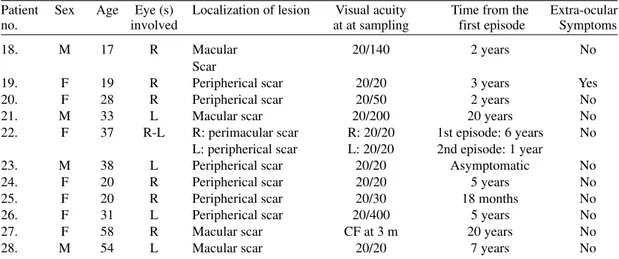
TABLE III
Clinical findings and outcome for eleven Colombian patients with chronic inactive ocular toxoplasmosis
Patient Sex Age Eye (s) Localization of lesion Visual acuity Time from the Extra-ocular no. involved at at sampling first episode Symptoms
18. M 17 R Macular 20/140 2 years No
Scar
19. F 19 R Peripherical scar 20/20 3 years Yes 20. F 28 R Peripherical scar 20/50 2 years No
21. M 33 L Macular scar 20/200 20 years No
22. F 37 R-L R: perimacular scar R: 20/20 1st episode: 6 years No L: peripherical scar L: 20/20 2nd episode: 1 year
23. M 38 L Peripherical scar 20/20 Asymptomatic No 24. F 20 R Peripherical scar 20/20 5 years No 25. F 20 R Peripherical scar 20/30 18 months No 26. F 31 L Peripherical scar 20/400 5 years No 27. F 58 R Macular scar CF at 3 m 20 years No
28. M 54 L Macular scar 20/20 7 years No
M: male; F: female; R: right; L: left; CF: counting fingers; Chorioret.: chorioretinitis; Cstd.: corticosteroids; P-S: pyrimethamine-sulfadoxine; severity score: normal 20/20, moderate: 20/30-20/100, severe > or = 20/200 (Snell’s visual acuity table).
TABLE IV
Serological results for ten Colombian patients with a first episode of ocular toxoplasmosis
Patient Inverse titers IgG IgM IgA IgE
no. IFAT IC IC IC
1. 64 8 2.5 0
2. 512 8 0 0
3. 512 9 0 0
4. 128 4.5 0 0
5. 256 10 1.5 0
6. 64 10 1.5 0
7. 128 12 12 1
8. 128 7 0 0
9. 512 10 0 0
10. 64 10 1 0
Total positives 6/10 1/10 1/10 (60%) (10%) (10%) Cutoff values: IgM > 9, IgA > 4, IgE > 1. positive results are shown in bold; IFAT: immunoflourescence antibody test; IC: immunocapture test.
TABLE V
Serological results for seven Colombian patients with a recidive of ocular toxoplasmosis
Patient Inverse titers IgG IgM IgA IgE
no. IFAT IC IC IC
11. 64 11 0.5 2
12. 64 0 0 0
13. 32 6.5 0 0
14. 128 8.5 0.5 0
15. 512 9.5 11 0
16. 128 5.5 0 0
17. 64 4 0 0
Total positives: 2/7 1/7 1/7 (28%) (14.2%) (14.2%)
Cutoff values: IgM > 9, IgA > 4, IgE > 1. positive results are shown in bold; IFAT: immunoflourescence antibody test; IC: immunocapture test.
or IgM plus IgE may be present in recurrent clini-cal episodes. Specific IgA without specific IgM has been reported in a case of reinfection in a French women who had an abortion (Fortier et al. 1991). Similarly, Pinon et al. (1995) described that in a group of HIV-infected patients with evidence of previous exposure to T. gondii, but no clinical manifestations, IgA and IgE were detected in 11% and 4% of cases. It would be important to deter-mine if recurrent episodes are due to endogenous reactivation of old infection or to a new reinfec-tion because in this way effective preventive
mea-sures may vary, like antibiotic prophylaxis in the case of endogenous reactivation or hygiene mea-sures for the case of exogenous reinfection.
compli-94 94 94 94
94 IgM, IgA, IgE in Ocular Toxoplasmosis Jorge Enrique Gómez-Marín et al.
cated with a persistent vitritis in our patient with serological evidence of recently acquired toxoplas-mosis. The study of Ronday et al. (1995) also found severe complications in six of eigth patients with presumed acquired ocular toxoplasmosis. It would be important to test whether Colombian strains of
T. gondii are different to strains from other geo-graphical regions and if different strains have dif-ferent pathogenic properties. In a national survey, 47% of the Colombian population possessed spe-cific IgG antibodies indicating a high exposition of population to T. gondii (Juliao et al. 1983), but we do not know the real impact of their pathogenic manifestations and studies about the prevalence of ocular toxoplasmosis in the general population are presently going on.
In conclusion, we found that 10% of patients with a first episode of ocular toxoplasmosis have serological findings of recently acquired infection (simultaneous presence of IgM, IgA and IgE). During recurrences specific anti-Toxoplasma IgM/ IgA and IgM/IgE were present and IgA alone was found in chronic setting. We need studies in order to determine the frequency of acquired ocular toxo-plasmosis, if the reinfections can trigger recur-rences (i.e. in the Colombian cases of recurrence with presence of IgM and IgA or IgM and IgE in serum) and the impact of strain diversity in the ocular presentation of human toxoplasmosis.
ACKNOWLEDGEMENT
To Erik Pisano for excellent technical assistance. To Jenny Sant for english revision of the manuscript.
REFERENCES
Akstein RB, Wilson LA, Teutsch SM 1982. Acquired toxoplasmosis. Opthalmology 89: 1299-1302. Bowie WR, King AS, Werker DH, Isaac-Renton JL, Bell
A, Eng SB, Marion SA 1997. Outbreak of toxoplas-mosis associated with municipal drinking water.
Lancet 350: 173-177.
Castaño JC, Gómez JE, Duque AM 1991. Toxoplasmo-sis ocular en el Quindio: características clínicas.
Biomédica 11 (Suppl. 1): 121
Frenkel JK 1990. Transmission of toxoplasmosis and the role of immunity in limiting transmission and illness. J Am Vet Med Acad 96: 233-240.
Fortier B, Aissi E, Ajana F, Dieusart P, Denis P, Martin De-Lasalle E, Lecomte-Hocke M, Vinatier D 1991. Spontaneous abortion and reinfection by Toxoplasma gondii (letter). Lancet 338: 444.
Gómez-Marín JE, Pinon JM, Bonhomme A, Guenounou M 1997. Does human toxoplasmosis involves an imbalance in T1/T2 cytokines? Med Hypotheses 48: 161-169.
Henderly DE, Gentsler AJ, Smith RE, Rao NA 1987. Chang-ing patterns of uveitis. Am J Opthalmol 103: 131-36. Holland GN, O’Connor GR, Belfort Jr R, Remington JS
1996. Toxoplasmosis. In JS Pepose, GN Holland & KR Wilhelmus (eds), Ocular Infection and Immu-nity, Mosby Yearbook, St Louis, p. 1183-223. Instituto Nacional de Salud 1981. Manual de Tecnicas
de Laboratorio, Imprenta Ministerio de Salud de Colombia, Bogota, p. 112-118.
Juliao O, Corredor A, Moreno GS 1983. Toxoplasmosis en Colombia, Imprenta Instituto Nacional de Salud, Bogota, p. 67.
Masur H, Jones TC, Lempert JA, Cherubini TD 1978. Out-break of toxoplasmosis in a family and documentation of acquired retinochoroiditis. Am J Med 64: 396-402. Montoya JG, Remington JS 1996. Toxoplasmic
chori-oretinitis in the setting of acute acquired toxoplas-mosis. Clin Infect Dis 23: 277-282.
Nussenblatt RB, Belfort R 1994. Ocular toxoplasmosis: an old disease revisited. JAMA 271: 304-307. Pinon JM, Foudrinier F, Mougeot G, Marx C, Aubert D,
Toupance O, Niel G, Danis M, Camerlinck P, Remy G, Frottier J, Jolly D, Bessieres MH, Richard-Lenoble D, Bonhomme A 1995. Evaluation of risk and diag-nostic value of quantitative assays for anti-Toxoplasma gondii immunoglobulin A (IgA), IgE, and IgM and analytical study of specific IgG in immunodeficient patients. J Clin Microbiol 33: 878-884.
Remington JS, McLeod R, Desmonts G 1995. Toxoplas-mosis. In JS Remington & JO Klein (eds), Infec-tious Diseases of the Fetus and Newborn Infant, 4th ed., WB Saunders, Philadelphia, p. 140-267. Ronday MJ, Luyendijk L, Baarsma S, Bollemeijer JG,
van der Lelij, Rothova A 1995. Presumed acquired ocular toxoplasmosis. Arch Opthalmol 113: 1524-1529. Rothova A, Suttorp-van-Schulten MSA, Fritts W, Kijsltra A 1996. Causes and frequency of blindness in pa-tients with intraocular inflammatory disease. Br J Opthalmol 80: 332-336.
Varela H, 1986. Uveitis. In F Chalem, N Escandon, A Campos & I Esguerra (eds), Medicina Interna, Edi-torial Norma, Bogota, p. 1081-1085.
TABLE VI
Serological results for eleven Colombian patients with chronic inactive ocular toxoplasmosis
Patient Inverse titers IgG IgM IgA IgE
no. IFAT IC IC IC
18. 64 4 0 0
19. 64 4.5 0 0
20. 64 7 0 0
21. 128 5.5 0 0
22. 64 5.5 4.5 0
23. 8 8 4 ND
24. 16 4.5 0 0
25. 64 8 0 0
26. 64 8.5 1 0
27. 8 8.5 0 0
28. 8 7.5 0.5 0