FACULDADE DE CIÊNCIAS
DEPARTAMENTO DE BIOLOGIA VEGETAL
Ana Paula Batista Elias
DOUTORAMENTO EM BIOLOGIA
(Especialidade em Biologia Celular)
2008
V
AS
CU
L
AR-ENDO
T
HE
LI
AL-G
R
OWTH-FA
C
TOR
S
-
P
L
I
-
C
ING
REGULATION OF
BY TISSUE MICROENVIRONMENT
A
L
-
T
E
- R
NATIVE
FACULDADE DE CIÊNCIAS
DEPARTAMENTO DE BIOLOGIA VEGETAL
Ana Paula Batista Elias
Tese orientada pelo Doutor Sérgio Jerónimo Rodrigues Dias,
sendo o responsável Institucional o Professor Doutor José
Manuel Gonçalves Barroso
DOUTORAMENTO EM BIOLOGIA
(Especialidade em Biologia Celular)
2008
V
AS
CU
L
AR-ENDO
T
HE
LI
AL-G
R
OWTH-FA
C
TOR
S
-
P
L
I
-
C
ING
REGULATION OF
BY TISSUE MICROENVIRONMENT
A
L
-
T
E
- R
NATIVE
The work described in this PhD thesis was developed under the supervision of Sérgio Dias (PhD) (Scientific supervisor) and Professor José Manuel Gonçalves Barroso (PhD) (Institutional supervisor) at the Angiogenesis Laboratory, Centro de Investigação de Patobiologia Molecular (CIPM), Instituto Português de Oncologia Francisco Gentil (IPOFG), in collaboration with the Instituto Gulbenkian de Ciência (IGC), between October 2003 and May 2008.
Se o “splicing” alternativo, estudado nesta tese, nos diz como a partir de um (gene) se
consegue fazer várias (proteínas), esta tese mostra também como é que várias
(pessoas/instituições) conseguem fazer uma (tese). Esta tese de Doutoramento traduz os esforços directos ou indirectos de muitas pessoas ao longo dos 4 anos.
Antes de iniciar o Doutoramento, o meu crescimento académico, que fez com que
estivesse preparada para desenvolver este trabalho, foi sem dúvida suportado tanto financeiramente como intelectualmente por toda a minha família. Também nesta fase, os conhecimentos tanto práticos como científicos transmitidos pela Doutora Joana Diamond durante o meu estágio de Licenciatura no Instituto Português de Oncologia Francisco Gentil (IPOFG) foram importantes para o meu desempenho neste trabalho.
Para me propor a fazer Doutoramento tive certamente uma ajuda essencial do
Doutor Sérgio Dias, actual orientador, e da minha família, especialmente dos meus Pais, do meu Irmão e do Telmo.
Para o desenrolar do trabalho contei sempre com:
A preciosa ajuda do Doutor Sérgio Dias, quer a nível prático, quer a nível teórico, essencial para o meu crescimento científico e intelectual. Com muita paciência e tolerância lá me fez levar a “água a bom porto”;
A disponibilidade do meu orientador da faculdade, Professor Doutor José Barroso, para o regular acompanhamento do trabalho ao longo destes 4 anos;
O interesse, disponibilidade e apoio clínico/prático de todos os elementos dos Serviços de Anatomia Patológica (em especial da Doutora Ana Félix) e Virologia (em especial da Doutora Carmo Ornelas e do Luís Martins) do IPOFG, sem os quais não teria conseguido ter acesso a amostras de doentes oncológicos;
Unidade de Biologia Celular do Sistema Imunológico do Instituto de Medicina Molecular (IMM), em especial do Doutor Luís Moita e do Pedro Alves, sem os quais não teria conseguido fazer os trabalhos com RNAi;
O interesse e ajuda do Nuno Morais na análise bioinformática;
A amizade, companheirismo e ajuda técnica/teórica de todos os elementos do grupo de Angiogénese com quem trabalhei durante estes anos e de todos os elementos dos restantes grupos do Centro de Investigação de Patobiologia Molecular - CIPM (Hematologia, Endocrinologia, Gastrenterologia, e Patologia) que me acompanharam nesta etapa ;
A amizade, o amor, os incentivos, o orgulho, a paciência, o apoio, o esforço, e tudo o que uma excelente família como a minha pode dar;
O espaço físico para execução deste trabalho, onde se incluem várias instituições: IPOFG, Instituto Gulbenkian de Ciência, IMM, Faculdade de Ciências da Universidade de Lisboa;
Financiamento por parte da Fundação para a Ciência e Tecnologia, e Liga Portuguesa Contra o Cancro, que permitiu a execução dos trabalhos experimentais e participação em Congressos Internacionais.
A todos, sem excepção, um muito obrigado por cada minuto da vossa vida/cada euro que foi gasto nesta tese. Tentei usar esses minutos/euros da melhor forma possível, espero que gostem do resultado...
Contents
Abbreviations ______________________________________________________ VII Resumo ___________________________________________________________ XV Abstract __________________________________________________________XIX
Chapter I - General Introduction ________________________________________ 1
1. Vascular system _______________________________________________________ 3
1.1. Blood vessels organization ___________________________________________________3 1.2. Blood vessels formation: vasculogenesis and angiogenesis __________________________5 1.2.1. Physiological vs. pathological angiogenesis __________________________________9 1.2.1.1. Tumor angiogenesis ________________________________________________9
2. Vascular Endothelial Growth Factor A___________________________________ 11
2.1. Gene and protein characterization_____________________________________________11 2.2. VEGF isoforms ___________________________________________________________16 2.3. VEGF expression in embryonic and adult tissues_________________________________20 2.3.1. VEGF isoforms expression pattern ________________________________________21 2.4. Regulation of VEGF expression ______________________________________________23 2.4.1. Regulation of VEGF isoforms expression___________________________________26 2.5. VEGF functions __________________________________________________________27 2.5.1. VEGF isoforms functions _______________________________________________28 2.6. VEGF-A receptors ________________________________________________________31 2.6.1. Genes and proteins characterization _______________________________________31 2.6.2. Responses mediated by VEGFR2 and VEGFR1______________________________34 2.6.3. VEGF receptors expression pattern________________________________________36
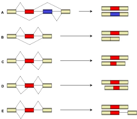
3. Alternative splicing ___________________________________________________ 37
3.1. Regulation of constitutive/alternative splicing ___________________________________38 3.1.1. SR proteins characterization _____________________________________________41
3.1.1.1. The functions of SR proteins_________________________________________44 3.1.2. hnRNP characterization ________________________________________________45 3.1.2.1. The fuctions of hnRNPs ____________________________________________46 3.1.3. Signaling pathways involved in alternative splicing ___________________________47 3.2. Alternative splicing in angiogenesis ___________________________________________48 Chapter II - Aims of the thesis__________________________________________ 51 Chapter III - VEGF isoforms pattern in physiologic and pathologic situations____ 55 Chapter IV - Microenvironment changes (in pH) affect VEGF alternative splicing 79 Chapter V - Screening for proteins that control VEGF alternative splicing_______ 99 Chapter VI - General Discussion ______________________________________ 117 Chapter VII - References_____________________________________________ 127
Figures Index
Figure I - 1. Differences and similarities between different types of blood vessel._________ 4 Figure I - 2. Differences in capillary vessels permeability. __________________________ 5 Figure I - 3. Two processes of blood vessels formation: angiogenesis vs. vasculogenesis. __ 6 Figure I - 4. The angiogenic switch in tumor development. _________________________ 10 Figure I - 5. Nucleotide and amino acid sequences of a bovine VEGF cDNA. __________ 12 Figure I - 6. Comparison of the bovine and human VEGF amino acids sequences. ______ 13 Figure I - 7. Important domains in VEGF and its receptors that mediate its binding. _____ 15 Figure I - 8. VEGF isoforms originated by alternative splicing. _____________________ 16 Figure I - 9. VEGF transcription from 2 different sites in the gene.___________________ 20 Figure I - 10. Mechanism of action of some VEGF expression regulators. _____________ 25 Figure I - 11. Role of each VEGF isoform in tumor angiogenesis.____________________ 30 Figure I - 12. VEGF-A receptors characterization. _______________________________ 33 Figure I - 13. Types of alternative splicing. _____________________________________ 38 Figure I - 14. Simplified view of the splicing process. _____________________________ 40 Figure I - 15. Schematic diagram of human SR proteins family. _____________________ 42 Figure I - 16. The exon-dependent and independent functions of SR proteins. __________ 45 Figure I - 17. Common structural domains shared by members of the hnRNP family. ____ 46 Figure I - 18. Alternative splicing affecting VEGF gene. ___________________________ 49
Figure II - 1. First aim of the thesis - If/which microenvironment changes are responsible for
VEGF alternative splicing. ________________________________________ 53
Figure II - 2. Second aim of the thesis - Which pathway(s) is(are) used to translate the
external signals into VEGF alternative splicing. _______________________ 54
Figure II - 3. Third aim of the thesis - Which proteins directly modulate the alternative
splicing of VEGF. _______________________________________________ 54
Figure III - 1. VEGF isoforms that arise from pre-mRNA alternative splicing.__________ 61 Figure III - 2. Different organs produce different VEGF isoforms. ___________________ 62 Figure III - 3. Murine estrus cycle.____________________________________________ 64 Figure III - 4. VEGF isoforms produced by murine ovary and endometrium after induction of
Figure III - 5. Human endometrial carcinoma samples classification and
immunohistochemistry for CD31. _________________________________ 68
Figure III - 6. VEGF isoforms produced by human endometrial carcinoma samples._____ 69
Figure IV - 1. VEGF isoforms that result from pre-mRNA alternative splicing. _________ 85 Figure IV - 2. VEGF isoforms expression pattern by RL95 cells in response to changes in the
microenvironment._____________________________________________ 86
Figure IV - 3. Correlation between VEGF isoforms pattern inversion and the activation of
stress signaling pathways in acidic conditions._______________________ 88
Figure IV - 4. SR proteins involved in the control of the VEGF splicing pattern. ________ 89 Figure IV - 5. Effect of SR proteins down-regulation in VEGF alternetive splicing in acidic
conditions. ___________________________________________________ 90
Figure IV - 6. Proposed model._______________________________________________ 92
Figure V - 1. VEGF isoforms that are produced by alternative splicing. ______________ 106 Figure V - 2. Putative binding sites for SR proteins and Tra2β in the VEGF sequence. __ 108 Figure V - 3. VEGF isoforms expression after downregulation of specific SR (related)
proteins. ____________________________________________________ 109
Figure VI - 1. Mechanisms that may coordinately regulate the expression of each VEGF
Tables Index
Table I - 1. Examples of regulatory factors of angiogenesis [16-18]. __________________ 8 Table I - 2. Human tumors overexpressing VEGF mRNA. __________________________ 22 Table I - 3. Changes in the VEGF isoforms pattern with tumor stage. _________________ 23
Table III - 1. Primers and probes used in real time RT-PCR for human/murine VEGF
isoforms. _____________________________________________________ 76
Table IV - 1. Primers and probes used in real time RT-PCR for VEGF isoforms. ________ 96
Abbreviations
IIICS Type III connecting segment
A Alanine
Ala Alanine
aFGF Acid fibroblast growth factor
Ang-1 Angiopoietin-1
AREs AU-rich elements
Arg Arginine
ASF Alternative splicing factor
Asn Asparagine
Asp Aspartate
BBP Branchpoint binding protein
BCR Breakpoint cluster region
bFGF Basic fibroblast growth factor
BHK fibroblasts Baby hamster kidney fibroblasts
BPS Branchpoint sequence
C Cysteine
cAMP Cyclic adenosine monophosphate
cDNA Complementary Deoxyribonucleic acid
CKII Casein kinase II
Cys Cysteine
D Aspartate
DMEM Dulbecco’s modified eagle’s medium
EC Endothelial cells
ECL Enhanced chemiluminescence
ECM Extracellular matrix
EDI Extra domain I
EDII Extra domain II
EGF Epidermal growth factor
ELISA Enzyme-linked immunosorbent assay
ELR motif Glutamic acid-leucine-arginine motif
EMBL European Molecular Biology Laboratory
EPC Endothelial progenitor cell
ERK Extracellular signal-regulated kinase
ESE Exonic splicing enhancer
ESS Exonic splicing silencer
F Phenylalanine
FBS Fetal bovine serum
FGF Fibroblast growth factor
Flk-1 Fetal liver kinase 1
FLT-1 Fms-like tyrosine kinase 1
FSH Follicle-stimulating hormone
G Glycine
G-CSF Granulocyte-colony stimulating factor
Gln Glutamine
Glu Glutamic acid
Gly Glycine
GM-CSF Granulocyte-macrophage colony stimulating factor
H Histidine
hCG Human chorionic gonadotropin
HGF Hepatocyte growth factor
HIF-1 Hypoxia-induced factor 1
His Histidine
HER Hypoxic response element
hnRNP Heterogeneous nuclear ribonucleoprotein
HPV Human papillomavirus
H-SIL High-grade squamous intraepithelial lesions
HuR Hypoxia-induced stability factor
I Isoleucine
Ig Immunoglobulin
IGF-I Insulin-like factor I
IL Interleukin
Ile Isoleucine
IRES Internal ribosome entry site
ISE Intronic splicing enhancer
ISS Intronic splicing silencer
K Lysine
KDa Kilo Daltons
KDR Kinase insert domain-containing receptor
KH K homology
L Leucine
Leu Leucine
LH Luteinizing hormone
Lys Lysine
M Methionine
mAb Monoclonal antibody
MAPK Mitogen-activated protein kinase
Met Methionine
min Minutes
ml Mililiters
mM Milimolar
MMP Matrix metalloproteinase
mRNA Messenger ribonucleic acid
N Asparagine
ng Nanogram
nM Nanomolar
NRP Neuropilin
NSCLC Non-small cell lung cancer
ORF Open-reading frame
P Proline
PAIP2 Poly(A)-binding protein-interacting protein 2
PD-ECGF Platelet-derived endothelial cell growth factor
PDGF Platelet-derived growth factor
PEDF Pigment epithelium derived factor
PGE1 Prostaglandin E1
PGE2 Prostaglandin E2
Phe Phenylalanine
PI3K Phosphatidylinositol 3-kinase
PKC Protein kinase C
PlGF Placental growth factor
Pro Proline
PTB Polypyrimidine tract-binding protein 1
PY Polypyrimidine tract
Q Glutamine
R Arginine
RBD RNA binding domain
RGG Arginine-Glycine-Glycine
RNAi RNA Interference
rpm Revolutions Per Minute
RQ-PCR Real time quantitative polymerase chain reaction
RRM RNA recognition motif
RS Serine-arginine residues
RT-PCR Reverse transcription polymerase chain reaction
S Serine
SAPK/JNK Stress-activated protein kinase/Jun-amino-terminal kinase
SDS-PAGE Sodium dodecyl sulphate-polyacrylamide gel electrophoresis
Ser Serine
SF1 Splicing factor 1
SFC Splicing factor compartment
shRNA Short hairpin RNA
siRNA Small interfering RNA
snRNA Small nuclear RNA
snRNP Small nuclear ribonucleoproteins
SRPK SR protein kinase
SRrp SR-related protein
ss Splice site
T Threonine
TAF Tumor angiogenesis factor
TGF-α Transforming growth factor α
TGF-β Transforming growth factor β
Thr Threonine
TIMPs Tissue inhibitors of metalloproteinases
TIS11b Tetradecanoyl phorbol acetate-inducible-sequence
TNF-α Tumor necrosis factor α
tPA Tissue-type plasminogen
Trp Triptofane
TSP Thrombospondin
TXA2 Thromboxane A2
Tyr Tyrosine
U2AF U2 auxiliary factor
uPA Urokinase-type plasminogen
UTR Untranslated region
V Valine
Val Valine
VEGF Vascular endothelial growth factor
VEGFR Vascular endothelial growth factor receptor
vHL von Hippel-Laudau
VPF Vascular permeability factor
W Triptofane Y Tyrosine µg Microgram µl Microliter µm Micrometer µM Micromolar
Resumo
O factor de crescimento do endotélio vascular (VEGF) tem sido um factor descrito como fundamental para a correcta formação da rede capilar durante a embriogenese, mas também para a formação de novos vasos sanguíneos durante a vida de um organismo adulto. O VEGF tem um papel activo nos dois processos que se sabe serem importantes para a formação de uma nova vasculatura, sendo eles a vasculogénese (formação de novos vasos a partir de progenitores endoteliais) e a angiogénese (formação de novos vasos a partir de vasos pré-existentes), e que no adulto ocorrem numa grande variedade de situações quer fisiológicas (por exemplo durante a cicatrização de uma ferida ou na reparação do endométrio após menstruação) quer patológicas (por exemplo na vascularização de um tumor ou num processo de artrite reumatóide). Para além de actuar ao nível das células endoteliais que constituem os vasos, este factor de crescimento também foi descrito como capaz de actuar em células tumorais, promovendo não só a sua sobrevivência mas também a sua migração.
O VEGF é codificado por um gene que após “splicing” alternativo entre os exões 5 e 8 do pré-mRNA, origina diversas isoformas (as isoformas VEGF121, VEGF145, VEGF165 e VEGF189 são as isoformas mais abundantes). A existência de splicing alternativo neste gene faz com que se formem diferentes isoformas com diferentes localizações, uma vez que os exões em causa codificam para domínios de ligação à heparina que se pode encontrar tanto na membrana celular como na matriz extracelular. A isoforma 121 do VEGF não contem os exões 6 e 7 e dessa forma é uma isoforma capaz de se difundir e actuar a longas distâncias. Ao contrário, a isoforma 189 tem na sua constituição quer o exão 6 quer o exão 7 e portanto encontra-se localizada junto à membrana da célula e retida na matriz extracelular, podendo no entanto ser clivada por uma protease e desta forma ter a mesma localização da isoforma 121. As isoformas 145 e 165, que tem o exão 6 e o exão 7 respectivamente, tem propriedades intermédias e podem sinalizar junto da membrana da célula ou a distâncias maiores. Para além da diferente localização, as várias isoformas têm consequentemente diferentes afinidades para os receptores do VEGF. Por exemplo
apenas a isoforma 165 do VEGF tem afinidade para se ligar ao receptor NRP1 (neuropilina 1). De acordo com as suas diferentes propriedades, o VEGF121 tem sido implicado no recrutamento dos vasos periféricos actuando desta forma a longa distância, e o VEGF189 parece ter um papel na ramificação dos micro-vasos no local da sua expressão. Mais uma vez as isoformas 145 e 165 parecem ter funções intermédias, podendo recrutar os grandes vasos e promover também a ramificação dos micro-capilares.
Apesar de existirem descritos vários trabalhos sobre o controlo ao nível da transcrição do VEGF (e.x. a hypoxia é um factor importante capaz de induzir a expressão do VEGF através da proteína HIF-1 que se liga à região promotora do VEGF), os mecanismos responsáveis pela regulação do “splicing” alternativo deste gene não são conhecidos. Sendo assim, os objectivos deste trabalho consistiram não só na caracterização dos sinais externos capazes de induzir uma alteração do padrão de isoformas do VEGF mas também na pesquisa das vias de sinalização e proteínas envolvidas na regulação do “splicing” alternativo deste gene em resposta a um sinal externo.
De uma forma resumida, neste trabalho demonstrou-se in vitro que uma mudança do pH extracelular de neutro para ácido era capaz de induzir uma alteração no padrão de isoformas do VEGF. Mais detalhadamente, a percentagem da isoforma 121 do VEGF nesta condição estava bastante aumentada quando comparada com a sua percentagem numa condição de pH neutro. Esta alteração do “splicing” alternativo do VEGF demonstrou-se estar associada à activação da via de sinalização da cinase p38 (a adição de um inibidor específico da via manteve o padrão de isoformas do VEGF igual ao controlo mesmo depois de se ter sujeitado as células a um pH ácido). A caracterização das proteínas capazes de controlar o “splicing” alternativo do VEGF em resposta ao pH ácido e à activação da via da cinase p38, não está ainda totalmente concluída, no entanto, através de métodos bioinformaticos e de técnicas de RNAi, a proteína Tra2β ( proteína reguladora do “splicing” alternativo relacionada com a família das proteínas SR que são caracterizadas por terem domínios ricos em serina/arginina e um domínio de ligação ao pré-mRNA) parece estar envolvida no
aumento especifico de expressão da isoforma 121 do VEGF. A inibição da expressão desta proteína, que se verificou ter possíveis locais de ligação nos exões 5 e 8, induzia uma diminuição da expressão da isoforma 121 do VEGF e pelo contrário, um aumento da isoforma 189. Este resultado poderá indicar que quando presente, a proteína Tra2β, liga-se aos exões 5 e 8 favorecendo assim o “splicing” dos exons 6 e 7 e originando desta forma a isoforma 121 do VEGF.
Outras proteínas já descritas como envolvidas no controlo do “splicing” alternativo, como é o caso das proteínas hnRNP (proteínas antagonistas das proteínas SR que se ligam a locais de silenciamento existentes tanto nos exões como nos intrões dos pré-mRNA), poderão estar a regular directamente a expressão das diferentes isoformas do VEGF. Para alem destas proteínas, também os RNAs não codificantes tem sido mais recentemente implicados no controlo do “splicing” alternativo dos genes e poderão também estar a afectar o VEGF.
Pela analise das isoformas do VEGF, tanto nos ovários como no útero, durante o ciclo éstrico de ratinho (angiogenese fisiológica), verificou-se que a isoforma 121 parecia ser a primeira a ser induzida numa condição de stress provocada pela ruptura do tecido/vasos (no ovário após ovulação e no endométrio aquando da descamação resultante da não fertilização do óvulo). De seguida a expressão da isoforma 165 e por fim da isoforma 189 parecia ser importante para a correcta formação e estabilização dos vasos. Este ciclo de expressão das isoformas do VEGF verificou-se não existir numa condição tumoral (angiogenese patológica). De acordo com os resultados obtidos em tumores do endométrio, o padrão de expressão das isoformas do VEGF é diferente do de uma condição não tumoral e notou-se um aumento na percentagem de todas as isoformas excepto da isoforma 165 que era a isoforma predominante nestes tecidos (chega a reduzir de 61% para 33%). Também por imunohistoquímica se verificou que na micro-vasculatura de tumores mais agressivos não havia uma correcta formação vascular o que poderia ser resultante da alteração do padrão de isoformas do VEGF.
Em suma, este trabalho veio clarificar a importância de alterações do micro-ambiente celular, nomeadamente da acidez (que se sabe ocorrer durante um processo tumoral em consequência do aumento da respiração anaeróbia por parte das células tumorais), no controlo do “splicing” alternativo do VEGF. Estes resultados são relevantes não só para o conhecimento básico da regulação génica do VEGF mas também poderá ter uma grande relevância no contexto da angiogenese patológica, uma vez que os defeitos na funcionalidade destes vasos, por exemplo em tumores, é muitas vezes responsável pela não eficácia dos tratamentos anti-tumorais (os vasos por serem muito permeáveis não são capazes de transportar correctamente as drogas administradas até ao seu alvo, o tumor). Sendo assim, o conhecimento pormenorizado da regulação deste gene pode ter um papel importante na abordagem terapêutica dos tumores.
Palavras chave: Carcinoma do endométrio, , Ciclo éstrico, “Splicing” alternativo,
Abstract
VEGF has been described to be important for new blood vessels formation in a huge number of situations. Besides acting on endothelial cells this growth factor can also act in tumoral cells, having in that case a role in cell survival and migration.
This gene encodes for several proteins generated by alternative splicing (VEGF121, 145, 165, and 189 being the most abundant ones). Due to its different properties, VEGF121 has been implicated in the recruitment of the peripheral vessels at long distances, and VEGF189 seems to have a role in the branching of the microvessels being limited to its expression site.
Although some previous work focused on the control of VEGF expression, the mechanisms that regulated the alternative splicing of VEGF was not known, so the aims of our work were to characterize the extracellular signal, signaling pathways and proteins involved in the regulation of this process.
Briefly, we showed in vitro that a change in extracellular pH (acidosis) could induce a significant increase in the percentage of VEGF121. This change was shown to be mediated by activation of the p38 stress signaling pathway. Finally, we speculate that the SR related protein, Tra2β, may have a role in VEGF121 up-regulation, in response to acidic conditions and p38 activation.
Interestingly, we observed that in a physiologic angiogenic process, VEGF121 seems to be the first isoform to be induced after vessels/tissue disruption. Than VEGF165 and finally VEGF189 seems to be important for the proper vessels formation and stabilization. This cyclic expression of VEGF isoforms was shown to be absent in the tumoral pathologic angiogenesis.
Taken together we were able to highlight the importance of microenvironment changes in the control of VEGF alternative splicing that could be of extreme importance in the context of pathologic angiogenesis.
Keywords: Alternative splicing, Endometrial carcinoma, Estrus cycle,
Chapter I
General Introduction
1.
Vascular system
Circulatory systems have evolved to transport respiratory gases, nutrients, waste products, hormones, antibodies, salts, and other materials, as well as blood and immune cells, to all tissues in our body [1]. Blood, the medium for transport of such materials, is a complex tissue containing many cell types. All circulatory systems comprise a main propulsive organ, usually a heart, which forces blood around in the body; an arterial system, which can act both to distribute blood and as a pressure reservoir; capillaries, in which transfer of materials occurs between blood and tissues; and a venous system, which acts as a blood reservoir and as a system for returning blood to the heart. A lymphatic system has evolved in conjunction with the high-pressure, closed circulatory system to recover fluid lost to tissues from the blood.
1.1. Blood vessels organization
A layer of endothelial cells (EC) lines the lumen of all blood vessels (see Figure I - 1). Peri-endothelial support cells are recruited to encase the endothelial tubes, providing maintenance and modulatory functions to the vessels; such cells include pericytes for small capillaries, smooth muscle cells for larger vessels, and myocardial cells in the heart [2].
The endothelium comprising the capillary wall is several orders or magnitude more permeable than epithelial cell layers, allowing substances to move with relative ease in and out of capillaries. However, the capillaries in various tissues differ considerably in permeability (Figure I - 2) [3]. The continuous capillaries, which are the least permeable, are located in muscle, nervous tissue, lung, connective tissue and exocrine glands. Fenestrated capillaries, which exhibit intermediate permeability, are found in the renal glomerulus, intestine, and endocrine glands. Sinusoidal or discontinuous capillaries, which are the most permeable, are present in the liver, bone marrow, spleen, lymph nodes, and adrenal cortex.
Figure I - 1. Differences and similarities between different types of blood vessel. Blood vessels are
divided into three groups: arteries, veins, and capillaries. All blood vessels (shown by cross sections with hematoxylin/eosin coloration and a schematic representation) have a layer of endothelial cells, which define the vessel lumen, and an elastic lamina. Arteries and veins are larger than capillaries and are surrounded by smooth muscle cells and pericytes (thicker layer in arteries), external elastic lamina and connective tissue (from inner to out side of the vessel). Blood vessels are thickest and their walls more complex in the immediate vicinity of the heart, where hydrostatic pressure is greatest. The
hematoxylin/eosin coloration image was available at the following webpage:
Continuous capillaries Fenestrated capillaries Sinusoidal capillaries
Figure I - 2. Differences in capillary vessels permeability. From the less to the most permeable
vessels we have the continues, the fenestrated and the sinusoidal or discontinuos capillaries. In
fenestrated capillaries, the endothelial cells are pierced by pores (fenestrations), which, extend through
its full thickness and provide channels across the capillary wall. In sinusoidal capillaries wide gaps could be present between the endothelial cells that permit leakage of material into and out of these vessels. There may be partial or complete absence of the basal lamina underlying the endothelium. Figure adapted from http://www.lab.anhb.uwa.edu.au/mb140/CorePages/Vascular/Vascular.htm.
1.2. Blood vessels formation: vasculogenesis and angiogenesis
In the embryo, the cardiovascular system is the first organ system to develop [3]. Blood vessels are constructed by two different processes: vasculogenesis and angiogenesis (Figure I - 3). During vasculogenesis, blood vessels are created de novo from the lateral plate mesoderm. In the first stage of vasculogenesis, groups of splanchnic mesoderm cells are specified to become hemangioblasts, the precursors of both the blood cells and the blood vessels [4]. These cells condense into blood islands in the extraembryonic yolk sac. The inner cells of these blood islands become hematopoietic stem cells (the precursors of all the blood cells), while the outer cells become angioblasts, the precursors of the endothelial cells lining the blood vessels. In the second stage of vasculogenesis, the angioblasts multiply and differentiate into endothelial cells. In the third stage, the endothelial cells form tubes and connect to form the primary capillary plexus, a network of capillaries.
In the second process, angiogenesis, this primary network will be remodeled and pruned into a distinct capillary bed, arteries, and veins [2, 5].
Figure I - 3. Two processes of blood vessels formation: angiogenesis vs. vasculogenesis. During
embryogenesis, the formation of the first blood vessels is dependent on the migration of the angioblast (the endothelium precursor cell) from the yolk sac to the embryo and its posterior differentiation in an endothelial cell, a process mediated by the growth factor VEGF and termed vasculogenesis. In adult, vasculogenesis also occur in situations of new blood vessel formation by recruitment of the endothelial progenitor cells from bone marrow. Again VEGF seems to be the most important factor in the control of this process in the adult. Angiogenesis, the formation of new blood vessels from pre-existing ones, occurs in embryo after vasculogenesis and once more was proven to be mediated at least by VEGF. In adult, angiogenesis is known to occur simultaneously to vasculogenesis after vessels destabilization (mediated by Ang-2). Several factors acting together (e.g. VEGF, FGF, EGF, IGF-1, CSF, Angiogenin, HGF, Ang-2) will induce the formation of new blood vessels and in a final step, Ang-1, TGF-β, and PDGF will work at vessels maturation (i.e. cover endothelial layer with extracellular matrix and pericytes).
Pardanaud et al. [6] , demonstrated that rudiments composed of mesoderm and ectoderm (brain, eyes, muscle, limb bud, thymus, kidney) are sites for angiogenesis, while mesodermal/endodermal rudiments undergo vasculogenesis (lung, pancreas, liver, heart, and spleen).
Three growth factors may be essential for initiating vasculogenesis. One of these, basic fibroblast growth factor (bFGF) is required for the generation of hemangioblasts from the splanchnic (visceral) mesoderm [7]. The second protein involved in vasculogenesis is vascular endothelial growth factor (VEGF). VEGF appears to enable the differentiation of the angioblasts and their multiplication to form endothelial tubes. VEGF is secreted by the mesenchymal cells near the blood islands, and the hemangioblasts and angioblasts have receptors for VEGF [8]. A third protein, angiopoietin-1 (Ang-1), mediates the interaction between the endothelial cells and the pericytes-smooth muscle-like cells they recruit to cover them [9-11].
For angiogenesis to occur, VEGF acts on the newly formed capillaries causing loosening of cell contacts and a degradation of the extracellular matrix at certain points. The exposed endothelial cells proliferate and sprout from these regions, eventually forming a new vessel. After the mature capillary network forms it is stabilized by transforming growth factor β (TGF-β, which plays a major role in regulating the extracellular matrix synthesis and accumulation) and platelet-derived growth factor (PDGF, which is necessary for the recruitment of the accessory pericyte cells) [12].
Until 1997, it was generally accepted that vessels in adult tissues could only grow by angiogenic mechanisms. After that, several studies have shown that endothelial progenitor cells (EPCs) also circulate postnatally in the peripheral blood and may be recruited from the bone marrow and incorporated into sites of active neovascularization [13-15], a process termed postnatal vasculogenesis.
Taken together, both the processes of vascular formation (vasculogenesis and angiogenesis) seem to occur in embryo and adult tissues although in the latter the angiogenic process has been more extensively characterized.
A broad spectrum of factors have been discovered to play a crucial role in the initiation and regulation of angiogenesis, and the switch to angiogenesis seems to be
regulated by a change in the local balance between angiogenic factors and their inhibitors. The list of regulators of angiogenesis includes [16]: cytokines, hormones and growth factors described in Table I - 1; hypoxia, nitric oxide and hypoglycemia; shear stress and stretch; components of ECM (e.g. laminin, fibronectin) and their receptors (integrins alpha V, alpha 5); matrix metalloproteinases (MMPs) and their tissue inhibitors (TIMPs); other proteases (e.g. urokinase-type and tissue-type plasminogen, uPA, tPA); fibrin; and inflammatory cells and pericytes.
Table I - 1. Examples of regulatory factors of angiogenesis [16-18].
Angiogenic factors Anti-angiogenic factors
Angiopoietins Angiostatin
aFGF, bFGF Thrombospondin 1 and 2 (TSP)
VEGF-A, -B, -C, -D, -E Endostatin
Placental growth factor (PlGF) TNF-α
PDGF Prolactin
Granulocyte-colony stimulating factor (G-CSF) Thromboxane A2 (TXA2)
Granulocyte-macrophage colony stimulating factor
(GM-CSF) TGF-β
Leptin Pigment epithelium derived factor (PEDF)
Tissue factor and factor V Vasostatin
Proliferin Soluble VEGF receptor 1
Insulin-like growth factor 1 (IGF-I) IL-12
TGF-α, TGF-β Arrestin
Human chorionic gonadotropin (hCG) IL-10
Estrogens Angiopoietin 2
Prostaglandin E1 (PGE1), PGE2 CXC chemokines without ELR motif (e.g. Platelet factor-4)
Tumor necrosis factor α (TNF-α) Canstatin
Hepatocyte growth factor (HGF) Interferon-α, -β, -γ
Platelet-derived endothelial cell growth factor (PD-ECGF)
Angiogenin
Epidermal growth factor (EGF) Angiotropin
CXC chemokines with ELR motif (e.g. Interleukin 8, IL-8)
1.2.1. Physiological vs. pathological angiogenesis
The development of a vascular supply is a fundamental requirement for organ development and differentiation during embryogenesis(described above) as well as for wound healing, post-ischaemic tissue restoration, immune response and reproductive functions (ovulation, development of the corpus luteum, repair of the menstruating uterus, and developmentof the placenta)in the adult [19-22].
The big difference between physiologic and pathologic vascularization is the fact that physiologic neovascularization is tightly regulated, both temporally and spatially, and pathologic vascularization is characterized by uncontrolled neovascularization (either excessive or deficient). Although generally focussing on tumour growth (solid and haematological tumors), increased vascular growth has been demonstrated in many other non-malignant diseases such rheumatoid arthritis,systemic lupus erythematosus, psoriasis, hemangiomas, proliferative retinopathy and atherosclerosis [23-25]. Also problems that may arise from abnormal vascular development have been described in bowel atresia, unilateralfacial atrophy and duodenalulcers [26, 27].
1.2.1.1. Tumor angiogenesis
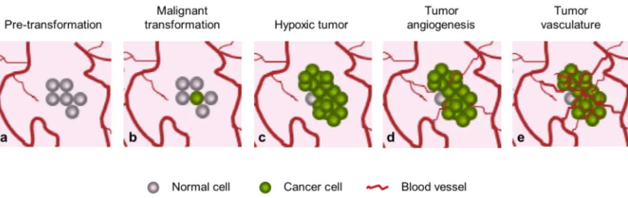
Virchow, the founder of pathological anatomy, drew attention to the huge number of blood vessels in a tumor mass back in 1865. Tumor vascularization was first studied systematically by Goldman, who described the vasoproliferative response of the organ in which a tumor develops as follows: “The normal blood vessels of the organs in which the tumor is developing are disturbed by chaotic growth, there is a dilatation and spiralling of the affected vessels, marked capillary budding and new vessel formation, particularly at the advancing border” [28].
In 1939, Ide et al. [28] was the first to suggest that tumors release specific factors capable of stimulating the growth of blood vessels. But the first tumor fraction responsible for angiogenesis with a molecular mass of about 10KDa was isolated by Folkman et al. [29] only in 1971. This active fraction was first called tumor
studied and numerous factors have been implicated in its positive or negative regulation (see Table I - 1). In solid tumor growth, changes in the relative balance of inducers and inhibitors of angiogenesis could induce a specific transition from the avascular to the vascular phase (called angiogenic switch, see Figure I - 4) [30]. Once it has developed an intrinsic vascular network, the tumor may grow indefinitely (unlike other forms of angiogenesis, tumor angiogenesis is not limited in time) both in
situ and at distant sites (metastasis) since this enables its cells to enter the vascular
bed and colonize other organs [31]. Tumor angiogenesis depends mainly on the release by neoplastic cells of growth factors that are specific for endothelial cells and that stimulate the growth of the host blood vessels.
Figure I - 4. The angiogenic switch in tumor development. Most tumors start growing as avascular
nodules (dormant) (c) until they get hypoxic and reach a steady-state level of proliferating and apoptosing cells. The 'angiogenic switch', has to occur to ensure exponential tumor growth (d). New blood-vessel formation and maturation will continue throughout tumor growth. Figure adapted from Berges, G; Benjamin, L; 2003 [32].
2. Vascular Endothelial Growth Factor A
2.1. Gene and protein characterization
In 1983, Senger et al. [33] purified a 34 to 42 KDa protein (Vascular permeability factor - VPF) that was responsible for the rapid increase in microvascular permeability that promotes accumulation of tumor ascites fluid.
In June of 1989 a growth factor for vascular endothelial cells was identified and purified, according to its heparin-binding property, from media conditioned by bovine pituitary follicular or folliculostellate cells [34]. This growth factor was a dimeric protein with a molecular mass of ~46 KDa, composed of two subunits of identical molecular mass (23 KDa). The purified growth factor had a maximal mitogenic effect on adrenal cortex-derived capillary endothelial cells. Further characterization of its bioactivity revealed that it exerts mitogenic effects also on vascular endothelial cells isolated from several districts but not on adrenal cortex cells, lens epithelial cells, corneal endothelial cells, keratinocytes or BHK-21 fibroblasts, indicating that its target cells specificity was unlike that of any previously characterized growth factor. Due to its apparent target cell selectivity, the name, vascular endothelial growth factor (VEGF or VEGF-A) was proposed.
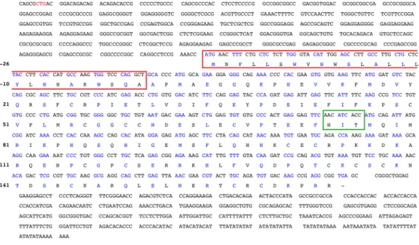
In December of the same year three different groups characterized, separately, the properties of VEGF [35], vasculotropin [36] and VPF [37] that by sequencing revealed to be the same protein. In opposite to FGFs, VEGF had a hydrophobic signal peptide required for the extracellular transport and induction of angiogenesis. This signal was encoded by the first 26 amino acids beginning with a methionine and the signal sequence cleavage site was represented by the sequence Ser-Gln-Ala (S-Q-A) at positions -3 to -1 (Figure I - 5). The total amino acids of bovine VEGF monomer was 190 which without the scretory signal sequence, referred above, gave rise to a protein of 164 amino acids with a molecular mass of ~19 KDa. The overall amino acid homology between human and bovine VEGF sequences exceeded 95%. Human VEGF had an additional amino acid (165) because of the insertion of a Gly in position 6 (Figure I - 6).
Figure I - 5. Nucleotide and amino acid sequences of a bovine VEGF cDNA. The amino acid
sequence of the hydrophobic signal peptide is in a red box. The protein sequence is numbered starting with 1 at the mature NH2-terminal alanine. The putative glycosylation site is shown in a green box.
ATG is in position -26 and the stop codon (TGA) in position 165. The other transcription initiation site (CTG) for the L-VEGF (see Figure I - 9) is pointed in red in VEGF sequence. Figure adapted from Leung, D, et al., 1989 [35].
Microsequencing revealed a unique N-terminal sequence of 21 amino acids that was localized immediately after the sequence that codify for extracellular transport of this growth factor. In the green box in Figure I - 5 is represented the glycosylation site which suggested that VEGF was a glycoprotein. Clusters of basic amino acids found in the VEGF sequence were at this time described as responsible for the binding of the molecule to heparin (which is a linear polysaccharide closely related to heparan sulphate that is found on the cell surface and in the extracellular matrix of all mammalian cells) by analogy with what had been proposed for FGF. Gitay-Goren et
al. in 1992 [38] demonstrated that cell surface-associated heparin-like molecules were
required for the interaction of VEGF165 with its cell surface receptors. The binding of VEGF was completely inhibited following digestion of endothelial cells with
heparinase and could be restored by the addition of exogenous heparin to the digested cells.
In 1994 the process of VEGF dimerization, known to occur since its discovery, was understood due to sequence blast and amino acids mutations. Eight of the cysteine residues found in VEGF and PDGF were conserved (Figure I - 6) [39]. Cysteines 2 and 4 appeared to be involved in intermolecular disulfide bridges analogous to the situation in PDGF. Cysteine 5 was also essential for VEGF dimerization and activity, while dimer formation and biological activity were only slightly affected by the mutation in cysteine 3. The covalent dimerization of VEGF was essential for effective receptor binding and biological activity.
In 1995, DiSalvo et al. [40] purified from GS-9L glioma cell line VEGF homodimers and a novel heterodimer composed of VEGF and PlGF subunits. This heterodimer was a mitogenic to vascular endothelial cell nearly as potent as VEGF homodimers. In 1996, Olofsson et al. [41] isolated and characterized a novel growth factor for endothelial cells, vascular endothelial growth factor B (VEGF-B) that formed cell-surface-associated disulfide-linked heterodimers with VEGF when coexpressed.
Figure I - 6. Comparison of the bovine and human VEGF amino acids sequences. Similar
sequences are boxed in red. All VEGF cysteines are numbered in the sequence and loop II acidic residues Asp63, Glu64, and Glu67 and loop III basic residues, Arg82, Lys84 and His86 are shown in green
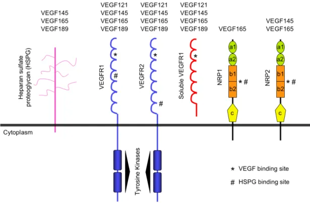
In the same year, the amino acids responsible for VEGF binding to its receptors, VEGFR1/fms-like tyrosine kinase 1 (FLT-1) and VEGFR2/kinase insert domain-containing receptor (KDR), were discovered [42]. VEGF dimerization leads to an elongated structure with 3 surface loops that cluster at each end of the molecule. Loops II (61 to 68 amino acids) and III (84 to 87 amino acids) in VEGF were involved in receptor recognition (Figure I - 6 and Figure I - 7A). The loop II acidic residues Asp63, Glu64, and Glu67 (present in exon 3) that mediated VEGF binding predominantly to VEGFR1, formed a negatively charged surface at one end of each monomer. In contrast, the loop III associated basic residues, Arg82, Lys84 and His86 (present in exon 4) that predominantly mediated binding to VEGFR2, cluster to form a positively charged surface at the other end of the VEGF monomer. A VEGF model based on PDGF indicated these positively and negatively charged regions were distal in the monomer but were spatially close in the dimer. The evidence of receptor binding sites at both ends of the dimer was consistent with the growth factors of the PDGF/VEGF family exhibiting bivalency. Bivalent binding of VEGF provided a mechanism for dimerization of the VEGFR2 and/or VEGFR1 tyrosine kinase receptors and subsequent tyrosine phosphorylation on endothelial cells (Figure I - 7).
In the same year, it was found that the removal of the carboxyl-terminal domain (111-165) of VEGF, whether it was due to alternative splicing of mRNA or to proteolysis, was associated with a significant loss in bioactivity [43].
Analyzing VEGF-VEGFRs binding it was observed that VEGF121 bound VEGFR2 with similar affinity as VEGF165 in the absence of heparin and a modest effect of heparin was the 3-4-fold increased capacity of VEGFR2 for binding VEGF. In contrast, VEGF121 bound VEGFR1 with 10-20 fold decreased affinity compared to VEGF165. This 55-amino acid domain appeared to enhance VEGFR1 binding in addition to the receptor binding determinants contained in 110-amino acid domain, but by itself the 55-amino acid domain could not compete with VEGF165 binding to VEGFR1. These results were also confirmed by Gitay-Goren et al. [44].They showed that VEGF121 bound selectively to the larger of the three VEGF receptors, VEGFR2, and like VEGF165, it was able to bind VEGFR2 in the absence of heparin. However,
heparin could potentiate the binding of VEGF165 and not VEGF121. They also showed that both VEGF165 and VEGF121 were susceptible to oxidative damage (although VEGF121 seemed to be somewhat more resistant than VEGF165 to oxidation) and that heparin restored the receptor binding ability of damaged VEGF165, but not the receptor binding ability of damaged VEGF121.
Since VEGF121 has no affinity for heparin, the modulation of its interaction with VEGFR1 must be mediated at the receptor level (see below in VEGF-A receptors, gene and protein characterization).
Figure I - 7. Important domains in VEGF and its receptors that mediate its binding. A) VEGF
loop II (Asp63, Glu64, Glu67) or loop III (Arg82, Lys84, His86) are essential for VEGF binding to the 2nd Ig-like loop in VEGFR1 or VEGFR2 respectively (B). VEGF binds to its receptors as an antiparallel covalent dimer and induce receptors dimerization and consequently its activation.
2.2. VEGF isoforms
In 1989 molecular cloning revealed shorter and a longer molecular species of human VEGF which display a deletion of 44 amino acids between position 116 and 159 and an insertion of 24 amino acids (KSVRGKGKGQKRKRKKSRYKSWSV) in position 116, respectively. Therefore, the mature proteins are expected to have 121 and 189 amino acids, respectively. The most likely explanation for this molecular heterogeneity was suggested to involve a process of alternative splicing of VEGF mRNA.
Figure I - 8. VEGF isoforms originated by alternative splicing. A huge amount of VEGF isoforms
have been described as a result of alternative splicing between the 8 exons that VEGF contain. Some of these isoforms have been tested for its functionality. The most studied ones are VEGF121, 145, 165, 189, 206, and VEGF165b. The first 5 isoforms differ in the heparin affinity, e.g. VEGF121 is not able to
bind heparin but in opposite VEGF189 has the high heparin affinity. VEGF165b have been described to
In 1991, it was discovered that the VEGF gene comprised eight exons (Figure I - 8) and it was confirmed that the various VEGF coding region forms arose by alternative splicing: the 165-amino-acid form of the protein was missing the residues encoded by exon 6, whereas the 121-amino-acid form was missing the residues encoded by exons 6 and 7 [45]. To summarize, the alternative splicing involved exons 6 and 7 and if neither exon was removed, VEGFl89 was generated.
In the same year, analysis of a variety of human cDNA libraries using primers that flank the insertion/deletion site indicated that VEGF165 was the predominantly expressed form in most of the libraries examined, although multiple transcript types were usually detected [46]. In the screening performed, a fourth form of VEGF, VEGF206, was discovered. This form contained a 41-amino acid insertion relative to VEGF165, and included the highly basic 24-amino acid insertion found in VEGF189. Like VEGF189, VEGF206 was predominantly cell and extracellular matrix (ECM)-associated and only very poorly secreted. This way, ECM represented an important source of VEGF and angiogenic potential [47].
The information supplied by alternative splicing had profound effects on the behavior of the translated proteins following secretion from the producer cell [48]. The shortest form, VEGF121 was secreted and freely soluble in the conditioned medium of the human embryonic kidney cell line CEN4 stably transfected with a VEGF121 expression vector. The transcript of VEGF165 contained an additional 44 codons in the carboxyl-terminal end of the protein that convert it to a basic polypeptide with heparin binding ability. This feature was responsible for the binding of 50-70% of this protein to the cell surface and ECM. VEGF189 that contained an additional 24 amino acids highly enriched in basic residues, bound much more tightly to heparin. Little or no VEGF189 was found in a freely soluble form in the conditioned medium of stably transfected CEN4 cells. Released of bound forms of VEGF165 and VEGF189 by treatment with heparin, heparan sulfate, or heparinase suggested that the binding site in these cell cultures involved a heparin-containing proteoglycan that were known to be constituents of the ECM. In melanoma cells it was seen that after treatment with heparinase, VEGF165 but not VEGF121 was able to restore its binding to VEGF
receptors after exogenous heparin addition [49]. These results suggested that cell-surface heparan sulfates could regulate the binding ability of the VEGFR of these cells to soluble isoforms of VEGF.
In 1993 during a screening for VEGF isoforms in human uterus another VEGF isoform of 145 amino acids was discovered [50]. The sequence of this isoform showed exons 1-6 and 8. In opposite to proliferative/secretory endometrium, myometrium and endometrial carcinoma cell lines, VEGF145 was not found in peripheral leukocytes (used as control) that indicated a possible tissue-specific splicing. In 1995 this isoform of VEGF was also found to be expressed in placenta and chorion [51] but only two years later the protein was characterized. VEGF145 induced the proliferation of vascular endothelial cells and promoted angiogenesis in
vivo [52]. This isoform shared with VEGF165 the ability to bind to the VEGFR2
receptor of endothelial cells and the affinity to bind to heparin.
VEGF183 [53], VEGF115 [54], VEGF148 [55] and VEGF162 [56] are VEGF isoforms expressed in human kidney, murine fibroblasts, human glomeruli and human ovarian carcinoma cells respectively (seeFigure I - 8). The first one is 6 amino acids shorter (18 nucleotides at the end of the exon 6a) than its closest relative, VEGF189. The second one differs from VEGF 120 by 37 amino acids at the carboxyl terminus. VEGF148 was considered a truncated form of VEGF165 due to a 35 bp deletion at the end of exon 7. This dictated a frame-shift and a premature stop codon at the start of the short exon 8. In this study it was found for the first time the presence of VEGF145 in a tissue that did not belong to the female reproductive system. None of these 3 new VEGF isoforms were characterized in terms of angiogenesis function. The latest one, VEGF162, is encoded by exons 1-5, 6A, 6B and 8 of the VEGF. This isoform binds less efficiently than VEGF145 but more efficiently than VEGF165 to basement membrane components due to the presence of the exon 6B that seems to interfere with the interaction of exon 6A with heparin. VEGF162 induces proliferation of endothelial cells in vitro and angiogenesis in vivo.
The VEGF isoforms described above resulted from alternative splicing with initiation at ATG 1039, but three independent groups [57-59] in 2001 demonstrated simultaneously the existence both in vitro and in vivo, of the L-VEGF isoform (VEGF precursor) initiated at the CTG 499, located 539 nucleotides upstream of the ATG codon (Figure I - 5). Start codons selection is controlled by two independent Internal Ribosome Entry Sites (IRES): IRES A is located within the 300 nucleotides upstream of the ATG and IRES B located at the 5’-end of the mRNA controls translation initiation of the L-VEGF at the CTG (Figure I - 9) [59]. This isoform was 206 amino acids longer than the classical ATG-initiated form and can be cleaved into two fragments; one NH2-specific fragment of 23 KDa (named N-VEGF) and a fragment with an apparent size similar to that of the classical ATG-initiated form. Intact L-VEGF and the N-L-VEGF were not secreted in contrary to the C-terminal cleavage product of L-VEGF and the ATG-initiated VEGF.
Encoded by the same gene, VEGF, and resulting from alternative splicing, another VEGF isoform called VEGF165b was discovered in 2002 (Figure I - 8); it also
contained 165 amino acids but had a different COOH-terminal [60]. In this isoform, the six amino acids usually encoded by exon 8 (CDKPRR) were replaced by six different amino acids (SLTRKD) encoded by 18 bases of mRNA spliced 66 bases downstream of the usual acceptor splice site for exon 8 (Figure I - 3). This new open-reading frame (ORF) was initially termed “exon 9”. However, there was no true “intron” between the two 18-base sequences and the more correct term for this ORF was “exon 8b”. The existence of this “exon 8b” predicted the existence of a family of sister isoforms with a novel COOH terminus. The putative family was termed VEGFxxxb where xxx was the amino acid number (e.g. VEGF121b, VEGF165b,
Figure I - 9. VEGF transcription from 2 different sites in the gene. A long transcript of VEGF
(L-VEGF) could be produced from CTG nucleotides by the control of Internal Ribosome Entry Site B (IRES B). L-VEGF is than cleaved into a N-VEGF and an m-RNA with the length of the ATG-initiated VEGF. If this cleavage does not occur the protein cannot be secreted.
2.3. VEGF expression in embryonic and adult tissues
In 1993, Jakeman et al. [61] showed that, during embryonic development, VEGF expression was detected within the first few days after implantation. At later developmental stages in the mouse or rat embryos, the VEGF mRNA was expressed in several organs, including heart, vertebral column, kidney, and along the surface of the spinal cord and brain.
Shifren et al., [62] analyzed the distribution of VEGF mRNA as well as the tissue- and cell-specific localization of VEGF peptide in the human midgestation (16-22 weeks) fetus. VEGF mRNA was detected in all fetal tissues studied and was most abundant in human fetal lung, kidney, and spleen; moderately abundant in heart, adrenal, pancreas, intestine, liver, testis, skin, muscle, and brain; and minimally
detected in thymus and placenta. Adult tissues also express VEGF, particularly these tissues with rapid vascular endothelial turnover, which are reproductive organs such as ovary [63] and uterus [64].
Despite the expression of VEGF during development or in adult tissues in physiologic conditions, in situ hybridization and RT-PCR studies demonstrated that the VEGF mRNA is markedly up-regulatedin the vast majority of human tumors (see Table I - 2), correlating with poor prognosis.
2.3.1. VEGF isoforms expression pattern
Until 1995, VEGF165/164 (human/rodent homologue) was often assumed to be the predominant isoform, although truly quantitative assessments were lacking. The quantification of VEGF mRNA forms in five rat tissues (brain, kidney, lung, spleen, and heart) make clear that VEGF mRNA splicing occurs in a tissue-specific manner [65]. VEGF188 was the predominant form (> 50% of total VEGF mRNA) in heart and lung, but was the least abundant form (6-15%) in all other samples. VEGF164 was lower (approximately 25%) in heart and lung, but was predominant (> 50%) in brain and kidney. VEGF164 and VEGF120 were present in equimolar amounts (each form approximately 46% of total) in the spleen. VEGF120 was also present in kidney (38%) and lung (27%) and was least abundant (approximately 15%) in brain and heart.
In more detail, the VEGF isoforms expression during mouse development and in adult tissues was analyzed and described in 2001 by Ng et al. [66]. Its expression patterns during organ development revealed a correlation between the origin of the vascular bed and the relative levels of VEGF isoforms. The highest levels of VEGF188 mRNA were detected in organs that are initially vascularized by vasculogenesis (e.g., lung, heart, and liver). On the other hand, organs vascularized primarily by means of angiogenesis including brain, eye, muscle, and kidney had higher levels of VEGF164 and VEGF120 mRNA.
Table I - 2. Human tumors overexpressing VEGF mRNA.
Tumors Tumor type Reference
Glioblastoma [67-69] Meningioma [69, 70] Intracranial tumors Capillary hemangioblastoma [69, 71] Esophageal carcinoma [72, 73] Gastric carcinoma [72, 74, 75]
Small bowel carcinoma [72]
Gastrointestinal tract tumors
Colorectal carcinoma [72, 76]
Kidney carcinoma [77, 78]
Urinary tract tumors
Bladder carcinoma [77]
Breast carcinoma [79, 80]
Ovarian carcinoma [81, 82]
Cervix carcinoma [83, 84]
Female reproductive tract tumors
Endometrial carcinoma [85, 86]
Germ cell tumor [87]
Male reproductive tract tumors
Prostate cancer [88, 89]
Acute myeloid leukemia [90-92]
Acute lymphoblastic leukemia [93]
Chronic myeloid leukemia [94]
Chronic lymphoblastic leukemia [95]
Multiple myeloma [96]
Hematopoietic tumors
Lymphomas [97]
Head and neck squamous cell carcinoma [98, 99]
Thyroid carcinoma [100, 101] Lung carcinoma [102, 103] Hepatocellular carcinoma [104] Angiosarcoma [105] Other tumors Melanoma [106]
In 2002, VEGF isoforms expression during rat muscle development was characterized [107]. The relative abundance of VEGF120 mRNA was evident, representing 35% of total VEGF mRNA on day 7 after birth and gradually decreasing with age to approximately 5% on day 37. In contrast, VEGF188 mRNA was very low in newborn rat, but increased to 40-50% on day 50. Neither VEGF144 nor VEGF164 mRNA showed significant changes in abundance after birth.
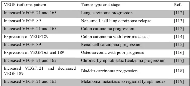
More recently, VEGF isoforms pattern have been characterized in different tumors at stages of tumor growth (see Table I - 3).
2.4. Regulation of VEGF expression
VEGF expression is regulated by a plethora of intrinsic and extrinsic factors. In 1992, hypoxia and hypoglycemia were classified as the major stimulators of VEGF expression [67]. Three years later the mechanisms of VEGF regulation by hypoxia was explained (Figure I - 10). Hypoxia-induced transcription of VEGF mRNA was mediated by the binding of hypoxia-inducible factor 1 (HIF-1) to a hypoxic response element (HRE) located in the VEGF promoter [108, 109]. In 1997, Jiang et al. [110] demonstrated that this transcription factor could be induced by the oncogene v-src independently of hypoxia and leading to increased expression of VEGF. Hypoxia could also act by increasing the half-life of VEGF mRNA. Levy et al. [111] described that the binding of a hypoxia-induced stability factor (HuR) increased the half-life of this mRNA three- to eight-fold.
Table I - 3. Changes in the VEGF isoforms pattern with tumor stage.
VEGF isoforms pattern Tumor type and stage Ref.
Increased VEGF121 and 165 Lung carcinoma progression [112]
Increased VEGF189 Non-small-cell lung carcinoma relapse [113]
Increased VEGF121 and 165 Colon carcinoma progression [112]
Expression of VEGF189 Colon carcinoma with liver metastasis [114]
Increased VEGF189 Renal cell carcinoma progression [115]
Expression of VEGF165 and 189 Osteosarcoma with poor prognosis [116]
Increased VEGF121 and 165 Chronic Lymphoblastic Leukemia progression [117]
Increased VEGF121 and decreased
VEGF 189 Bladder carcinoma progression [118]
In 1996 and 1998 two different studies demonstrated that VEGF was up-regulated in response to glucose deprivation [120, 121]. Remarkably, not only lack of glucose but also high glucose levelsresult in an upsurge of VEGF mRNA [122].
Another metabolic stress described to regulate VEGF expression was acidic extracellular pH (Figure I - 10). In 2002 Xu et al. [123] showed in human glioblastoma cells that acidic pH (pH 6.6) activates Ras and the ERK1/2 MAPK pathway to enhance VEGF transcription via AP-1, leading to increased VEGF production.
Several cytokines have been described to also modulate angiogenesis by regulating VEGF expression (Figure I - 10). The factors that could potentiate VEGF production and this way stimulate angiogenesis include epidermal growth factor [124], platelet-derived growth factor (PDGF) [125], fibroblast growth factor 4 (FGF-4) [126], tumor necrosis factor α (TNF-α) [127], transforming growth factor β (TGF- β) [128], TGF-α [129], insulin-like growth factor I [130], interleukin 1β (IL-1β) [131] and IL-6 [132]. In turn, other cytokines such as IL-10 and IL-13 could inhibit the release of VEGF [133].
Figure I - 10. Mechanism of action of some VEGF expression regulators. VEGF expression is
up-regulated by hypoxia and the oncogene v-src via HIF-1, by TNF-α, IL-1β, FGF, and PDGF via Sp1, by TGF-α via AP-2 and by acidic pH via AP-1. All these transcription factors have binding sites at the VEGF promoter: HIF-1 and AP-1 bind to 5'-TACGTGGG (-975 to -968 bp) and 5'-TGACTAA (-937
to -931 bp) respectively and AP2 and Sp1 bind to a sequence between 88 and 65bp and between
-85 and -50 bp respectively [125, 134-136]. In contrast the tumor suppressor genes, vHL and p53 inhibit VEGF expression by inhibiting HIF-1 and Sp1 action at the VEGF promoter level. Hypoxia also control VEGF expression by stabilization of its mRNA acting through HuR.