Ewa Piskadło
Dissertation presented to obtain the Ph.D degree in Molecular Biology
Instituto de Tecnologia Química e Biológica António Xavier | Universidade Nova de Lisboa
Research work coordinated by:
Oeiras, September, 2017
chromosome architecture
by condensin I
tains a microscopy image of a live mitotic Drosophila embryo; chromatin is marked by histone-RFP (orange) and centromeres by CID-EGFP (cyan). Knit-ting pattern of chromosomes can be found in Appendix 11.
Graphics authors: Ewa Piskadło and Raquel A. Oliveira
Maintenance of metaphase chromosome architecture by condensin I. Ewa Piskadło
mosome Structure Is Dynamically Maintained by Condensin I-Directed DNA
(De)catenation.eLife 6 (2017): e26120.
Piskadlo, Ewa, and Raquel A Oliveira. Novel Insights into Mitotic Chromosome
Condensation.F1000Research 5 (2016): F1000 Faculty Rev-1807.
Declaration: This dissertation is a result of my own research carried out between July 2013 and July 2017 in the laboratory of Dr. Raquel A. Oliveira, Instituto Gulbenkian de Ciência in Oeiras, Portugal, within the PhD Programme in Inte-grative Biomedical Sciences (edition 2013).
Declaração: Esta dissertação é o resultado do meu próprio trabalho desenvolvido entre Julho 2013 e Juhlo 2017 no laboratório do Doutora Raquel A. Oliveira, Ins-tituto Gulbenkian de Ciência em Oeiras, Portugal, no âmbito do Programa de Doutoramento em Integrative Biomedical Sciences (edição 2013).
Financial support: This dissertation had the financial support from Fundação para a Ciência e a Tecnologia and European Social Found, through grant
SFRH/BD/52172/2013 awarded to Ewa Piskadło, and Fundação Calouste Gulben-kian.
Apoio financeiro: Esta dissertação teve o apoio financeiro da Fundação para a Ciência e a Tecnologia e do Fundo Social Europeu no âmbito do Quadro Comu-nitário de apoio através da bolsa de doutoramento
The completion of this thesis would not be possible without the enormous sup-port from multiple people and institutions. The most enormous thanks must be directed to my supervisor Raquel A. Oliveira. I am massively grateful for giving me an opportunity to grow as a scientist and as a person by her endless supply of guidance, support, and encouragement.
To each and every current and former member of the Chromosome Dynamics Group I owe giant thanks for all the moments inside and outside of the lab. I was extremely privileged to work with a group of amazing people, where we could always count on each other. I wish you all the best for the future.
I would like to express my gratitude towards the Instituto Gulbenkian de Ciência for allowing me to perform my research and creating an outstanding environ-ment for engaging in science. Especially, I would like to thank the Director and Assistants of the IGC’s PhD program, Élio, Ana, and Manuela, for making par-ticipation in the program such a great experience.
I would also like to thank members of my Thesis Committee, Alekos and Ivo, for their help and guidance during last four years, that help this thesis gain its current shape. My gratitude also goes to fantastic people in IGC’s Fly, Imaging, and Electron Microscopy facilities and the Zhen Ho wing, who helped me a great deal during the course of my project.
And last, but not least, thank you to my family and friends for an infinite amount of support in all forms you have given me throughout the years. I couldn’t have done it without you.
F
aithful segregation of the genome into two daughter cells is one of the most fundamental events for every living organism. In each round of the cell cycle, cells need to orchestrate a sequence of complex steps to replicate their genetic material, pack it neatly into mitotic chromosomes and perform their pre-cise separation when all the prerequisites are met. One of the most fascinating questions in biology is to understand the internal organization of mitotic chro-mosomes. Even though mitotic chromosomes were first described around 140 years ago, how exactly interphase DNA molecules are packed to become mitotic chromosomes is still a mystery. Despite the lack of precise details about chromo-some condensation mechanisms, it is believed that in the heart of this process lies a group of protein complexes called condensins. The mechanism by which con-densins are able to enforce or guide the condensation process is yet unknown. In this thesis, we will present our advances in understanding condensin’s function in maintaining mitotic chromosome compaction and internal architecture.Condensin’s role in mitosis was extensively studied using mutants for its sub-units or by slow depletion approaches. Those methods were nonetheless not pre-cise or fast enough to permit accurate studies of condensin’s role in maintaining chromosome’s structure. In the search for an acute tool that would allow decisive studies of fast processes, such as specific stages of mitosis, we have developed a
Drosophila melanogastersystem for condensin I inactivation based on Tobacco
Etch Virus (TEV) protease cleavage. The steps performed to build this system are the subject of Chapter 2. We show that it is possible to inactivate condensin I in the context of a developing organism with great efficiency and time control.
Having generated a tool to study condensin I with an unprecedented temporal resolution, we have endeavored to explore condensin I’s role in the maintenance of metaphase chromosome architecture, as described in Chapter 3. Based on our data we propose that condensin I works in collaboration with topoisomerase II constantly throughout mitosis to ensure a correct amount of links between DNA molecules. Removing functional condensin I breaks this balance resulting in an increased number of erroneous entanglements introduced de novo by topoi-somerase II. Such entanglements increase chromatin density leading to
cooperation with topoisomerase II, and in the broader context of chromatin dy-namics.
Mitotic chromosomes are constantly subjected to various forces acting on them. Chapter 4 contains preliminary data showing that soon after the destruc-tion of the mitotic spindle, scattered chromatids rapidly congress back together. These findings suggest that other factors besides the mitotic spindle can arrange the location of the chromosomes. We hypothesize that such inwards forces may influence the surfaces of chromosomes, which can exacerbate the overcom-paction phenotype described in Chapter 3, observed after inactivation of con-densin I. We further speculate what factors could cause the observed phenomena. In this thesis, we explore condensin I’s role in mitosis using a novel system for condensin I inactivation in Drosophila embryos. We propose that condensin I governs the physical properties of chromosomes and their internal structure by imposing control over the amount of inter-DNA intertwines throughout mitosis. We thus uncover a fascinating highly controlled dynamics of the chromosome catenation state and provide new knowledge valuable for the full understanding of mitotic chromosome condensation and architecture.
A
correcta segregação do genoma recém-duplicado para ambas as células fi-lhas é um evento fudamental para qualquer organismo vivo. Em cada ciclo celular, as células têm de coordenar uma sequência bem estabelecida de eventos complexos que lhes permitem realizar a replicação do DNA, a compactação do mesmo em cromosomas mitóticos e a sua separação precisa. Cada um destes eventos é supervisionado por mecanismos de controlo que apenas permitem a passagem ao próximo passo após todos os pré-requisitos terem sido garantidos.Uma das questões mais fascinantes em biologia é comprender a organização interna dos cromossomas mitóticos. De facto, os cromossomas mitóticos foram descritos há cerca de 140 anos, mas o mecanismo exacto que permite ás células compactarem o seu material genético em cromossomas durante a mitose conti-nua a ser desconhecido. Existe um grupo de proteínas que é fundamental para este processo, as condensinas, no entanto o exacto modo de acção destas pro-teínas na promoção e/ou manutenção da condensação do material genético está ainda por esclarecer. Nesta dissertação serão descritos os avanços que fizemos para a compreensão do papel da condensina I na manutenção da estrutura in-terna e compactação dos cromosomas em mitose. Tradicionalmente, os estudos da função da condensina foram feitos recorrendo a mutantes para as diferentes su-bunidades da proteína ou por deplecção da mesma por períodos longos, contudo esta abordagem não permite ter resolução temporal suficiente para investigar de forma precisa o papel da condensina. Para colmatar esta limitação, desevolve-mos um sistema de inactivação da condensina I baseado na clivagem da proteína pela protease TEV (Tobacco Etch Virus). Este sistema, cujo desenvolvimento e implementação constam no Capítulo 2, tem a vantagem de permitir inactivar rapidamente a condensina I nos diferentes momentos da mitose. A resolução temporal sem precendentes, é assim decisiva para compreender a função da pro-teína em processos que são naturalmente rápidos.
Para além da rapidez na inactivação da condensina, demonstramos ainda que o sistema funciona no contexto de desenvolvimento dum organismo com grande eficiência. Após validação do nosso sistema, utilizámos esta abordagem para ex-plorar a função da condensina na manutenção da arquitectura dos cromosomas
constante durante a mitose para assegurar a quantidade certa de ligações entre as moléculas de DNA. Ao remover funcionalmente a condensina I, verifica-se um desequilíbrio entre as duas proteínas o que resulta no aumento dos emaranhados no DNA introduzidos de novo pela topoisomerase II. Estas ligações promovem o aumento da densidade da cromatina o que provoca uma hipercondensação dos cromossomas durante a metafase. No final, discutimos estes resultados no con-texto dos modelos propostos para o mecanismo de acção da condensina I., espe-cificamente no que diz respeito á sua cooperação com a topoisomerase II e no contexto mais amplo da dinâmica da cromatina.
Os cromosomas mitóticos estão constantemente sujeitos á acção de várias forças. No Capítulo 4 constam resultados perliminares que mostram que logo após a destruição do fuso mitótico, as cromátides mitóticas, já em anafase, vol-tam a convergir para o centro da célula. Esta observação sugere que a localização dos cromosomas/cromátides em mitose é determinada por outros factores para além do fuso mitótico. Perante isto, formulámos a hipótese que estas forças em direcção ao centro influenciam a superfície dos cromossomas o que pode exacer-bar o fenótipo de hiper-compactação observada após inactivação da condensina I, descrito no Capítulo 3. Adicionalmente, especulamos sobre outros factores que estão potencialmente envolvidos neste fenómeno.
Em conclusão, neste trabalho exploramos o papel da condensina I em mitose usando para isso um sistema novo, rápido e eficaz para a inactivação da proteína em embriões de Drosophila. Pela interpretação dos nossos resultados propomos que a condensina I regula as propriedades físicas e estrutura interna dos cromo-somas através do controlo da quantidade de emaranhados nas moléculas de DNA durante a mitose. Este estudo permitiu desvendar que a dinâmica do estado de catenação dos cromossomas é um processo altamente controlado e fascinante. Contribuímos assim com novo e fundamental conhecimento para a compreensão da condensação e arquitetura dos cromossomas em mitose.
1 General introduction 1
1.1 Cell cycle and mitosis . . . 2
1.1.1 Cell cycle . . . 2
1.1.2 Mitosis . . . 3
1.2 Architecture of mitotic chromosomes . . . 3
1.2.1 Morphology of mitotic chromosomes . . . 4
1.2.2 Models of mitotic chromosome folding . . . 5
1.3 Factors shaping mitotic chromosomes . . . 9
1.3.1 Condensins . . . 9 1.3.2 Topoisomerase II . . . 10 1.3.3 Kif4 . . . 10 1.3.4 Histone modifications . . . 11 1.4 Condensins . . . 11 1.4.1 SMC complexes family . . . 11 1.4.2 Architecture of SMC complexes . . . 16 1.4.3 Discovery of condensins . . . 18
1.4.4 Enzymatic activity of condensins . . . 19
1.4.5 Spatial and temporal localization and ratio of condensin I and II . . . 21
1.4.6 Condensins in chromosome compaction . . . 23
1.4.7 Condensins in sister chromatids resolution . . . 26
1.4.8 Regulation of condensins . . . 28
1.5 Topoisomerase II in mitosis . . . 29
1.5.1 Topoisomerase II and sister chromatid resolution . . . . 30
1.5.2 Topoisomerase II and chromosome compaction . . . 31
1.5.3 Topoisomerase II and biophysical properties of chromo-somes . . . 33
2 Development of an acute system for condensin I inactivation 59
2.1 Introduction . . . 61
2.2 Results . . . 64
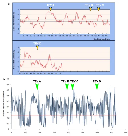
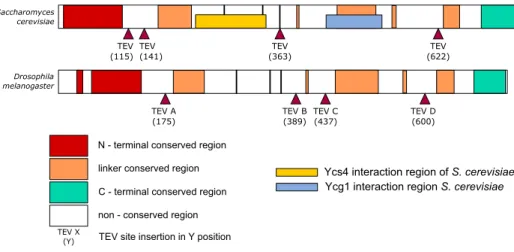
2.2.1 Selecting TEV protease cleavage sites for Barren subunit of condensin I . . . 64
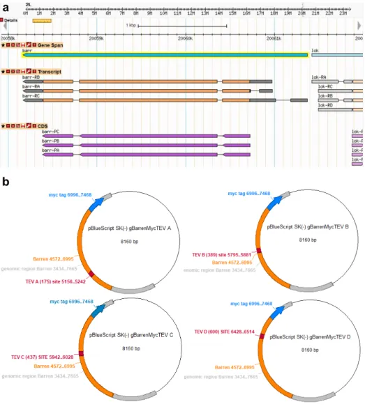
2.2.2 Cloning TEV sites into Barren . . . 65
2.2.3 Transient expression of BarrenTEV A-Dconstructs in DL2 cells . . . 68
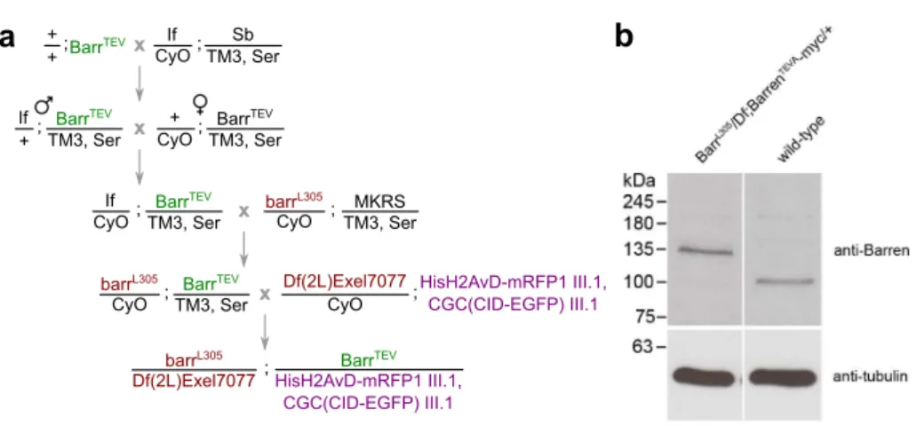
2.2.4 Generating Drosophila melanogaster strains surviving only on TEV-cleavable version of Barren subunit . . . . 69
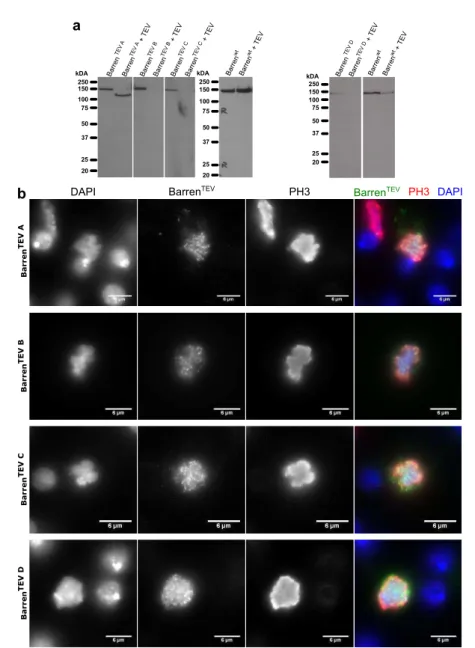
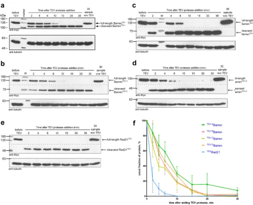
2.2.5 Testing efficiency of BarrenTEVA-D proteins cleavage in vitroand in vivo. . . . 71
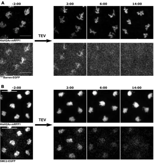
2.2.6 Inactivation of condensin I prior to mitosis. . . 74
2.3 Discussion . . . 77
2.4 Materials and Methods . . . 81
2.4.1 Generation of recombinant plasmids . . . 81
2.4.2 Constructing BarenTEVEGFP plasmids . . . . 82
2.4.3 Constructing genomic BarrenTEVMyc plasmids . . . . . 82
2.4.4 Fly strains . . . 83
2.4.5 Western blotting . . . 84
2.4.6 Transient transfection of DL2 cells . . . 85
2.4.7 Immunofluorescence of DL2 cells . . . 85
2.4.8 Microscopy . . . 86
2.4.9 Microinjections . . . 86
2.4.10 Protein purification . . . 87
2.4.11 mRNA expression of BarrenTEVMyc in Drosophila em-bryos . . . 87
2.4.12 Multi-sequence alignment of Barren subunit of condensin I and basic structure prediction . . . 88
References . . . 89
3.1 Introduction . . . 97
3.2 Results . . . 98
3.2.1 Centromere impairment following condensin I removal. 98 3.2.2 Measuring chromosome compaction in absence of con-densin I. . . 99
3.2.3 Simultaneous inactivation of condensin I and topoiso-merase II abolishes chromosome overcompaction. . . . 102
3.2.4 Does condensin I inactivation lead to re-entanglement of DNA in vivo? . . . 109
3.2.5 Testing the proximity effect. . . 109
3.2.6 Assessing levels of de novo catenations in condensin I-inactivated embryos. . . 113
3.3 Discussion . . . 117
3.4 Materials and Methods . . . 123
3.4.1 Constructing and testing UbcH10S114C (wt)plasmid . . . 123
3.4.2 Fly strains . . . 123
3.4.3 Microscopy . . . 123
3.4.4 Microinjections . . . 124
3.4.5 Protein purification . . . 124
3.4.6 Electron microscopy of Drosophila embryos . . . 125
3.4.7 Quantitative analysis of compaction of chromosomes . . 126
3.4.8 Quantitative analysis of chromatids movements in time . 126 3.4.9 Statistical analyses . . . 126
References . . . 128
4 Exploring the external forces acting on mitotic chromosomes 133 4.1 Introduction . . . 135
4.2 Results . . . 136
4.2.1 Isolated chromatids rapidly congress upon disruption of mitotic spindle. . . 136
4.3 Discussion . . . 139
4.4 Materials and Methods . . . 144
4.4.3 Microinjections . . . 145 4.4.4 Protein purification . . . 145 References . . . 146 5 General Discussion 149 References . . . 157 Appendix i
.1 List of all BarrenTEVfly lines. . . . iii
.2 Scheme of cloning of cDNA BarrenTEVA-DEGFP constructs . . . iv
.3 Scheme of cloning of genomic BarrenTEVA-DMyc constructs . . v
.4 Script for statistical analysis of centromere distance after induced anaphase using linear mixed models. . . vi .5 Script for statistical analysis of histone profiles after induced
anaphase using linear mixed models. . . viii .6 Legends of the movies. . . xi .7 Source data files legends . . . xiv .8 Publication 1: Piskadlo, Ewa, and Raquel A Oliveira. Novel
In-sights into Mitotic Chromosome Condensation. F1000Research
5 (2016): F1000 Faculty Rev-1807. . . xvi .9 Publication 2: Piskadlo, Ewa, Alexandra Tavares, and Raquel
A Oliveira. Metaphase Chromosome Structure Is Dynamically
Maintained by Condensin I-Directed DNA (De)catenation.eLife
6 (2017): e26120. . . xxvii .10 Outreach movie of the work . . . l .11 Knitting pattern of chromosomes presented on the cover of the
thesis . . . li
Condensation.F1000Research 5 (2016): F1000 Faculty Rev-1807.
1.1
Cell cycle and mitosis
1.1.1 Cell cycle
T
hecell cycle is a highly conserved and ordered process. It allows the cre-ation of a genetically identical copy of a cell and is the basis of cell mul-tiplication, growth, and differentiation into specialized units. Most simply, cell cycle can be divided into two parts – mitosis, when genome segregation takes place, and interphase, which is typically the longest part of cell cycle. During interphase, cell’s functions are focused on growth, metabolizing nutrients and producing all the necessary proteins needed to sustain life. In multicellular or-ganisms, cells in interphase perform their specialized functions in the context of the whole organism. The progression through all the stages of cell cycle is under control of protein regulators, mostly cyclins and cyclin-dependent kinases that synchronize the processes and help to perform quality control over the events, activating checkpoint mechanisms in case of disruptions to prevent faulty divi-sion. Interphase period comprises few consecutive phases that are focused on cell vitality functions and preparing the genome for the subsequent division. Just after the previous division cells enter into G1 phase (or G0, if they halt their pro-liferation permanently or temporarily) to intensively grow, rearrange organelles, increase transcription and translation in preparation of next stages. Once the quality conditions are met, cells enter the S phase. Then each molecule of DNA is copied exactly once via a semiconservative mechanism and cell cycle moves to G2 phase. In this phase cell resumes intensive metabolic activity and growth and mitochondria need to supply enough energy for mitotic division. G2 phase is followed by entry to mitotic division and creation of two daughter cells, each starting its own new cell cycle.1.1.2 Mitosis
Mitosis has been first described in the 19th century and has captivated genera-tions of scientists ever since. This fascinating process comprises the assembly of interphase chromatin into individual chromosomes and subsequently the equal separation of the genetic material between two daughter cells. Mitosis is un-doubtedly an extremely complex operation that needs to be precisely conducted and controlled under the penalty of dismantling genome integrity. Mitosis can be divided into few stages. The first one – called prophase – is when chromatin begins to condense and rearrange to form compacted, rod-shaped chromosomes. At the same time sister chromatids begin their resolution into separate units (Na-gasaka et al. 2016). Around the nucleus microtubules are reorganized to form a network of microtubules originating from two centrosomes (or microtubule organizing centres) that move towards opposite poles of the cell to form the mitotic spindle. Later in prophase the nuclear envelope formed around eukary-otic nucleus is disintegrated in a process known as nuclear envelope breakdown (NEBD). Chromosomes are then captured and bioriented by microtubules from the opposite poles in a process called ‘search and capture’ (Heald and Khod-jakov 2015). In the next stage, metaphase, chromosomes reach almost the full condensation and are captured by the microtubules and the correct, bioriented at-tachments are stabilized (Sarangapani and Asbury 2014). This causes all chromo-somes of the cell to be aligned on the so called metaphase plate, which is a plane equidistant to both centrosomes. Such arrangement is able to trigger anaphase stage, in which chromosomes reach their maximal compaction and when sister chromatids separate, allowing microtubules to segregate DNA molecules to the opposite poles (Kamenz and Hauf 2017). In the final stage of mitosis, telophase, nuclear envelope is reformed around two freshly separated sets of chromosomes reconstituting nuclei and cytokinesis is triggered to separate the mother cell into two entities (Hetzer 2010).
1.2
Architecture of mitotic chromosomes
Mitotic chromosomes are striking structures in a cell and were of the first de-scribed already in the 19th century. Mitotic chromosome assembly, although
poorly understood at the molecular level, fulfils three major tasks essential for faithful chromosome segregation: Firstly, it ensures chromosome compaction, making cell division feasible within the cell space. Secondly, it provides chro-mosomes with the right mechanical properties (e.g. bendiness and rigidity) to facilitate their drastic movements during mitosis. Lastly, it ensures the resolution of the topological constrains that exist between the two sister DNA molecules, as well as between neighbouring chromosomes (chromosome individualization), a key requisite for efficient chromosome partitioning. Despite the utmost im-portance of chromosome condensation for the fidelity of mitosis, the molecular mechanisms that drive this process remain very unclear.
1.2.1 Morphology of mitotic chromosomes
Chromatin is a structure composed of DNA and various proteins and RNAs in-teracting with it. To ensure that DNA molecules fit inside of a human interphase nucleus, they need to be compacted 200-1000 times compared to their stretched, linear length (Lawrence et al. 1990). Importantly the condensation in interphase cannot be too restrictive to permit access to transcriptional, replication and reg-ulatory sites, allowing cell to perform its genetic program. The first level of compaction is wrapping the DNA around histones to form nucleosomes. His-tones are extremely conserved proteins and they have many variants (Biterge and Schneider 2014). Some of them, so called core histones H2A, H2B, H3, and H4, form an octamers around which DNA is wrapped 1.67 times in a left-handed turn. Regions of DNA between octamers and bound to H1 histone to stabilize the nucleosome structure. This basic nucleosome strand can be further compacter to reach desired compaction. Modifications of histone post-translational modifica-tions helps regulating local compaction of the chromatin (Bowman and Poirier 2015; Wilkins et al. 2014; Kruitwagen et al. 2015). Also specific histones mark certain regions of chromatin to change properties of chromatin, such as CENP-A binding to centromeres to allow kinetochore assembly.
To ensure that cell division is feasible within the cell space, vertebrate cells compact their DNA around 2-3 times more than in interphase, as estimated by chromatin volume measurements (Martin and Cardoso 2010; Mora-Bermudez et al. 2007) and Förster resonance energy transfer (FRET)-based assays between
histones (Llères et al. 2009). Spatial compaction, however, is not the only impor-tant outcome of condensation. The structural reorganization during condensation leads to the separation of the identical sister chromatids from each other (known as sister chromatid resolution). Several topological constrains arise throughout interphase (most notably during DNA replication) that result in the entanglement of the two DNA molecules. The resolution of such intertwines (i.e., individual-ization) is crucial for efficient and faithful chromosome segregation during mi-tosis. Condensation of chromatin into sturdy chromosomes is also necessary to establish proper physical properties. Chromosomes must be stiff, resilient and elastic enough to withstand forces coming from pulling microtubules and cyto-plasmic drags during mitosis to prevent damage and breaks caused by external tensions.
Centromeres, morphologically visible as constrictions in the X-shaped chro-mosomes, are specialized regions of chromosomes to which sister chromatids are connected until anaphase in majority of animals. They also allow the assembly of the kinetochore, a proteinaceous structure to which microtubules are binding dur-ing mitosis and are crucial for successful chromosome segregation. Centromeres are enriched in α-sequences and a specific variants of histone, CENP-A, a variant of core histone H3 (Schalch and Steiner 2017). Most importantly, centromeres constitute a chromatin scaffold on which kinetochores assemble in order to an-chor spindle microtubules to chromosomes (Nagpal and Fukagawa 2016). Kine-tochores are complex structures that comprise of multiple proteins of various functions, such as structural (i.e. CENP-B), motor (i.e. dynein) or checkpoint proteins (ie. Mad2, BubR1) (Nagpal and Fukagawa 2016). The main function of kinetochores is to ensure polarity of the division and ensuring biorientation of chromosomes before segregation and transmitting dragging forces to chromatids once separation occurs.
1.2.2 Models of mitotic chromosome folding
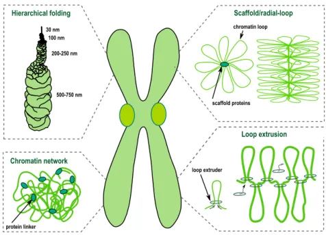
Over the past decades detailed characterization of metaphase chromosomes, us-ing different cytological approaches, has led to the proposal of several models for mitotic chromosome assembly (Figure 1.1).
Hierarchical folding Chromatin network Scaffold/radial-loop Loop extrusion scaffold proteins chromatin loop protein linker loop extruder 30 nm 100 nm 200-250 nm 500-750 nm
Fig. 1.1. Schematic represenation of several possible models of mitotic chromosome folding.
Hierarchical folding
Classical views on chromosome organization postulate that mitotic chromosomes result from chromatin fibre folding. DuPraw suggested that fibre folding occurs randomly, transversely and longitudinally, with no intermediate levels of com-paction (DuPraw 1966). However, mitotic chromosomes fold into a reproducible structure in every mitosis, at least to some extent. Mitotic chromosomes acquire a reproducible length and display an invariable signature pattern of bands after staining with specific dyes, such as Giemsa. Moreover, specific DNA sequences occupy a reproducible position along the longitudinal and transverse axes of the chromosome (Baumgartner et al. 1991). Although some degree of randomness was observed within chromosomal domains (Dietzel and Belmont 2001; Strukov and Belmont 2009), chromosome assembly cannot be explained as a purely ran-dom process.
Alternatively, it has been suggested that metaphase chromosomes result from helical coiling events (helical-coiling model). The nucleo-histone fibre is pro-posed to be coiled up into a helix which is hierarchically wound up into larger
helices to achieve the compactness of the mitotic chromosome (Sedat and Manue-lidis 1978; Belmont 1987). This model has been widely accepted as lower levels of chromatin organization were long postulated to result from hierarchical fold-ing: wrapping of DNA around nucleosomes forms an 11-nm bead-on-a-string structure that coils up into a 30nm fibre. However, the existence of 30 nm fibre
in vivois yet to be confirmed and has been recently highly debated(Maeshima et
al. 2011; Joti et al. 2012; Razin and Gavrilov 2014). A strong argument against existence of an ordered hierarchical architecture of mitotic chromosomes was recently presented using a ChromEMT method. This approach merges electron microscopy tomography imaging and special labelling enhancing the DNA con-trast, combined with mild treatment to preserve native structure of chromatin, in contrast to standard electron microscopy assays (Ou et al. 2017). High resolution imaging of human epithelial cells using this method failed to uncover any signs of discrete higher-order chromatin fibres. The only motif found was unordered flexible chains of various length and 5- to 24 nm in diameter, that are packed to different density depending of the cell cycle stage, with the highest packing density reached in mitosis.
Scaffold model
Using EM studies, Paulson and Laemmli (Paulson and Laemmli 1977) brought a novel view on chromosome organization. Upon histone removal, chromosomes revealed a scaffold or core that has the shape of intact chromosomes, surrounded by loops of chromatin attached to this central core (Adolph et al. 1977; Earn-shaw 1983). These and subsequent studies lead to the consolidation of the scaffold/radial-loop model which argues that radial DNA loops extend out from a protein element or scaffold positioned along the central axis of the chromatid. However, this model has been challenged by studies that evaluate the compo-nents for chromosome continuity (see below). Moreover, major compocompo-nents of the chromosome scaffold were shown to display a highly dynamic association with mitotic chromatin (Gerlich et al. 2006; Oliveira, Heidmann, et al. 2007; Christensen et al. 2002; Tavormina et al. 2002), arguing against the existence of a stiff schaffold anchoring DNA loops.
Chromatin network model
Analysis of the biophysical properties of mitotic chromosome has challenged the idea that the continuity of mitotic chromosomes depends on its proteina-ceous core, in contrast to what the chromosome scaffold would predict. Taking advantage of the highly elastic behavior displayed by mitotic chromosomes, in
vitroelasticity measurements revealed that the elastic response of mitotic
chro-mosomes is lost after DNA digestion (Poirier and Marko 2002). Mild protease treatment, in contrast, does not impair a reversible elastic response, despite a progressively reduced force constant (Poirier and Marko 2002; Pope et al. 2006). This led to the proposal of the chromatin-network model in which chromatin it-self is proposed to be the mechanical contiguous component of the mitotic chro-mosome.
Loop extrusion
Loop extrusion is a relatively new model of how mitotic chromosomes can com-pact and be organized. It can be viewed as a specific variant of chromatin network model in some aspects and it has rapidly gained a great recognition in the chromo-some field. The general idea is that instead of chromochromo-some loops being anchored to a stiff scaffold in the chromosomal axis, the loops are generated by constant, dynamic pulling of DNA through a specialized ring-like motor proteins that cause organization and compaction at the same time. This model first emerged in 1990 as ‘DNA reeling mechanism’ to explain proposed existence of loop-based organization of chromosomes (Riggs 1990). Later, the idea was raised by several researchers who pointed at SMC complexes (namely cohesin and condensin) to be possible loop extruding factors (Nasmyth 2001; Alipour and Marko 2012). In this model mitotic chromosomes would be loop-based structures. In contrast to a standard loop/scaffold model, loop extrusion-based condensation does not re-quire any stiff scaffold, and the loops would be very dynamic, regulated by loop extrusion protein factors. The loop extrusion was shown in polymer dynamics models to be sufficient to explain mitotic chromosome compaction and individu-alization of even a mammalian-sized chromosomes (Goloborodko, Marko, et al. 2016; Goloborodko, Imakaev, et al. 2016; Naumova et al. 2013; Alipour and Marko 2012; Fudenberg et al. 2016).
Other models of chromosome folding
More recent ideas for the internal folding of chromosomes have that mitotic chro-mosomes are arranged into stacks of 6nm layers (Daban 2015). Those layers would be perpendicular to the chromosome axis and contain around 1 Mb of consequent DNA. Such arrangement of chromosomes has the advantage of ex-plaining properties of G-bands and geometry of chromosome translocations in a better way than other models.
Despite the differential contributions for chromatin/protein components within the chromosome organization, these models might not be mutually exclusive and stacks, coils and radial extruded loops may co-exist within a less ordered struc-ture.
1.3
Factors shaping mitotic chromosomes
Despite the several unknowns on the precise molecular details of chromosome assembly, some key components are believed to be crucial for chromosome or-ganization.
1.3.1 Condensins
Condensins are a conserved group of multi-subunit proteins fulfilling many roles in chromatin organization throughout the cell cycle, but their most prominent function is to ensure efficient chromosome segregation (reviewed in Hirano 2012, Piazza, Haering, et al. 2013, and Hirano 2016). They were first isolated from
Xenopuseggs extract and it was suggested that this protein complex is required
for proper chromosome condensation in vitro (Hirano and Mitchison 1994; Hi-rano, Kobayashi, et al. 1997). However, subsequent studies have challenged the view for condensin’s requirement in chromosome condensation, as chromo-somes do condense to a certain degree upon condensin’s inactivation in several
in vivostudies (Hudson et al. 2003; Gerlich et al. 2006; Steffensen et al. 2001;
Oliveira, Coelho, et al. 2005; Hagstrom et al. 2002; Lavoie, Hogan, et al. 2002). In addition to chromosome compaction, several studies revealed other roles for condensin in mitotic chromosome organization: maintenance of chromosomal structural integrity (Gerlich et al. 2006; Oliveira, Coelho, et al. 2005; Ribeiro
et al. 2009) and resolution of topological DNA entanglements (Oliveira, Coelho, et al. 2005; Ribeiro et al. 2009; Steffensen et al. 2001; Hagstrom et al. 2002; Hud-son et al. 2003). Condensins’ function in mitosis and beyond it are discussed in more details later in this Chapter.
1.3.2 Topoisomerase II
Topoisomerase II can introduce several changes in the topology of DNA molecules by driving both supercoiling and relaxing the supercoils, and also the catenation and decatenation of DNA molecules (Schoeffler and Berger 2005). Although some of these reactions can be brought about by topoisomerase I, only topoiso-merase II can promote the resolution of catenated sister-DNA molecules. Topoi-somerase II is able decatenate intertwined DNAs by transiently cutting both strands of a DNA molecule, which are then resealed after passage through an-other DNA duplex. It is therefore essential for sister chromatid resolution and their efficient separation at the end of mitosis. Topoisomerase II is also a major component of the chromosome scaffold (Earnshaw 1985) and it has long been debatable whether or not this enzyme is promoting chromosome compaction in addition (or in parallel) to sister chromatid resolution (see more detailed discus-sion in Chapter 3 and Chapter 5).
1.3.3 Kif4
Kif4 is a motor protein able to bind to mitotic chromosomes. Studies in ver-tebrate cells reveal that Kif4 contributes to the establishment of a correct mor-phology and structure of chromosomes (Mazumdar et al. 2004; Samejima et al. 2012).It is proposed to cooperate or work alongside condensin in shortening the lateral axis of chromosomes, possibly by creating loops of chromatin (Samejima et al. 2012), although little is known about the molecular mechanisms in this process. Kif4 was also reported to play an important role in mouse meiosis seg-regation (Camlin et al. 2017), suggesting that Kif4 assist in both kinds of cell division. Interestingly, it was recently observed that condensin I is associating with Kif4 in human cell extracts (Takahashi et al. 2016). This binding, as well as Kif4 motor activity, are necessary for precise axial localization of condensin I to mitotic chromosome axis and granting mitosis chromosomes their biophysical
properties. These findings highlight the tight cooperation between condensin I and Kif4 in establishing mitotic chromosomes.
1.3.4 Histone modifications
During mitosis and concomitantly with chromosome condensation, the land-scape of histone modifications is altered. Histone H1, the linker histone, is hyper-phosphorylated during mitosis (Fischer and Laemmli 1980; Boggs et al. 2000) and it was initially thought to directly participate in condensation. How-ever, subsequent studies suggest that histone H1 phosphorylation is not necessary for condensation (Guo et al. 1995; Shen et al. 1995) but nevertheless changes the overall chromatin structure (Maresca et al. 2005; Fan et al. 2005). Another key mitotic histone modification is phosphorylation of serine 10 residue of histone 3 (H3 S10), by the mitotic kinase Aurora B (Hendzel et al. 1997). The role for this modification in chromosome condensation has also been controversial (Van Hooser et al. 1998; Wei et al. 1999; Hsu et al. 2000) although recent evidence propose that it drives recruitment of deacetylase Hst2 which, in turn, induces deacetylation of lysine 16 of histone 4. This change in the properties of histone 4 tail promotes interaction with histones H2A and H2B from other nucleosomes (Wilkins et al. 2014), thereby shortening the distance between neighbouring nu-cleosomes. This would thus support that histone modifications can alone pro-mote the condensation of chromosomes. It should be noted that several histones and histone modifications were also described to be a chromosomal ’receptor’ for condensin binding (Ball et al. 2002; Kim et al. 2009; Liu et al. 2010; Tada et al. 2011). Thus, some histone modifications may not be a direct contributor for chromosome compaction but rather a facilitator, by promoting the binding of specialized proteins that model DNA topology.
1.4
Condensins
1.4.1 SMC complexes family
The name SMC is an abbreviation of Structural Maintenance of Chromosomes and the name reflects well on the major common task of those complexes. These
well-conserved proteins are necessary for various aspects of chromatin architec-ture thought the cell cycle, including (but not limited to) chromosome condensa-tion, sister chromatid cohesion, regulation of interphase chromatin interactions and DNA damage repair.
Cohesin
The canonical role of cohesin in proliferating cells is holding together two strands of identical replicated DNA after replication. This assures even distribution of DNA to the daughter cells in anaphase by allowing chromosome biorienta-tion by mitotic spindle towards the opposite cell poles. Cohesin forms a ring large enough to fit two strands of naked DNA, therefore it can encircle two DNA molecules and bind them together (Haering, Löwe, et al. 2002; Gruber et al. 2003; Ivanov and Nasmyth 2005; Haering, Farcas, et al. 2008). During replication cohesin is loaded onto freshly replicated DNA to establish cohesion. Replicated DNA is kept tightly together by cohesin along all the chromosome length. In yeast, cohesin is kept this way until the very beginning of anaphase, at which point cohesin is rapidly removed to allow segregation of sister chro-matids (Uhlmann, Lottspeich, et al. 1999). In higher eukaryotes, however, co-hesin is removed from the arms of chromosomes in prophase by regulated open-ing of the cohesin ropen-ing that allows DNA to release DNA from its topological em-brace, but the cohesin is kept around centromeric region (Haarhuis et al. 2014; Mirkovic and Oliveira 2017). This allows mitotic chromosomes to establish its well-known X-shaped morphology in the next stage of mitosis, metaphase, with sister chromatids separated along their arms and connected mostly around cen-tromeric region. The rest of cohesin is released once anaphase is initiated, by rapid proteolytic cleavage of cohesin’s kleisin subunit by protein named sepa-rase to facilitate segregation of the DNA (Uhlmann, Wernic, et al. 2000; Hauf et al. 2001; Oliveira, Hamilton, et al. 2010).
Besides its role in segregation fiedelity, cohesin is implicated in regulation of genome in interphase. For example, cohesin contributes to gene regulation by changing long-range DNA contacts in cis (Hadjur et al. 2009; Nativio et al. 2009; Zuin et al. 2014; Sofueva et al. 2013) and is proposed to act as a major factor for higher order organization of interphase nucleus (reviewed in Barrington et al.
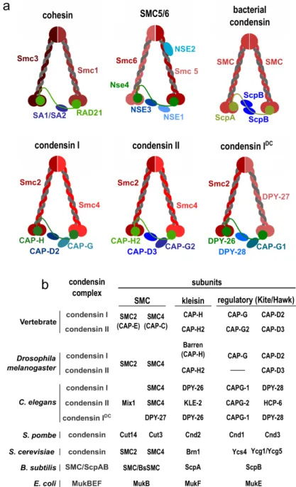
Fig. 1.2. Structural Maintenance of Chromosomes complexes. (a)Cartoon represen-tation of main SMC complexes. (b) Table of subunit composition of condensin complexes in various organisms.
2017).
SMC5/6
SMC5/6 complex, still unnamed in contrast to cohesin and condensin, has been mostly studied for its role in DNA repair. Most likely SMC5/6 is involved in ho-mologous recombination needed for resolving replication products and for DNA damage repair. Mutations in SMC5/6’s subunits lead to hypersensitivity when challenged with agents causing DNA damage or replication forks stalling, such as UV light, ionizing radiation or hydroxyurea (Lehmann et al. 1995; De Piccoli et al. 2006; Ampatzidou et al. 2006; Zhao and Blobel 2005; McDonald et al. 2003). Besides facilitating DNA repair, SMC5/6 complex is proposed to also play a role in maintaining sister chromatin cohesion in yeast and chicken and human cells (Gallego-Paez et al. 2014; Stephan et al. 2011; Almedawar et al. 2012).
Eukaryotic condensins
Condensins, as their name suggests, were believed to be a major driver for mi-totic chromosome condensation (Hirano, Kobayashi, et al. 1997; Freeman et al. 2000). Since obtaining its name the role of condensin in promoting efficient com-paction of chromosomes has remained rather controversial (Bhat et al. 1996; Stef-fensen et al. 2001; Hagstrom et al. 2002; Hudson et al. 2003; Oliveira, Coelho, et al. 2005). Along with its precise role, the mechanisms of action of condensins on mitotic chromatin still remain enigmatic. Eukaryotic condensins are a small group of protein complexes that are quite conserved and necessary to support live in nearly all known eukaryotes. Almost all eukaryotes possess condensin I, and majority also have condensin II (Hirano 2012), that vary in their non-SMC subunit composition (Figure 1.2b).
Besides the mitotic roles, which will be described in more details later in this Chapter, condensins were also found to influence interphase organization of chromatin. Condensin II, thanks to its association to chromatin during inter-phase, plays several roles outside mitosis in several organisms. In Drosophila condensin II was shown to antagonize transvection (Hartl et al. 2008), which is a process of influencing transcriptional activity of certain alleles by the action of
the corresponding allele on the homologous chromosome. Such activity could be explained by condensin II’s ability to restrict trans interactions between homol-ogous chromatids. Probably through the same ability of disrupting long-range interactions in trans, condensin II was also implicated in dispersing polytene chromosomes in Drosophila’s ovarian nurse cells. Polytene chromosomes have multiple copies of chromosomes that align with their homologs creating massive chromosomes. During oogenesis in Drosophila polytene chromosomes must be dispersed, and mutations of condensin II is preventing unpairing and polytene chromosomes cannot be disassembled (Hartl et al. 2008). Condensin II has also an influence of gene transcription during interphase in C. elegans, Drosophila, mouse, and human cells (Kranz et al. 2013; Dowen et al. 2013; Longworth et al. 2012; Wallace et al. 2015; Yuen et al. 2017). Also yeast were shown to con-trol their transcription via condensin complex, such as clustering tRNA genes (Haeusler et al. 2008; D’Ambrosio et al. 2008). Moreover, condensin is required in order to compartmentalize chromosomes in interphase into discrete chromo-some territories, deciding on interphase chromatin architecture in Drosophila (condensin II), C. elegans (condensin IDC), and fission yeast (Bauer, Hartl, et al.
2012; Lau et al. 2014; Iwasaki et al. 2016) .
Besides condensin I and II, a third eukaryotic condensin variant was iden-tified in C. elegans, named condensin IDC after ’dosage compensation’, which
accurately describes its main function (Csankovszki et al. 2009). Condensin IDC differs from condensin I only by replacing SMC4 by it’s another version,
called DPY-27. In contrast to cohesin and the other condensins that work glob-ally, condensin IDCis associating to X chromosome only in order to equalizing
transcription of X chromosome in hermaphrodites.
Prokaryotic SMCs
Three families of SMC complexes were identified in many bacteria and archaea up to date, MukBEF, SMC-ScpAB, and MksBEF, and they play a vital role in chromosome segregation. The prokaryotic organisms proven to be a great tool in SMC complexes research. Thanks to small size of their genome and simple ma-nipulation on SMC proteins they provided important insights for understanding the mechanistic processes governing SMCs.
MukBEF is the first ever described SMC complex, and it can be encountered in enterobacteria and some γ-proteobacteria (Niki, Jaffé, et al. 1991; Hiraga et al. 2000). Mutating MukBEF complex in E. coli results in chromosome con-densation problems, segregation anomalies, as judged by anucleated cells and sharp decrease in colony viability (Niki, Jaffé, et al. 1991; Yamanaka et al. 1996; Wang, Mordukhova, et al. 2006), suggesting that MukBEF serves a similar role in mitosis as eukaryotic condensin.
Similar problems in genome division were observed when the second group of prokaryotic SMC complexes, named SMC-ScpAB. SMC-ScpAB are found in many bacteria and archaea not possessing MukBEF complexes. When SMC-ScpAB were mutated in bacteria normally expressing SMC-SMC-ScpAB, namely B.
subtilis and C. crescentus, it give rise to chromosome compaction and
segre-gation defects (Britton et al. 1998; Wang, Tang, et al. 2014), confirming their condensin-like role. SMC-ScpAB bears much closer similarity to condensin and cohesin of eukaryotes than other prokaryotic SMC groups (Cobbe and Heck 2004). Recent publication uncovered a mechanism by which SMC-ScpAB is able to condense and segregate DNA in B. subtilis by loop extrusion mechanism. It proposes that SMC complex is loaded on the circular chromosome on the parS site by ParB protein (Wang, Brandão, et al. 2017) followed by translocation of the prokaryotic condensin via active loop extrusion to travel through the circu-lar genome, juxtaposing the arms of the chromosome by multiple sliding SMC rings, leading to compaction (Wang, Brandão, et al. 2017).
The third SMC prokaryotic complex was found through bioinformatics anal-ysis which identified novel proteins resembling MukBEF, therefore newly dis-covered complexes family was named MksBEF (MukBEF-like SMC proteins) (Petrushenko et al. 2011). This protein is not highly conserved and it can be found in large variety of proteobacteria, and can also be present in one organ-ism together with other MksBEF, MukBEF or SMC-ScpAB (Petrushenko et al. 2011). TThe exact roles MksBEF complex are not yet fully explored.
1.4.2 Architecture of SMC complexes
SMC protein family are a group of complexes built on a similar structural plan (Figure 1.2a). The core of the complex are SMC protein dimers. Each SMC
sub-unit protein is 1000-1500 amino acid long and has three distinct parts. Firstly, a head of SMC, which is a globular domain containing ABC-type ATPase includ-ing Walker A/B motifs responsible for enzymatic abilities of SMC complexes. On the opposite side of the protein there is a hinge domain that is responsible for proper folding of the protein, interacting with its partner during dimerization, and other functionalities of the holocomplex. Those two parts are connected by a ∼50nm coiled coil. In eukaryotes they are always heterodimers, namely SMC1-SMC3 for cohesin, SMC2-SMC4 for condensins, or SMC5-SMC6, while in prokaryotes SMCs (SMC, MukB, and MksB) subunits form homodimers.
Two SMC proteins are directly interacting by their hinges and the heads of SMCs are connected by another protein, called kleisin after Greek word ’closure’ (κλεíσiµo or kleisimo). Prokaryotic kleisins include ScpA from SMC/ScpAB, MukF from MukBEF and MksF from MksBEF complexes. Analysis of kleis-in/SMC interphases showed that N-terminus of kleisin is binding to the lower part of coiled coil of first SMC subunit via its helix-turn-helix motif (Onn et al. 2007; Bürmann et al. 2013; Gligoris et al. 2014), and opposite end of kleisin is connecting to the bottom part of globular ATPase head of the other SMC protein via its winged-helix domain (Bürmann et al. 2013; Haering, Schoffnegger, et al. 2004; Onn et al. 2007). Kleisin subunit is connecting two SMCs to form a closed ring-like structure that is believed to be a key feature in organizing chromatin, as it allows topological entrapment of DNA inside of the SMC complex ring.
Peripheral subunits are believed to modulate the behavior of a given SMC complex. Those subunits bind to the kleisin and can belong to either Kite or Hawk group of proteins (Palecek and Gruber 2015; Wells et al. 2017). Prokary-otic SMC complexes and eukaryProkary-otic SMC5/6 contain peripheral subunits belong-ing to Kite family, eukaryotic cohesin and condensins use Hawk proteins. In general, all those subunits are important to support function of the holocomplex. In particular Hawk subunits of eukaryotic condensin and cohesin were shown to be crucial for regulation of their respective complexes. Pds5 and Scc3, reg-ulatory subunits of cohesin, play a major role in regulating cohesin’s ability to encircle DNA. In case of eukaryotic condensins it was shown that Hawks sub-units are necessary to support condensin function in yeast, Xenopus and human cells (Lavoie, Hogan, et al. 2002; Piazza, Rutkowska, et al. 2014; Kinoshita et al.
2015; Bhalla et al. 2002; Sutani et al. 1999), probably due to the elastic nature of HEAT repeat motifs that are able to regulate dynamics of binding to DNA and influence rate of ATP hydrolysis of the complex depending on its environment (Forwood et al. 2010; Kinoshita et al. 2015).
1.4.3 Discovery of condensins
The first gene encoding a protein belonging to the SMC family was described in E. coli. A mutation of mukB gene caused generation of anucleated bacteria (Niki, Jaffé, et al. 1991). Soon after that a genetic screen in buddying yeast led to discovery of SMC1 (stability of minichromosomes) protein that was crucial for chromosome segregation, as mutation of smc1-1 gene lead to large increase in minichromosome nondisjunction rate (Strunnikov AV, Larionov VL 1993). The same study predicted that SMC1 gene is conserved in evolution both in prokaryotes and eukaryotes, and its protein product represents a novel protein family. This followed by fission yeast studies describing SMC2 (cut14) and SMC4 (cut3) subunits if condensin that proved to be necessary for chromosomes segregation and condensation (Saka et al. 1994).
Parallel studies of mitotic structure of human cells revealed that when mi-totic chromosomes are stripped of histones in particular condition, the protein scaffold is holding radial DNA loops, keeping the general shape of the chromo-somes (Adolph et al. 1977; Earnshaw 1983). The subsequent analysis identified ScII (SMC2), closely related to SMC1 just discovered in yeast, to be the major component of such scaffold (Saitoh et al. 1994).
At the approximately the same time biochemical analysis of Xenopus egg extracts uncovered that sperm chromosome condensation in this system requires not only histones, but also a set of other proteins associating to the chromatin. Those proteins were identified to be topoisomerase II and C and XCAP-E, later known generally as SMC4 and SMC2. These two proteins were pro-posed to form a heterodimer and due their sequence were qualified to belong to the SMC family (Hirano and Mitchison 1994). Further analysis of Xenopus egg extracts revealed that this mysterious complex was not a heterodimer, but rather a pentamer containing XCAP-C, XCAP-E, XCAP-D2, XCAP-G, and XCAP-H (which was described just a year before in Drosophila melanogaster to be
neces-sary for chromosome segregation fidelity (Bhat et al. 1996)) This freshly defined complex was named condensin, as it was believed to be a main driver of chro-mosome condensation (Hirano, Kobayashi, et al. 1997). Later it was shown that there are multiple versions of condensins. Besides the canonical condensin com-plex described by Hirano’s group (Hirano, Kobayashi, et al. 1997), condensin I, some organisms were shown to possess different variants of condensin. Almost a decade after identifying condensin I, condensin II was described to exist be-sides condensin I in HeLa cells, which shared SMC2 and SMC4 subunits, but had its own regulatory subunits and displayed a significantly different behavior in the cell (Ono et al. 2003). Another different form of condensin was identified in Caenorhabditis elegans, which besides condensin I and II also has a unique condensin IDCthat plays an important role in dosage compensation (Csankovszki
et al. 2009).
1.4.4 Enzymatic activity of condensins
The exact reactions of condensin complex in chromosomal context and how its enzymatic activity affects chromosome condensation is not clearly understood.
In vitro studies have brought some clues of what are the basic reactions
per-formed by condensin complexes. These studies, described below, shed some light on possible modes of action, helping to build and test models of condensins loading and action.
Condensin was first shown to able to introduce positive supercoiling in cir-cular DNA plasmids in presence of ATP and topoisomerase I (Kimura and Hi-rano 1997). Supercoiling is only possible when all the subunits of condensin are present (Kimura and Hirano 2000), so this process requires the whole intact complex, in contrast to some other condensin’s enzymatic activities. Condensin’s positive supercoiling activity is tightly regulated in a cell cycle dependent man-ner, as condensins from Xenopus extracts, human cells and yeast require phos-phorylation by Cyclin-dependent kinase 1 (Cdk1) and Polo kinase to accelerate their supercoiling activity (Kimura 1998; Kimura, Cuvier, et al. 2001; St-Pierre et al. 2009). Interestingly, condensins are able to change the global topology of DNA, introducing vast amount of positive supercoil(Kimura, Rybenkov, et al. 1999; Stray et al. 2005). Mechanistic insight of condensin-mediated supercoiling
in vivoand how it influences mitotic chromosomes are nonetheless still missing.
The next reaction, closely related to positive supercoiling, is decatenation, which means disentangling two topologically linked fragments of DNA. Although the only enzyme in eukaryotes that is able to change catenation state in such way is topoisomerase II, condensin has been implicated in aiding in this process indirectly. Condensin’s ability to introduce positive supercoiling in catenated substrates would be driving topoisomerase II’s activity towards decatenation of entangled DNA, which was shown in vivo in yeast minichromosomes (Baxter, Sen, et al. 2011; Charbin et al. 2014; Sen et al. 2016), which is thought to be crucial for chromosome condensation and segregation.
Another of enzymatic reaction of condensin observed in vitro is an ability to reanneal separated strands of double-stranded DNA (Sakai et al. 2003). Re-naturation of single-stranded DNA does not require the whole complex. Instead, SMC2-SMC4 heterodimer alone was shown to be more efficient in strand an-nealing than the entire condensin (Sakai et al. 2003). It may be explained by a high affinity of hinge domain to bind to single-stranded DNA (Hirano and Hirano 2006; Griese et al. 2010; Akai et al. 2011; Niki and Yano 2016), which may un-derlie the condensin’s loading process, explaining why the dimer association to single-stranded DNA is particularly high. It was also proposed that, thanks to its reannealing activity, condensin might work in vivo as a ‘mitotic cleanser’, facil-itating removing unwanted proteins or transcripts from the unwounded (single-stranded) fragments of DNA and reforming double-stranded DNA for mitotic process (Niki and Yano 2016). However, there are no direct proofs for this hy-pothesis.
Recently condensin was shown to be able to translocate along DNA molecules in an ATP-hydrolysis dependent manner (Terekawa et al. 2017). This ability is one of necessary qualification needed to qualify as a hypothetical loop extruder in the loop extrusion model of chromatin organization. Loop extrusion-like pro-cess by prokaryotic condensin SMC-ScpA in vivo was described in B. subtilis. SMC complex is loaded onto a single site, parS, and is subsequently traveling the chromosome by actively enlarging the loop as it travels towards the opposite end of the circular DNA (Wang, Brandão, et al. 2017). Crystallography data of prokaryotic SMC complex suggests that SMC might perform loop extrusion by
capture-merging cycle thanks to its ability to switch between open and closed state upon ATP hydrolysis (Diebold-Durand et al. 2017).
1.4.5 Spatial and temporal localization and ratio of condensin I and II
There are two main condensin complexes in animal cells and they differ signifi-cantly in their localization throughout the cell cycle. Condensin I in interphase is restricted to cytoplasm and only allowed to enter the nucleus in early mitosis, and in contrast, condensin II is bound to chromatin both in interphase and mito-sis (Hirota et al. 2004; Ono 2004). In mitomito-sis both condensins are accumulated in the longitudinal chromatids’ axes, but do not tend to overlap perfectly (Ono et al. 2003). Temporal studies showed that condensin II is first to localize to the axes, and condensin I binds slightly later. Those observations appear to sup-port the hypothesis of two step compaction of mitotic chromosomes, where two subsequent folding actions are required for condensation (Hirano 2005; Poirier and Marko 2002; Naumova et al. 2013). In such model condensins would be good candidates to drive various modes of compaction – condensin II, already present in nucleus since interphase, could induce the first changes, followed by condensin I binding and its action as a second step. In vivo studies seem to sup-port this idea. Chicken cells depleted of either condensin I or condensin II show different phenotypes of disruption of mitotic chromosomes, therefore their func-tion in generating and organizing chromosomes are not redundant (Green et al. 2012). The authors of this publication, based on microscopy and other data, pro-pose that condensin II is responsible for axial stacking of DNA loop and their long range and more stable interaction, followed by condensin I introducing fre-quent, dynamic, short range loops for higher order organization. The mode of binding to chromatin is quite different between condensin I and II. Condensin I is very dynamic, associating and dissociating from the mitotic chromosomes with recovery time after photobleaching of very few minutes for HeLa cell and
Drosophilaembryos (Gerlich et al. 2006; Oliveira, Heidmann, et al. 2007), while
condensin II is much more stably bound to chromatin, with very weak recovery after photobleaching (Gerlich et al. 2006).
resem-bles more condensin I in its function. Interestingly, condensin in fission yeast S.
pombeis excluded from nucleus during interphase and only binds to chromatin
in mitosis (Sutani et al. 1999), closely resembling mammalian condensin I, while in buddying yeast S. cerevisiae condensin localizes to the chromatin regardless of cell cycle stage (Freeman et al. 2000), suggesting that condensins in various organisms can be fine-tuned to perform slightly different roles.
Interestingly, even if a given organism does express both condensin I and con-densin II, their relative proportion and importance is varying between the species. In Xenopus egg extracts the ratio of condensin I and II is about 1:5, 1:10 is found in chicken cells, and 1:1 in HeLa cells (Ono et al. 2003; Shintomi and Hirano 2011; Ohta et al. 2010). Whether the relative abundance plays a role in shaping the chromosomes was addressed in Xenopus eggs extracts and in chicken cells. It was shown that condensin I and II are not redundant and that depletion of one of the condensins (changing the ratio to 1:0 or 0:1) leads to different phenotypes – depleting condensin I makes chromosomes shorter and wider, while removing condensin II is leading to too long and thin chromosomes (Ono et al. 2003; Hi-rota et al. 2004; Green et al. 2012). A more precise tool to assess the importance of precise controlled ratio was developed in Xenopus extracts. Rather than de-pleting completely one complex, it allowed changing condensin I to condensin II ratio from 1:5 to 1:1 causing a change in the morphology of chromosomes to become shorter and thicker than the control situation, showing that the ratio between the two complexes indeed is important, not only binary matter of their presence or absence (Shintomi and Hirano 2011).
Interestingly, in case of Drosophila functions of condensin I and II are even more separated. Mutating condensin II subunits CAP-H2 and CAP-D3 produce viable flies, although with male sterility problems (Savvidou et al. 2005; Hartl et al. 2008), whereas removing condensin I subunits is embryonic lethal, suggesting that development is strongly biased for condensin I, and condensin II is more important for germline development and interphase functions (Hartl et al. 2008; Hirano 2012)).
1.4.6 Condensins in chromosome compaction
Condensin was proclaimed to contribute to mitotic condensation process since the first experiments in cell-free extracts of Xenopus laevis eggs, where it was shown to be necessary to trigger formation of chromosome-like structures from decondensed chromatin (Hirano, Kobayashi, et al. 1997). Further exploring the
Xenopus extract system in more detail confirmed the need of condensin in
es-tablishing chromosome condensation, by removing particular subunits of con-densin and observing failure in obtaining chromosomes (Hirano and Mitchison 1994; Hirano, Kobayashi, et al. 1997). More recently, the necessity of condensin in Xenopus system was confirmed by another in vitro study that pinpointed just six purified factors that are needed to condense egg chromosomes, and one of them was condensin I primed by Cdk1 phosphorylation (Shintomi, Takahashi, et al. 2015). Also in some other systems chromosome condensation was dis-rupted once cells were deprived of condensin. For example in yeast S. cerevisiae and S. pombe loss of condensin leads to condensation defects (Freeman et al. 2000; Sutani et al. 1999; Saka et al. 1994; Petrova et al. 2013; Lavoie, Tuffo, et al. 2000; Kruitwagen et al. 2015). In particular quantitative microscopy analy-sis proved that condensin is responsible for long range compaction in buddying yeast (Kruitwagen et al. 2015), supporting a direct role of condensin in imposing compaction. In addition increasing amounts of condensin II in interphase cells in Drosophila leads to overcondensation of their chromatin (Buster et al. 2013) that could imply that condensins have an intrinsic ability to generate chromatin compaction. At the same time in vitro studies of naked DNA stretched by mag-netic tweezers allowed to observe directly that condensin is able to compact the DNA. This approach revealed that purified condensin complexes isolated from E.
coli, S. cerevisiae, and X. laevis can induce shortening of DNA molecules in an
ATP-dependent manner (Strick et al. 2004; Cui et al. 2008; Eeftens et al. 2017). However it is not certain how closely this artificial model can be translated onto histone-based chromatin in vivo, especially that histones may constitute a bar-rier for condensin loading onto DNA (Toselli Mollereau et al. 2016) and likely change DNA bending properties.
Whether condensin really induced compaction per se has been questioned since depletion of condensin in some organisms leads to very mild phenotype
in chromosome condensation, with much more severe problems observed in seg-regation efficiency. For example C. elegans embryos with SMC-4 silenced by RNAi can reach high levels of compaction of chromosomes in mitosis, but their morphology is faulty (Hagstrom 2002). Also chromosomes of chicken cells de-pleted of condensins display a relatively normal morphology, but they are very sensitive to external factors that shows that they lack internal structural integrity (Hudson et al. 2003). In Drosophila chromosomes did not exhibit major conden-sation problems in mutants of condensin’s subunits CAP-H/Barren (Bhat et al. 1996; Oliveira, Coelho, et al. 2005; Coelho et al. 2003), SMC4 (Steffensen et al. 2001), or CAP-G (Dej et al. 2004). Instead, centromere stiffness is impaired, sug-gesting underlying disruption of chromosome architecture (Oliveira, Coelho, et al. 2005). In contrast, newer studies studies in DT40 chicken cells, but this time with more precise conditional SMC2 subunit knockout, showed that condensin-depleted chromosomes reached only 60% of compaction level of their wild type counterparts (Vagnarelli et al. 2006), exposing a stronger condensation pheno-type. These defects are accompanied by a strong impairment of stiffness of cen-tromeric regions of chromosomes established in such cells (Ribeiro et al. 2009), as in case of Drosophila mutants. Metaphase chromosomes in HeLa cells deple-tion of condensin I subunits does not affect metaphase compacdeple-tion level, but it clearly impairs mechanical stability of centromeres, as they experience excessive stretching, unable to resist spindle forces (Gerlich et al. 2006). Mouse oocytes require condensin II (and condensin I, to a smaller extent) both for condensation of meiotic chromosomes and to confer their rigidity (Houlard et al. 2015).
The parsimonious conclusion of those depletion/inactivation experiments is that condensin’s main responsibility is to organize internal structure of mitotic chromosomes rather than inducing compaction per se. Absence of condensin is therefore probably affecting the inner architecture of chromosomes, which in turn may lead to mechanical and compaction problems, such as condensa-tion issues, wrong morphology and severely diminished resistance to perturba-tions. Mechanical disruption in centromeric region in response to condensin removal is particularly evident, as distance between centromeres or kinetochores in metaphase is clearly increased (Oliveira, Coelho, et al. 2005; Gerlich et al. 2006; Ribeiro et al. 2009; Samoshkin et al. 2009). This can be explained by the
fact that centromeres, being attached to kinetochore components, are subjected directly to strong forces and it is easier to compromise this region comparing to chromosome arms. It is proposed that condensin-made loops are crucial for creating specific spring-like structure of centromeres and ensuring proper physi-cal properties to allow withstanding spindle forces, and achieving bioorientation by responding to kinetochore attachment state (Stephens et al. 2013; Lawrimore et al. 2015).
How could condensins impose chromatin compaction and/or organization? One of probable solutions was that condensin is able to cause bringing together two distant regions of a DNA molecule and create a loop by supercoiling and/or topological entrapment (Cuylen, Metz, et al. 2011; Cuylen and Haering 2011; Baxter and Aragón 2012; Samejima et al. 2012).Since the compaction would happen only within a single DNA molecule, it also facilitates individualization of sister chromatids in mitosis, separating the molecule from its sister and other DNAs. Some speculated that efficient compaction via looping would require oligomerization or other kind of condensin complexes clustering in order to con-gregate DNA loops and further promote their spatial compaction (Swedlow and Hirano 2003; Hirano and Hirano 2006; Strick et al. 2004). Such cooperative be-havior also explains the localization of condensin inside the chromosomal axis in metaphase chromosomes to hold the loops. The oligomerization may be a strategy of some bacterial SMC complexes (Cui et al. 2008; Matoba et al. 2005), but was not proven so far in eukaryotic organisms.
As described before, condensin is able to introduce positive supercoiling into a naked DNA, and generating globally great amount of supercoiled struc-tures. This activity was proposed as a possible way to achieve chromosome compaction by condensin (Bazett-Jones et al. 2002). Surprisingly, very recent data using DNA of different topology controlled by magnetic tweezers showed that yeast condensin isolated from S. cerevisiae is still able to compact nicked DNA, in which it is not possible to create positive supercoils since one strand is broken (Eeftens et al. 2017). That results might be interpreted as that condensin’s action is not dependent of on introducing new supercoils and condensin is rather stabilizing already existing topological structures.
condensin may be a loop extrusion factor. Condensin would be able to bind to a single site of the DNA and progressively extrude DNA to create a loop. Multiple condensins extruding loops along the chromosome in theory would be enough to drive efficient condensation alone, as loop extruding factors are able to lead to chromosome-like compaction, condensation kinetics and segregation in sev-eral biophysical models (Alipour and Marko 2012; Goloborodko, Marko, et al. 2016; Goloborodko, Imakaev, et al. 2016). One most compelling feature of the loop extrusion model for mitotic condensation is that condensin can only cre-ate loops within a single DNA molecule as the starting point is a single point on a chromosome. This allows to avoid creating erroneous links between sister chromatids or different chromosomes, that would lead to segregation problems, especially in anaphase. In alternative models, evoking that condensin produces loops via binding to two distinct sites and joining them together, this was a ma-jor caveat, as it would be difficult to explain how condensin would always create links between the same chromatid in a crowded nucleus. In addition, the loop extrusion model can explain accumulation of condensin molecules within the axis of chromosomes, with DNA loops spread around it (Alipour and Marko 2012; Goloborodko, Marko, et al. 2016) without a need of complex oligomer-ization, as loops can only be extruded as long as loop extrusion factor doesn’t encounter some barrier, for example another condensin extruding its loop. This would cause condensins stacking together or relatively close to each other at the central axis.
1.4.7 Condensins in sister chromatids resolution
Another important aspect of condensin’s function in mitosis is resolving sis-ter chromatids before anaphase onset. The recurring phenotype of removing condensins from cells or organisms was severe problems in genome segrega-tion. Those issues ranged from lack of chromatids resolution before anaphase to anaphase bridges and lagging chromosomes upon anaphase onset (Saka et al. 1994; Bhat et al. 1996; Sutani et al. 1999; Steffensen et al. 2001; Hagstrom et
al. 2002; Oliveira, Coelho, et al. 2005; Ono 2004). Replicated chromosomes are topologically entangled during interphase mostly during the nature of replication forks action. Those links between DNA molecules need to be eliminated before