UNIVERSIDADE DE LISBOA
FACULDADE DE CIÊNCIAS
DEPARTAMENTO DE BIOLOGIA VEGETAL
OPTIMIZATION OF LOW SERUM HUMAN
UMBILICAL CORD MATRIX MESENCHYMAL STEM
CELL (ucmMSC) CULTURE FOR CONDITIONED
MEDIA PREPARATION: IN VITRO AND IN VIVO
WOUND HEALING APPLICATIONS
RELATÓRIO FINAL
Elysse Cebola Filipe
MESTRADO EM BIOLOGIA MOLECULAR E GENÉTICA
2011
UNIVERSIDADE DE LISBOA
FACULDADE DE CIÊNCIAS
DEPARTAMENTO DE BIOLOGIA VEGETAL
OPTIMIZATION OF LOW SERUM HUMAN UMBILICAL
CORD MATRIX MESENCHYMAL STEM CELL (ucmMSC)
CULTURE FOR CONDITIONED MEDIA PREPARATION: IN
VITRO AND IN VIVO WOUND HEALING APPLICATIONS
DISSERTAÇÃO ORIENTADO PELA:
PROFESSORA DOUTORA GABRIELA RODRIGUES – FCUL DOUTORA JOANA MIRANDA – iMED-FFUL
Elysse Cebola Filipe
MESTRADO EM BIOLOGIA MOLECULAR E GENÉTICA
2011
Acknowledgments
First and foremost, I would like the thank the teams CBT-iMED and ECBio, namely Profª Matilde Castro, Dr Helder Cruz and Dr Pedro Cruz for making this thesis project possible, having known that stem cell research has been an ambition since the day I knew all it entailed. To Joana Miranda, my thesis supervisor at CBT-iMED, and to my un-official supervisors at ECBio, Rita Barcia and Miguel Santos; Thankyou. Without your guidance and patience, I would have never made it to where I am and what I know today.
To Professoras Gabriela Rodrigues e Rita Zilhão who, although crazy busy, always made the time to see me, guide me and help solve my (bureaucratic) problems.
I would also like to thank all my fellows at the CBT-iMED laboratory in no particular order: Ana Sofia Fernandes, Prof. Nuno Oliveira, Madalena, Inês, Sandrina, Patrícia, Joana and Ana Sofia Coelho; and to my collegues at ECBio laboratory: Joana, Mariana Filipe, Mariana Teixeira and Zé Paulo. Thankyou for all your help, which without, this learning curb would have been a much steeper one to climb.
I must not go without thanking the team at the Centro de Investigação de Patobiologia Molecular for their contribution, who lent their expertise in in vivo testing. And of course to the team at the Centro de Estudos em Ciência Animal at ICETA, University of Porto for all their northern hospitality, upon allowing me to actively participate in the in vivo assay.
To my family, who always have and always will be by my side and whom for I strive to make proud. To my friends, who are always there with an outstretched helping hand, thank you. And to my Miguel Ângelo, you’ve made life a painting worth living.
Index
1.1. Abstract i 1.1.2 Key words i 1.2. Resumo ii 1.2.1. Palavras Chave vi 2. Introduction 1 2.1. Stem Cells 12.2. Mesenchymal Stem Cells 2
2.2.1. Trophic Effects of Mesenchymal Stem Cells 5
2.3. Skin Healing 6
2.3.1. Inflammation 6
2.3.2. Proliferation 7
2.3.3. Remodeling 7
3. Materials and Methods 9
3.1. ucmMSC Isolation and Culture 9
3.2. Conditioned Media from ucmMSCs and bmMSCs 10
3.3. In vitro Scratch Assay 11
3.4. In vivo Wound Healing Assay in the Mouse Model 12
3.5. Development of an Ovine Model for Wound Healing 12
3.6. Statistical Analysis 13
4. Results and Discussion 14
4.2. Conditioned Media Preparation 15
4.3. In vitro Scratch Assays 18
4.3.1. Primary Human Dermal Fibroblasts 19 4.3.2. HaCaT - Human Keratinocyte Cell Line 21 4.4. In vivo Wound Healing Assay in the Mouse Model 24 4.5. Development of an Ovine Model for Wound Healing 28
5. Conclusion 30
6. Bibliography xiii
7. Annexes xii
7.1. Protein Quantification of Conditioned Media xii
7.2. SDS-PAGE of Conditioned Media xii
7.3. In vitro Scratch Assay in B16-F10 Mouse Melanoma Cell Line xiii 7.4. Flow Cytometry of Peripheral Blood in Mouse Model xiv
7.5. Wound Healing in Ovine Model xv
i
1.1. Abstract
Research in the area of mesenchymal stem cells (MSCs) has come to prove the extensive therapeutic value of these cells. Increasing evidence supports that MSCs play a role in tissue regeneration not by means of cellular engraftment and differentiation, but through the secretion of soluble trophic factors which enhance and assist in the repair and regeneration of damaged tissues by paracrine activation of surrounding cells. This thesis aims to evaluate the therapeutic value of conditioned media prepared from umbilical cord matrix derived MSC (ucmMSC) culture on cellular migration and wound regeneration. For this purpose an in vitro migrational scratch assay in human dermal fibroblasts and in human keratinocytes; and an in vivo wound healing assay in the mouse model were performed. A novel ovine model for the wound healing assay was also developed. Conditioned media containing 0% and 2% FBS were prepared with confluent or growing ucmMSCs cultures and growing bmMSC and applied to the models mentioned above. Media supplemented with 0% and 2% FBS that were not in contact with cells were herein used as controls. Overall, it could be observed that conditioned media enhanced cellular migration when compared to the respective controls. Moreover, it was concluded that growing MSCs secreted a richer protein profile which resulted in increased cellular migration and consequent wound healing, further validating the therapeutic value of MSCs in cutaneous wound healing via secretion of soluble trophic factors. Since the use of ucmMSCs represents an inexhaustible source of easily isolated MSCs from a non-controversial origin, future mass production of MSC trophic factors could be achieved in an off-the-shelf manner. The prospective of a commercially available concentrate of MSC secreted factors which accelerates the healing of cutaneous injuries upon topical application would be a momentous achievement, reducing the invalidity and morbidity associated with cutaneous wounding and excessive scarring.
1.1.1. Key Words
Umbilical cord matrix derived Mesenchymal Stem Cells; Conditioned media; Trophic factors; Wound regeneration; Scratch assay; Wound healing assay
ii
1.2. Resumo
Durante todo o desenvolvimento embrionário, desde do oócito fertalizado até ao feto desenvolvido, as células atravessam vários estádios de potência. Desde a célula totipotente que existe até à 3ª divisão celular [1], à celula pluripotente existente no botão embrionário do blastocisto e que após isolamento é conhecido como célula estaminal embrionária [1-2], até às células progenitoras dos diversos orgãos que permanecem durante toda a vida do organismo [1].
A célula estaminal mesenquimatosa, em particular, é um subtipo de célula estaminal multipotente, capaz de se diferenciar em vários tecidos da linhagem da mesoderme. Estas células existem em vários tecidos extra-embrionários, fetais e adultos, tais como: o sangue, fígado e medula óssea fetais [3]; o saco e líquido amnióticos, a placenta [4-5], o sangue do cordão umbilical [6] e a matriz do cordão umbilical [7] e; em vários tecidos adultos como a medula óssea, o tecido adiposo, o timo, o cérebro, o fígado, o pulmão, o rim, a artéria aorta e o músculo, entre outros [1, 8-9]. Por definição, uma célula estaminal mesenquimatosa é aderente, expressa os marcadores de superfície CD105, CD73 e CD90 e não expressa os marcadores CD45, CD34, CD14 ou CD11b, CD79α ou CD19 e HLA-DR, e é capaz de se diferenciar em osteoblastos, condrócitos e adipócitos [10]. As células estaminais mesenquimatosas contam com inúmeras características particulares que fazem com que estas sejam alvo, cada vez mais, de investigação na área da terapia celular. Estas células são fáceis de manter em cultura, nomeadamente por dispensarem o uso de feeder-layers, obrigatório na cultura de células estaminais embrionárias [11], e apesar de não terem uma capacidade de replicação infinita, que geralmente leva à instabilidade genómica, são capazes de produzir um grande pool de células genéticamente estáveis [12], essencial para a aplicação em terapia celular humana. A terapia génica é também uma área onde a utilização destas células é promissora, uma vez que, as mesmas têm provado ter a capacidade de ser transfectadas com ADN exógeno [13-14]. Uma das vantagens mais importante das células estaminais mesenquimatosas é a facilidade de isolamento a partir de uma variedade de tecidos extra-embrionárias e adultas, como tal, insenta da controvérsia que rodeia o uso de células estaminais embrionárias. Outras características destas células que as tornam cada vez mais interessantes são a sua capacidade de modular o sistema imunitário e assim reduzir a inflamação e, no caso de um transplante, induzir a tolerância às células administradas [15-16]; a tendência destas células de migrarem para tecidos lesados onde interagem com as células inflamatórias, diferenciam e libertam factores que promovem a regeneração do tecido [11]; e
iii
a sua extensa capacidade de diferenciação que vai além dos tecidos da mesoderme obrigatórias para a sua caracterização, como sejam os cardiomiócitos [17], e células da endoderme e ectoderme tais como os hepatócitos [18], as células produtoras de insulina [19] e os neurónios [20]. Uma outra característica cada vez mais evidente das células estaminais mesenquimatosas é a sua acção em lesões, onde a presença destas células leva à regeneração do tecido sem evidências de diferençiação [21]. De facto, pensa-se que as células estaminais mesenquimatosas libertam factores tróficos que activam, por acção parácrina, as células circundantes levando a uma regeneração do tecido mais rápida e eficiente [21]. Foi a partir destas descobertas mais recentes que este projecto foi desenhado, tendo como objectivo principal desvendar o papel de células estaminais mesenquimatosas isoladas a partir da matriz do cordão umbilical em lesões cutâneas, procurando também saber as condições de cultura mais favoráveis à libertação destes factores tróficos.
A pele é um orgão que desempenha funções homeostáticos e de protecção imprescindíveis à nossa sobrevivência, por isso a sua capacidade de regeneração é tão importante. A reacção que ocorre no local da lesão é um processo dinâmico que requer a interacção de diversos tipos celulares. É controlado por uma gama de factores de crescimentos e de citocinas secretadas no local [22-23]. A migração de diversos tipos célulares, tais como os queratinócitos e fibroblastos da derme, representam, igualmente, passos fulcrais à correcta produção do tecido de granulação e posterior reorganização em tecido cutâneo regenerado.
Neste trabalho a acção parácrina das células estaminais mesenquimatosas foi avaliada a partir de um concentrado de meio de cultura que teve em contacto com células estaminais mesenquimatosas isoladas a partir da matriz do cordão umbilical ou, alternativamente, da medula óssea, como referência. Este concentrado, daqui em diante designado de “meio condicionado” foi testado em células cultivadas in vitro e também in vivo em dois modelos animais. O meio condicionado foi obtido após uma sucessão de passagens a concentrações descrescentes de soro, de modo a obter um meio condicionado com baixa quantidade de proteínas do soro, e uma maior percentagem de proteínas secretadas pelas células estaminais mesenquimatosas em cultura. Foi obtido meio condicionado a partir de culturas de células estaminais mesenquimatosas isoladas a partir da matriz do cordão umbilical: i) em crescimento em meio suplementado com 2% de soro bovino fetal (FBS) e ii) confluentes em meio suplementado com 0% e 2% de FBS. Foi igualmente preparado meio condicionado a partir de culturas de células estaminais mesenquimatosas isoladas da medula óssea em crescimento em meio suplementado com 2% de FBS. Meio suplementado com 0% e 2% de
iv
FBS que não esteve em contacto com células em cultura foi usado como controlo. Todos os meios condicionados foram posteriormente concentradas em concentradores de proteína, quantificadas e sujeitas a uma electroforese num gel desnaturante de poliacrilamida de modo a permitir uma comparação do perfil proteico das amostras.
Por forma a estudar o efeito dos diversos meios condicionados na migração celular foi realizado o ensaio in vitro da ferida, que avalia a capacidade de migração de diferentes tipos de células após contacto com um determinado composto. Este ensaio consiste em efectuar um risco de ≈0,5mm numa monocamada de células, que provoca a interrupção de ligações célula-a-célula e induz assim a migração das células no bordo do risco de modo a criar novos contactos célula-a-célula [24]. Neste sentido, e admitindo que as células estaminais mesenquimatosas secretam factores tróficos favoráveis à regeneração de uma lesão, é de esperar que a migração celular seja tanto maior quanto maior for a concentração de meio condicionado utilizada no ensaio.
Tendo em consideração o papel fundamental dos queratinócitos e fibroblastos da derme na regeneração de feridas cutâneas, o ensaio in vitro da ferida foi realizado em culturas primárias humanas de fibroblastos da derme e na linha celular HaCaT de queratinócitos humanos. Os resultados obtidos demonstraram que a migração das células é, de facto, dependente da concentração de proteína no meio. Os resultados mais promissores foram obtidos com a linha HaCaT possivelmente devido à natureza de crescimento lento destas células que permite avaliar melhor a migração celular sem que esta seja confundida com crescimento celular. Foi também possível ver que os meios condicionados obtidos a partir de células estaminais mesenquimatosas em crescimento induziam uma maior migração do que os meios condicionados obtidos a partir de células confluentes.
O efeito dos factores tróficos dos meios condicionados foram de seguida testados em modelos animais com o ensaio in vivo da ferida, que consiste na remoção das camadas epiderme e derme de uma secção da pele que é posteriormente tratado com um composto. A velocidade de fecho da ferida e a qualidade do tecido regenerado são indicadores do efeito benéfico do composto na regeneração da pele. Este efeito é avaliado por observação macroscópica da dimensão da ferida e microscópica de cortes histológicas das feridas.
O primeiro ensaio in vivo da ferida foi realizado no murganho, no Centro de Investigação de Patobiologia Molecular, onde foram testados os diversos tipos de meios condicionados administradas por injecção sub-cutânea no local da ferida durante 10 dias. Os resultados
v
demonstram, mais uma vez, que os meios condicionados obtidos a partir de células estaminais mesenquimatosas em crescimento induzem uma regeneração mais acelerada em comparação com os controlos aos dias 3 e 7. Os resultados permitem assim confirmar o efeito benéfico do meio condicionado que é dependente dos factores tróficos secretados pelas células estaminais mesenquimatosas.
Apesar das vantagens da utilização do modelo murganho, nomeadamente a acessibilidade, fácil manuseio e baixo custo, o seu uso no model in vivo da ferida não é ideal devido à sua forma característica de cicatrização cutâneo que se processa de forma muito diferente dos humanos. Deste modo, em colaboração com o Centro de Estudos em Ciência Animal de ICETA da Universidade do Porto, foi desenvolvido um novo modelo in vivo para o ensaio da ferida, o modelo ovino. O ensaio preliminar no novo modelo ovino teve por objectivo identificar pontos cruciais do procedimento, nomeadamente o tipo de ferida (Ex.: de incisão ou excisão) a efectuar, o suporte físico (matriz) para a aplicação das amostras, a concentração das amostras e também o tipo de penso de protecção utilizado. Pontos estes que devem ser optimizados de forma a obter resultados proporcionais à acção do meio condicionado e das células. Neste primeiro ensaio preliminar foram ainda testados o meio condicionado obtido a partir de células estaminais mesenquimatosas em fase de crescimento; e células estaminais mesenquimatosas aplicadas directamente na ferida de tipo excisão. Para todos os grupos a testar, bem como os seus controlos, a aplicação foi tópica e exequível pela adição uma matriz de carboximetil celulose às amostras que aumentou a viscosidade destas. O desenvolvimento do processo e a análise do tamanho da ferida foi monitorizado ao longo dos 13 dias. Neste ensaio preliminar foi visto que a matriz de carboximetil celulose provocou uma reacção granulocítica local, o que retardou os efeitos benéficos do meio condicionado e das células. Esta manifestação de incompatibilidade ao carboximetil celulose realça a importância do veículo utilizado para a aplicação tópica e a necessidade desta ser um composto inerte e de fácil absorção pelas células. Por fim, a utilização de pensos absorventes para a protecção da ferida aberta mostrou também ser uma possível fonte de erro, que devido à sua natureza absorvente, possívelmente limitou a quantidade de proteína em contacto directo com a ferida. Neste sentido, num trabalho futuro pretende-se avaliar outras matrizes (Ex.: FloSeal), pensos e concentrações das amostras.
Os resultados aqui apresentados vão de encontro ao descrito por outros autores, que sugerem um papel parácrino essencial das células estaminais mesenquimatosas na correcta e rápida regeneração de lesões cutâneas. É também interessante verificar que as células estaminais
vi
mesenquimatosas isoladas a partir da matriz do cordão umbilical, em particular, demonstaram ter uma potencial aplicação terapêutica visto que estas apresentam inúmeras vantagens em relação às células estaminais mesenquimatosas isoladas da medula óssea. Ao contrário da medula óssea, o cordão umbilical é um tecido rico em células estaminais mesenquimatosas, a sua colheita é não invasiva e está sujeito a menor variabilidade entre dadores. Finalmente, esta capacidade das células estaminais mesequimatosas de libertar inúmeros factores de crescimento e citocinas torna possível a perspectiva futura de uma terapêutica comercialmente disponível que permite uma rápida regeneração de lesões cutâneas perante uma simples aplicação tópica.
1.2.1. Palavras Chave
Células Estaminais Mesenquimatosas isoladas a partir do cordão umbilical; Meio condicionado; Factores tróficos; Lesão cutânea; Ensaio da ferida
1
2. Introduction
2.1. Stem cells
Stem cell biology is at the forefront of novel translational research. The prospects which come hand in hand with the immense potential of these cells are what has propelled the scientific community to pursue this still quite obscure area of research. The day the haze lifts on stem cell biology, we will have effective treatment and even perhaps cure to a number of pathologies currently untreatable, not to mention the scientific value of better understanding the human body and its development.
Defining a stem cell requires fulfillment of three fundamental aspects. Firstly, a stem cell must show the ability of indefinite self-renewal, or better said, ability to maintain a population of undifferentiated stem cells. This may be achieved by asymmetrical division of the stem cell into an identical daughter stem cell and a second, more restricted daughter cell which no longer has the differentiating capacity of the originating cell and which will eventually become a mature terminally differentiated cell. Secondly, a stem cell must have the ability to re-populate the tissue in which it resides, necessary in cases of tissue damage or injury; and thirdly, multi-lineage differentiating ability, which varies depending on the level of stemness [25]. The first cells to arise in embryonic development are totipotent stem cells, which are formed as early as the fertilization of the oocyte by the spermatozoid, and persist until the stage of 4-8 cells. These totipotent stem cells are capable of giving rise to a whole organism, including the support tissues [1], fact which was demonstrated by Willadsen and Polge in 1981 with the separation of eight-cell blastomeres and posterior development of full-term calves [26]. Pluripotent stem cells, on the other hand, no longer have the potential to organize into an embryo, although they are able to differentiate into cells from any of the three germ layers: endoderm, ectoderm and mesoderm. Pluripotent stem cells can be isolated from the blastocyst, more specifically, the inner cell mass which if left untouched, would give rise to the fetus and fetal membranes. These cells are more commonly known as embryonic stem cells [1-2, 27]. Following determination of these pluripotent stem cells, process also known as gastrulation, these cells become committed to one of the three germ layers mentioned above, and as the embryonic development continues the differentiation potential of the stem cells ultimately decreases, as cells begin to specialize into tissues and differentiate to terminally differentiated cells [1]. In the post-natal animal, stem cells do not cease to exist; they do however have a considerably limited differentiation ability, generally thought able to give rise
2
to cells from one particular organ and therefore adequately named oligo/unipotent [1]. This last matter has been continuously tested over the years, as evidence shows a certain plasticity among post-natal stem cells, especially among mesenchymal stem cells (MSCs), a subset of less determined post-natal stem cells isolated from a number of neonatal and adult tissues, such as: fetal blood, liver and bone marrow [3], amniotic sac and fluid, placenta [4-5], umbilical cord blood [6], umbilical cord matrix [7]; and adult bone marrow, thymus, brain, liver, lung, kidney, aorta, muscle, spleen [28] and adipose tissue [8]. A number of articles describe the engraftment and trans-differentiation of bone marrow derived stem cells into a variety of tissues, such as brain, liver, muscle, kidney, pancreas and lung, amongst others [1, 9]. Although, as frequently pointed out by a number of more skeptical scientists, it must not be ignored the possibility of stem cell fusion with differentiated cells, portraying a terminally differentiated cell identity whilst the cell maintains its potency at heart.
2.2. Mesenchymal stem cells
Mesenchymal stem cells are characterized by the fulfillment of three criteria defined by the International Society for Cellular Therapy (ISCT) [10]. The position paper published in 2006 by the ISCT states that the isolated cells must display: a) plastic adherence when maintained in standard two-dimensional culture; b) specific surface protein expression, typically confirmed by flow cytometry where a minimum of 95% of the cell population must portray the expression of surface markers CD105, CD73 and CD90, whereas the markers CD45, CD34, CD14 or CD11b, CD79α or CD19 and HLA-DR should show a less than 2% expression among the isolated cells; and c) tri-lineage differentiation capacity of the isolated cells (i.e., these cells must be capable of differentiation into osteoblasts, chondrocytes and adipocytes when adequately stimulated with the correct differentiation factors in vitro ). In vivo it is accepted, although still not proven, that mesenchymal stem cells constitute a reservoir of multipotent stem cells, capable of forming a variety of mesenchymal tissues in response to tecidular damage including bone, cartilage, fat, muscle, tendons and ligaments, amongst others. The scope and performance of this pool of stem cells significantly impacts the capacity of one’s body to successfully remodel, repair and regenerate when necessary [12]. Interestingly, it has been hypothesized and consequently proven that the number of MSCs decreases with age and infirmity [12, 29] which coincides with the general knowledge that the sick or elderly tend to regenerate at a much slower rate.
3
Mesenchymal stem cells present a number of unique characteristics which make these cells an ever increasing source of scientific interest. One of the main advantages of MSCs is the ease of isolation from non-controversial sources at a relatively low cost, as oppose to the use of embryonic stem cells where the isolation is a complex, expensive procedure requiring the sacrifice of a human embryo. This inevitability brings up a number of ethical questions which has hindered scientific advances in this area and driven investigation into the field of mesenchymal stem cells. The maintenance of MSCs in culture does not require the use of feeder layers or high serum concentrations as do embryonic stem cells [11], and although MSCs are not an infinite cell source, they do show an impressive replicative capacity in vitro which is of extreme importance, considering the large number of cells necessary for cellular therapies.
More recently, mesenchymal stem cells have generated a great deal of interest within the scientific community due to their ability to display immune-modulatory activities, although the specific means by which MSCs induce peripheral tolerance is still under investigation. It has been shown that the MSC mediated immune-suppression is not contact dependant, and relies on a number of secreted molecules. The immunosuppressive functions of MSCs are activated by the secretion of IFN-γ alone or in conjunction with TNFα, IL-1α or IL-1β secreted by activated T cells and NK cells. This activation in turn leads to the secretion of a variety of compounds which combined, permit the induction of tolerance to the administered cells [15-16]. This complex regulation requires the input of a selection of compounds which includes indoleamine 2,3-dioxygenase (IDO); Prostaglandin E2; IL-6; HLA-G5 and to a lesser
extent in human MSCs, Nitric Oxide. The combined effect of these molecules leads to inhibition of proliferation of T and B-lymphocytes through arrest in G0/G1 phase of the cell
cycle; suppression of T-lymphocyte and NK cell cytotoxicity; and inhibition of monocyte maturation into dendritic cells, as observed by the reduced level of MHC-II and co-stimulatory molecules. This in turn decreases the activation of T cells which is dependent on antigen presentation [15-16]. The immune-modulatory effects of MSCs are therefore a combined effort by all the secreted molecules as none of these molecules alone would be sufficient to induce peripheral tolerance [15]. As has already been mentioned, the immune-modulatory effects of MSCs are of great interest and may open doors to novel applications of MSCs, such as the co-infusion with bone marrow transplants to reduce the likelihood of graft versus host disease, a common complication of such intervention [16].
4
Whilst the exact molecular mechanisms of immune-modulation by MSCs are still under study, the prospects of such characteristic are momentous: the necessity of HLA matching in cellular therapy would be abolished, providing such therapies to all citizens in a prompt manner, independent of their blood type or HLA haplotype. This scenario holds a huge advantage over the use of embryonic stem cells for cellular therapies as it has been estimated that a cell bank of a staggering 150 embryonic stem cell lines would be necessary to ensure sufficient compatibility for most of the population, whilst less than 20% would benefit from a complete match of HLA haplotypes [30].
Increasing evidence has shown that mesenchymal stem cells have the ability to migrate to specific sites of injury or tissue regeneration where they may employ immune modulation, differentiation or paracrine effects [11]. This homing property is of significant importance for future clinical applications of MSCs, as it permits a less invasive, systemic administration of these cells which subsequently home to sites of injury by migrating towards inflammatory cytokines. The process by which MSCs home to specific sites of injury is still poorly understood, although it is thought to resemble the leukocyte homing cascade. This is supported by the fact that MSCs present a large spectrum of cell adhesion molecules and cytokine receptors, as do leukocytes [31-33]. Not only does this homing ability permit localized engraftment of MSCs into sites of injury, but there are also a number of groups that are using MSCs as a method of localized delivery vehicle for therapeutic compounds [34]. An increasing number of published works report MSC differentiation not only into osteoblasts [35], chondrocytes [36] and adipocytes [37] as required for mesenchymal stem cell characterization, but also into other cells of mesodermal origin such as myoblasts [38] and cardiomyocytes [17]; and even into cells of different embryonic lineages, such as hepatocytes [18] and insulin-producing cells [19] from the endodermal germ layer, and neurons [20], from the ectodermal germ layer. This trans-differentiation ability of MSCs vastly expands the prospectives for mesenchymal stem cell therapies. Mesenchymal stem cells have been shown to differentiate not only in vitro , but also in vivo; after localized or systemic administration of undifferentiated MSCs, these cells home to a specific site of tissue damage and differentiate into cells of the target tissue. A number of studies have reported the use of undifferentiated MSCs in the treatment of osteogenesis imperfecta [14, 39], Parkinsons disease [40], Crohn’s disease [41], isquemic disease [42], renal failure [43] and autoimmune encephalomyelitis [44], just to name a few. An extensive list of current clinical trials which use mesenchymal stem cells are reviewed in the article by Salem and Thiemermann [33].
5
The receptibility of MSCs to exogenous DNA without weakening the mesenchymal stem cell properties has been consistently reported by a number of groups, allowing a range of new applications in the field of gene therapy. Such is the case of osteogenesis imperfecta improvement by the administration of autologous MSCs which were targeted for mutated COL1A1 gene knock out [13]. Another distinct applicability is the possible tracking of these cells in vivo, by transfection of MSCs with marker genes [14].
The MSCs used in this project were isolated from the umbilical cord matrix (ucmMSCs), which pose a number of important advantages over other sources of MSCs such as the commonly used bone marrow aspirate. One of the main differences lies in the fact that ucmMSCs are isolated from a neonatal tissue, containing a more primitive MSC expressing Oct-1, Oct-4 and Nanog markers which are characteristic for embryonic stem cells. The isolation of MSCs from the umbilical cord matrix is also significantly easier, inexpensive, less invasive, non-controversial and with a higher success rate than other sources of embryonic or adult MSCs. The isolation of MSCs from a tissue of human origin shows a substantial advantage over commonly used murine MSCs, since many of the results obtained from murine MSC experiments cannot be correctly extrapolated to human MSCs due to the considerable inter-species variability that is very evident with regards to stem cell technology [12, 49].
2.2.1. Trophic effects of Mesenchymal stem cells
The in situ engraftment and differentiation of MSCs into sites of tissue damage has been demonstrated in a number of experiments, as mentioned above, yet an increasing amount of evidence also shows that MSCs have the ability to induce tissue regeneration without local engraftment nor differentiation [21]. Such are the examples of in vivo testing in a Middle Cerebral Artery Occlusion Model (inducing brain ischemia) where MSCs administered directly or systemically mediate a number of positive responses leading to the regain of coordinated function [21, 45]; and in a Meniscus Regeneration Model where details of total medial meniscus regeneration are reported in contrast to the control animal, for which the authors state massive cartilage erosion and osteophyte formation [21, 46]. In both of these examples, the extent of MSC engraftment was minimal or even absent, and therefore the observed tissue regeneration begs for an alternative explanation. It is hypothesized by Caplan and Dennis [21], that the MSC secreted agents enhance and assist in the repair and regeneration of damaged tissues via paracrine activation of surrounding cells.
6
The ability of MSCs to mediate local cellular dynamics via the secretion of bioactive factors is referred to as a trophic effect. Such bioactive factors may induce proliferative, anti-inflammatory/immune-regulatory, angiogenic and anti-apoptotic effects [47]. The paracrine secretion of trophic factors has the potential to induce profound effects on the MSCs local environment, as is suggested by the cytokine-mediated control of hematopoiesis which is thought to be regulated by MSCs in the vascular niche of bone marrow [21, 47-48]. It has therefore been proposed that in response to tissue damage, MSCs in vivo may act in two distinct manners: by in situ differentiation into mature cell types; or alternatively by the secretion of such trophic factors, modulating the behavior of surrounding cells without generating newly differentiated cells [21].
In this context, the aim of this project was to investigate the role of MSCs in cutaneous tissue damage and discover whether these cells secrete trophic factors which lead to efficient regeneration in the event of skin trauma.
2.3. Skin healing
The human skin is the main organ of the integumentary system, consisting of the skin and its derivatives such as hair, nails, sweat and oil glands. The skin plays a number of essential roles such as homeostatic regulation of body temperature and hydration; water resistance; protection of underlying tissues from exterior chemical, biological, mechanical and thermal aggressions; storage of fat and water; vitamin D synthesis; and sensation, providing us with specialized nerve endings which permit sensory perception, also an important defense mechanism [50]. This organ’s ability to regenerate in response to mechanical insult is therefore essential. The healing process is a dynamic procedure mediated through a complex feedback of growth factors secreted by a variety of cells types. This process is divided into three phases, overlapping in time: Inflammation, Proliferation and Remodeling.
2.3.1. Inflammation
In the event of cutaneous injury, disruption of dermal blood vessels generally occurs, leading to leakage of blood, immediate coagulation and consequent formation of a fibrin clot. Platelets immobilized in the fibrin clot in turn release soluble mediators, namely PDGF, which recruits and activates inflammatory cells and surrounding dermal fibroblasts. The first cells at the scene are neutrophils, which respond by clearing the damaged area of invading
7
microorganisms and foreign particles, concomitantly releasing pro-inflammatory cytokines that further activate dermal fibroblasts and keratinocytes [22]. The arrival of circulating monocytes and their subsequent activation leads to continued phagocytosis of contaminating bacteria with release of PDGF and VEGF, initiating the formation of granulation tissue. Subsequent secretion of CSF-1 begins a positive feedback loop for the survival of macrophages which continue secreting a battery of cytokines, namely TNF- α, α, TGF-β, IL-1 and IGF, all of which play essential parts in cutaneous wound healing [22-23].
2.3.2. Proliferation
The reepithelialization of wounds through keratinocyte migration initiates within the first few hours of wound healing. The hemidesmosome attachments between the basal lamina and keratinocytes are disrupted, allowing free movement of the cells at the wound edges to migrate and lay a new lawn of progenitor keratinocytes. These keratinocytes are later stimulated to proliferate, providing the wounded area with a new stratified epidermis with an underlying basal lamina [22-23, 51].
In an early phase of wound healing, growth factor and cytokine secretion by inflammatory cells induce neighboring dermal fibroblasts to proliferate and migrate into the defected area. The secretion of fibrin, fibronectin and hyaluronic acid by these dermal fibroblasts gives rise to the provisional matrix, named granulation tissue. Neovascularization of the granulation tissue also occurs, providing support for the reepithelialization that is occurring in parallel. An intimate and sensitive feedback loop is in play between keratinocytes and fibroblasts, as both secrete a number of cytokines and growth factors which in turn, stimulates the other, allowing for an organized and successful wound healing [22-23, 51-52].
2.3.3. Remodelling
At a later stage of wound healing, fibroblasts are stimulated to initiate a collagen synthesis/catabolism phase, allowing substitution of the granulation tissue for scar tissue. For final wound closure, fibroblasts assume a myofibroblast phenotype allowing exertion of contractile forces and connective tissue compaction [23, 51-52].
All of the referred articles regarding cutaneous wound healing mentioned the flawless skin regeneration seen in embryos, and suggest the existence of fundamental differences between embryonic and adult wound healing. Considering the tight regulation of this process by
8
secreted cytokines and growth factors, coupled with the MSC’s ability to secrete trophic factors and the inverse relationship between MSC numbers and age; the fundamental difference observed between embryonic and adult wound healing is perhaps due to the increased number of MSCs at this developmental stage and their proposed ability to conduct perfect wound healing via the secretion of soluble mediators. This above supposition, alongside a number of published articles in this field which exemplify the MSC interaction in cutaneous wound healing, is the basis of this master’s thesis project.
In order to study the effect of ucmMSC secreted trophic factors on cellular migration and consequent cutaneous wound healing, a migrational in vitro scratch assay was performed in two skin related cell types. An in vivo would healing assay was also performed on the mouse model followed by the development of a novel animal model for wound healing in the sheep. The secreted trophic factors of MSCs were collected via media contact with MSCs for a defined period of time which was subsequently concentrated, producing a final concentrate of media containing MSC secreted trophic factors, herein named conditioned media. A novel comparison was also performed between conditioned media obtained from growing and confluent MSCs in an attempt to understand differences in the secreted protein profile between these two stages of the cell cycle and the effect these differences may have in cellular migration and cutaneous regeneration.
One of the chief objectives of partnership between the CBT-iMED laboratory and the research and development company ECBio S.A. is to obtain a final product containing ucmMSC secreted trophic factors for the treatment of cutaneous wounds. The use of secreted trophic factors in the form of a conditioned media poses significant advantages over cell based therapies for future EMA (European Medicines Agency) approval. Therefore the ultimate aim is to make feasible the mass-production of a topically applied gel or cream containing ucmMSC conditioned media for future off-the-shelf commercialization.
9
3. Materials
andMethods
3.1. ucmMSC isolation and culture
Umbilical cords from full-tern natural or caesarian births were obtained with informed consent. Cells were isolated from the umbilical cord matrix as described in the international patent (WO/2009/044379) Optimized and defined method for isolation and preservation of precursor cells from human umbilical cord submitted by Medinfar laboratories, but developed by ECBio S.A. in 2008 [53] with a number of minor alterations. In summary, after 24 hours in contact with a saline solution containing antibiotics and antimycotics, umbilical cords were cleaned of excess blood clots and cut into transverse sections of 2,5cm. These pieces were subsequently treated with collagenase and trypsin for 4h at 37C after which cells were allowed to adhere to the lower surface of a Poly-D-Lysin coated t-flask for 30 minutes. After this time, the liquid media was collected and centrifuged at 200xg at 4C for 5 minutes, supernatant was collected and returned to the original t-flask and the pellet was discarded. The isolated cells were maintained in a humidified chamber at 37C and 7% CO2, cells were
subject to regular medium replacements and maintained until confluence. The MSC phenocyte was confirmed by flow cytometry, as described in section 2.2. Detection was performed with the following antibodies and their respective isotypes (all from BioLegend® unless stated otherwise): PE anti-human CD105 (eBioScience®); APC anti-human CD73; PE anti-human CD90; APC anti-human CD44; PerCP/Cy5.5 anti human CD45: FITC anti-human CD34; FITC anti-human CD31; PerCP/Cy5.5 anti-human CD14; Pacific Blue anti-human CD19 and pacific-blue anti-human HLA-DR. MSCs derived from umbilical cord matrix (ucmMSC) were maintained in culture in α-MEM (Minimum Essential Medium Eagle Alpha Modification, Sigma-Aldrich®) with 2mM glutamine, supplemented with 20% FBS (Heat Inactivated Fetal Bovine Serum, Invitrogen™) at 37C in a 5% CO2, humid environment. For
cell passage, PBS (Dulbecco’s Phosphate Buffered Saline, Sigma-Aldrich®) was used prior to trypsinization to wash cells, followed by contact with TrypLE™ Select (Invitrogen™), a serum independent trypsin. TrypLE™ Select was inactivated by addition of PBS and subsequent centrifugation at 200g for 10 minutes (Sartorius, Model: 2-6). Seeding density was 1 x 10⁴ cells/cm2, unless indicated otherwise. Cell viability was evaluated using the trypan blue (Sigma-Aldrich®) exclusion method and observed under an Olympus CK30 inverted microscope
10
3.2. Conditioned media from ucmMSC and bmMSCs
ucmMSCs were used as freshly isolated or otherwise thawed at a low passage, whereas the bmMSCs (bone marrow derived MSC), purchased from Innoprotwere thawed and maintained under identical culture conditions as those described for ucmMSCs. For production of conditioned media, cells were first passaged into α-MEM supplemented with 5% FBS and 2mM glutamine. At confluence MSCs were again trypsinized and inoculated into α-MEM supplemented with 2% FBS and 2mM glutamine, the cells are again maintained until 80-90% confluence. At this point, the protocol followed one of three possibilities, each resulting in different conditioned media:
a) “Growing 2%”” - At confluence, MSCs were trypsinized and inoculated into α-MEM supplemented with 2% FBS and 2mM glutamine. Cells were kept at 37C in a 5% CO2,
humid environment either until confluence or a maximum of 7 days, unless stated otherwise.
b) “Confluent 0%” - Cell culture medium was discarded and replaced with α-MEM containing only 2mM glutamine and left for 72 hours at 37C in a 5% CO2, humid
environment, unless stated otherwise.
c) “Confluent 2%” - Cell culture medium was discarded and replaced with α-MEM supplemented with 2% FBS and 2mM glutamine and left for 72 hours at 37C in a 5% CO2, humid environment, unless stated otherwise.
Control samples consisted of a) α-MEM without FBS, contaning 2mM glutamine (Control 0%) and b) α-MEM supplemented with 2% FBS and 2mM glutamine (Control 2%) which had never come into contact with cells.
Conditioned media and the control samples were concentrated in spin concentrators with a cut-off of 5kDa (Agilent Technologies) as per manufacturers recommendations. Conditioned media and control sample concentration was followed by protein quantification via the Bradford method (Bio-Rad) or BCA™ protein quantification (Thermo Scientific) in order to calculate overall protein content of the conditioned media (see table 3). A SDS-PAGE electrophoresis in 10% precise™ protein gels (Thermo Scientific) was performed to also allow for comparison of the protein content between samples.
11 3.3. In vitro Scratch Assay
In vitro scratch assays were performed in order to assess the migration inducing capacity of the various conditioned media. Since the aim of this thesis was to evaluate the effect of MSC conditioned media on skin regeneration, this assay was done with three different cell types of cutaneous origin: Primary Human Dermal Fibroblasts (Zen-bio), B16-F10 Mouse Melanoma cell lines (ATCC®Primary Cell Solutions™). and HaCaT human keratinocyte cell line (Cell-Line-Service). Dermal Fibroblasts were cultured as a monolayer in 24-well plates at a seeding density of 2,5 x 10⁴ cells/cm2, with dermal fibroblast growth medium (Zen-bio). Once the cells reached 80% confluence, cell media was replaced with dermal fibroblast basal medium (Zen-bio) for 24 hours as so to ensure cells were no longer in division. Next, the scratches were methodically executed on the monolayer of dermal fibroblasts with a sterile pipette tip leaving a gap of ≈ 0,5mm in width. Immediately after, wells were washed with PBS and kept in a final volume of 200µL of basal media supplemented with the different groups of concentrated conditioned media at different concentrations. The width of the scratch was measured at 0h and 20h post-scratch. In the case of B16-F10 cell line, wells were inoculated with 2,5 x 10⁴ cells/cm2 in DMEM (Sigma-Aldrich®) with 4,5g/L glucose (Sigma-Aldrich®), 4mM L-Glutamine (Sigma-Aldrich®), 100mU/mL Penicillin, 100µg/mL Streptomycin (Sigma-Aldrich®) and 10% FBS. At 80% confluence, medium was replaced with serum free media for 24h, time after which cells were scratched and treated as described above for dermal fibroblasts. The width of the scratch was measured at 0h and 15h post-scratch. HaCaT cells were treated in a similar manner, having been inoculated at 4,0 x 10⁴ cells/cm2 per well and left until 60-70% confluence in high glucose DMEM (Sigma-Aldrich®) supplemented with 2mM L-Glutamine and 10% FBS, after which medium was replaced with serum free medium for 24 hours. Monolayers were scored in an identical fashion, and width of scratch was measured at 0h and 44h. Photographs were taken at an amplification of 40x on a Nikon Eclipse Ti-U inverted microscope with a conjugated Nikon DS-Qi1Mc camera. Cellular migration was analyzed in the NIS-Elements BR 3.10 program by calculation of wound closure, given as the total scratch area occupied by the cells after contact with conditioned media, which was calculated in relation to the initial scratch area at 0h.
12
3.4. In vivo Wound Healing Assay in the Mouse Model
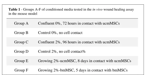
The in vivo wound healing assay was performed according to the guidelines of the ethics committee of the Instituto Gulbenkian e Ciência (IGC). 18 BalbC mice were subjected to two full-thickness wounds with a diameter that ranged from 0,62-0,96cm. The wounds were treated by sub-dermal injection every 24h for 10 days with 50µL of the conditioned media samples described in section 3.2. There were 6 test groups, each consisting of 3 mice shown in table 1.
Table 1 - Groups A-F of conditioned media tested in the in vivo wound healing assay
in the mouse model
Group A Confluent 0%, 72 hours in contact with ucmMSCs Group B Control 0%, no cell contact
Group C Confluent 2%, 96 hours in contact with ucmMSCs Group D Control 2%, no cell contact%
Group E Growing 2%-ucmMSC, 8 days in contact with ucmMSCs Group F Growing 2%-bmMSC, 5 days in contact with bmMSCs
For a time-course analysis of the assay, photographs of the mice were taken on day 0, 3, 7 and 11, followed by macroscopic measurement of the wound boundaries. One wound per mouse was excised on day 7 for histological analysis; biopsies were fixed in formalin and processed for histological evaluation. Hematoxylin-Eosin stained sections were examined by light microscopy. The distance between the borders of the epidermis at the margin of the wound was assessed using the software Image J. The number of Hematopoietic (Sca1+) and Endothelial (Sca1/Flk+) progenitor cells in circulation was also analyzed by means of flow cytometry of peripheral blood on days 0, 3, 7 and 11. This assay was performed by the lab members at the CIPM laboratory under the supervision of Dr. Sérgio Dias.
3.5. In vivo Wound Healing Assay in the Ovine Model
This in vivo wound healing assay was performed according to the recommendations of FELASA, and approved by the ethics committee of ICBAS of the University of Porto. Each sheep was subjected to 14 circular full thickness excision wounds of a diameter of 2cm, which were then treated by topical administration of 200μL of a CMC (carboxymethyl cellulose,
13
Sigma-Aldrich®) gel-type mixture containing either conditioned media from growing ucmMSCs or ucmMSCs themselves. These mixtures were applied every 24 hours for the first 5 days, followed by 4 more applications at 48h intervals. The assay was performed on two sheep, giving a total of 28 wounds. The groups are as shown in table 2.
Table 2 - Groups A to E of the in vivo wound healing assay in the ovine model Group A No treatment
Group B Growing 2%-ucmMSC in 6% CMC Group C Control 2% in 6% CMC
Group D Non-concentrated α-MEM in CMC at 6% Group E Live ucmMSCs in 6% CMC
After application of the CMC mixes, the wounds were covered with compressing pads and bandages. On each day of application, photographs were taken and the dimensions of the wounds were annotated. Once the wounds were completely healed, on day 20, the wound scars were surgically removed. Routine histological treatment with Hematoxylin-Eosin staining was performed. Nine fields per biopsy were thoroughly analyzed at 400x amplification for the presence of granulocytes, lymphocytes, plasma cells, macrophages and giant cells; evidences of necrosis, neo-vascularisation, fibrosis and fatty infiltrate were also registered. A score was attributed to each biopsy as tabulated by the International Organization for Standardization in Part 6 of the ISO-10993. This assay was performed at the Department of Clinical Veterinary, CECA – ICETA, University of Porto, under the supervision of Drª Ana Colette Maurício.
3.6. Statistical Analysis
All data were analyzed using the statistical program GraphPad Prism® 5.03, results are presented with standard errors of the mean (SEM) where possible. Further analysis of statistical significances was performed by means of the Mann-Whitney U test.
14
4. Results
andDiscussion
Recent reports suggest that in wound repair, MSCs act essentially through the secretion of soluble factors, which enhance the regeneration of injured cells, promote angiogenesis, stimulate surrounding cell proliferation and differentiation, reduce inflammation and hamper immune reactions at sites of tissue damage [21, 54]. Conditioned media collected from bone marrow derived MSCs by Chen et. al. was shown, through q-PCR and ELISA, to contain 79 different cytokines and growth factors, 24 of which are up-regulated when compared to conditioned media from dermal fibroblasts, such as EGF, KGF, IGF-1, FGF 4/6/7/9, VEGF-α, PDGF-ββ, EPO and HGF [55-56]. Considering the complex nature of cutaneous wound healing and the extensive list of soluble factors in play, it seems logic to propose that conditioned media may well play a role in promoting a more efficient and rapid cutaneous tissue regeneration. Moreover, trophic factors released by these cells may be more effective when compared to factors secreted by other, less primitive MSCs, such as bmMSCs.
The inclusion of bone marrow derived MSCs in the in vitro and in vivo assays was considered as a reference, given their recognized potential in cutaneous would healing and current status as the golden standard for MSC isolation. The unveiling of bmMSC trophic factor effects in conditioned media could possibly open doors in the future to autologous treatment of wounds for individuals without a private bank of ucmMSCs. In contrast to previous studies using MSCs, conditioned media was obtained not just from MSCs at confluence, but from MSCs in their growth phase. The testing of conditioned media from both of these stages was an effort to understand protein differences at these two distinct phases of the cell cycle. It was thus hypothesized that growing MSCs would secrete a protein profile more favorable to tissue regeneration, considering that in vivo, MSCs at sites of tissue damage are also in an active cell cycle, and not arrested in G0 as are cells at confluence [57].
4.1. Mesenchymal stem cell isolation from umbilical cord matrix
Mesenchymal stem cells were isolated from umbilical cord matrix using the technique described in section 3.1. This technique releases plastic adherent MSCs which, after approximately 9 days in culture, yields an average of 3x10⁵ MSCs per cm of umbilical cord treated. Mesenchymal stem cell phenotype was confirmed by flow cytometry where markers CD105, CD73, CD90 and CD44 showed expression on more than 95% of isolated cells, whereas the markers CD45, CD34, CD31, CD14, CD19 and HLA-DR were expressed on less
15
than 2% of the population (see fig. 1). The
tri-lineage differentiation ability of the isolated cells was also separately confirmed by members of ECBio S.A. (data not shown). The results from the surface marker analysis and the demonstration of tri-lineage differentiation by the mesenchymal stem cells isolated from the umbilical cord matrix, as required by the ISCT [10], confirm that the cells isolated from the umbilical cord matrix are in fact, mesenchymal stem cells.
4.2. Conditioned media preparation
The unraveling of the unique characteristics of mesenchymal stem cells (described in section 2.2) such as immune modulation, homing ability, differentiation capacity, and newly discovered trophic effects have made these cells increasingly popular for cellular and gene therapies. Despite the invasive nature of bone marrow aspirate collection and the low yield of MSCs from this source, which ranges from 0,001% to 0,01% of all mononuclear cells [58], the isolation of MSCs from bone marrow is still considered as the golden standard. This tendency is however changing, owing to the increasing evidence which proves that umbilical cord matrix is in fact a better source of MSCs due to a higher and non-invasive yield of cells [58-59].
Consequently, it was decided that alongside the conditioned media collected from umbilical cord derived MSCs, conditioned media from commercially available bone marrow derived MSCs would also be tested as a comparison. It was proposed that the conditioned media to be obtained from these two sources should ideally contain the least possible amount of FBS proteins for a number of reasons, namely, to reduce the risk of bovine disease transmission and xeno-immune reaction but also to reduce the serum proteins which may mask the effects of proteins secreted by the mesenchymal stem cell in culture. Thus, the MSCs were subjected to growth conditions of diminishing FBS concentrations in order to adapt these cells to a final conditioning period at low serum concentrations (see fig. 2).
Fig. 1 - Percentage of positive ucmMSCs for each of the tested surface markers at
16
Two main groups of conditioned media were collected: from MSCs in their growth phase (see fig. 2b) and from MSCs at confluence (see fig. 2a). The collection of conditioned media from both growing and confluent cells was an effort to understand protein secretion differences at these two distinct phases of the cell cycle.
Throughout the adaptation period (steps 0-2) it was noticed that cells changed morphology, becoming more stretched and with increasingly irregular surfaces with decreasing serum concentrations (see fig. 3) and, as expected, took longer to reach confluence. The conditioning step (step 3) was performed at a final serum concentration of 2% for both groups growing 2% and confluent 2% and at 0% for group confluent 0%. A conditioning step at 0% FBS was attempted for cells in growth, but a reduced cell adhesion was observed with almost no cell duplication, yielding a very low protein concentration (data not shown). For this reason, coupled with the possibility of toxic factor secretion by cells in such conditions of stress, conditioned media from growing MSCs at 0% FBS was no longer considered.
After conditioned media collection, the media was concentrated in protein concentrators with a 5kDa cut-off, followed by protein quantification. Two controls were equally treated: α-MEM supplemented with 2% FBS (Control 2%), as a control for groups growing 2% and confluent 2%; and α-MEM without FBS (Control 0%) as a control for group confluent 0%. Both controls never came into contact with cells. The average protein concentration of each different media is shown in table 3.
As shown in table 3, it is obvious the difference in protein concentration between control 0% and group confluent 0%, where the protein content of the latter is more than twenty-fold of Fig. 2 - Scheme of MSC conditioned media preparation: Steps 0-2: Growth stages where cells were cultured in
media with the decreasing percentages of FBS until confluence. These steps enable a better adaptation of the cells to low serum concentrations. Step 3: Conditioning period of a- Confluent MSCs and b- Growing MSCs. The blue arrow represents a trypsinization step, whereas the red arrow represents a medium replacement of confluent cultures.
a b
17
that of the control 0%. This indicates that during the 72 hours in culture, the confluent umbilical cord derived MSCs secreted a significant amount of protein into the media; although it must not be ignored the possibility that a considerable amount of this protein is likely to be reminiscent FBS proteins that were not washed away before medium replacement with α-MEM without FBS. Regarding the three groups of conditioned media containing 2% FBS (Growing 2% - ucmMSC/bmMSC and confluent 2%), a total protein content similar to that of the control 2% was observed. It is important to remember that the control medium sample never came into contact with cells and therefore still contained all FBS proteins; whereas the conditioned media samples (both growing and confluent) were in contact with cells for several days and therefore subject to the FBS protein consumption by the cells in culture. In fact, the comparison of the protein content of confluent and growing ucmMSCs reveals the lower protein content of the latter. Such results may be due to a combination of the extended period in culture, and therefore higher consumption of FBS proteins, coupled with the lower total number of cells/cm2 in culture and thus lower total number of trophic factor secreting cells. Lastly, upon comparison of the protein concentrations of conditioned media obtained from ucmMSCs and bmMSCs, it is clear the higher protein content of conditioned media from bone marrow derived MSCs. This observation may be a consequence of the larger number of bmMSCs/cm2 when compared to ucmMSCs/cm2, resulting in a higher number of total soluble factor secreting cells.
As previously mentioned (Section 3.2.), an SDS-PAGE of the conditioned media samples and their respective controls was performed, in order to permit qualitative comparisons of the protein profiles between different samples. Equal concentrations of protein were applied in all wells. Upon analysis of the SDS-PAGE gel shown in fig. 13, it can be observed in all conditioned media samples (lanes a-d) a 69,3kDa band corresponding to bovine
serum albumin
(www.uniprot.org/uniref/Uniref100_P02769)
due to FBS contamination. The still significant
Fig. 3 - Photographs of ucmMSCs at the different
stages of conditioned media preparation at an amplification of 100x. a- ucmMSCs at confluence after growth in media supplemented with 20% FBS;
b- ucmMSCs 3 days post inoculation in media
supplemented with 5% FBS; c- ucmMSCs 5 days post inoculation in media supplemented with 2% FBS; d- ucmMSCs 72 hours post medium replacement into media without FBS (producing conditioned media for confluent 0%); e- ucmMSCs 96 hours post medium replacement into media supplemented with 2% FBS (confluent 2%); f- ucmMSCs 8 days post inoculum into media supplemented with 2% FBS (growing 2%-ucmMSC)
18
presence of albumin in the confluent 0% group (lane a of fig. 13) validates the above hypothesis that much of the quantified protein content of this group is, in fact, consequence of the reminiscent FBS proteins. Comparing the protein profiles of lanes a to d with their respective controls, it can be observed the presence of a number of bands in conditioned media which are absent in the latter (see boxes in fig. 13), suggesting that the MSCs in culture do indeed secrete a number of unique proteins into their culture medium. The protein profiles of the confluent (lanes a and b of fig. 13) and growing (lanes c and d of fig. 13) groups do not show as many differences as expected, although this may be due to the low sensitivity of the method and high concentration of albumin which mask the variability among less abundant proteins of each sample.
The conditioned media from groups confluent 0%, confluent 2%, growing 2%-ucmMSC, growing 2%-bmMSC and respective controls were all used in the following in vitro scratch assay (section 4.3.) and in vivo wound healing assay in the murine model (section 4.4.). For the in vivo wound healing assay performed in the ovine model (section 4.5.) only conditioned media from group growing 2%-ucmMSC and control 2% were used.
4.3. In vitro Scratch Assays
A commonly used method for evaluating cellular migration capacity is the in vitro scratch assay. This assay relies on the observation that when a monolayer of confluent cells is interrupted by a newly created gap, cells will generally migrate to fill in the gap creating new cell-cell contacts [24]. Considering the substantial cell migration which is necessary for skin wound healing, an in vitro assay which evaluates such behavior seemed ideal. The adaptability of this assay into 24-well plates allowed for a large screening of samples at different concentrations, which made possible a preliminary analysis of the various conditioned media, previous to their application in in vivo wound healing assays.
This assay was optimized using the B16-F10 mouse melanoma cell line and performed in two distinct skin related cell types: primary dermal fibroblasts and the spontaneously immortalized human keratinocyte HaCaT cell line. The migration inducing capacity of the various conditioned media was quantified via calculation of the area occupied by the cells after a given time in contact with conditioned media. In order to calculate such area, the area of the scratch was measured at time 0h, and again at 15h, 20h and 44h for the B16-F10,
19
primary dermal fibroblasts and HaCaT cell lines, respectively; the difference between these two areas was considered an appropriate measurement of cellular migration.
The cellular migration of B16-F10 mouse melanoma cells in contact with conditioned media did not show any significant differences when compared to respective control (see fig. 14).
4.3.1. Primary Human Dermal Fibroblasts
The use of a primary culture of adult human dermal fibroblasts for this in vitro migrational assay was based on the importance of dermal fibroblasts migration into the wound site, as explained in section 2.4, and a correct dermal fibroblast interaction for successful wound healing and scar formation.
Results show that the conditioned media induced migration of dermal fibroblasts. This is particularly evident in figures 4a and 5b with regards to the conditioned media from group confluent 0%, where the induced migration effect on dermal fibroblasts is superior to that of control 0%, at all concentrations tested. At 500µg/mL of protein, the migration of dermal
Fig. 4 - In vitro scratch assay with primary human dermal fibroblasts. Graphs represent the average area
occupied by the cells after 20 hours in contact with different concentrations of conditioned media from a- Group confluent 0% (hatched black line) and group control 0% (solid black line); b- Group confluent 2% (hatched gray line); group control 0% (solid black line) and group control 2% (solid gray line); c- Group control 0% (solid black line); control 2% (solid black line); group growing 2%-ucmMSC (dotted black line); and group growing 2%-bmMSC (dotted gray line) d- Group control 0% (solid black line); confluent 2% (hatched gray line); group growing 2%-ucmMSC (dotted black line); and group growing 2%-bmMSC (dotted gray line).
d c
b a
20
fibroblasts in contact with confluent 0% was sigificanly higher, by 1,5 times that of the control.
This trend, however, was not evident with conditioned media from confluent 2% when compared with the respective control 2% (see fig. 4b and 5a). Such results may be explained by the fact that the control sample of 2% FBS contains a considerably larger amount of cytokines and growth factors than the conditioned media, given it never came into contact with cells.
The considerably increased amount of growth factors and cytokines in the control 2% sample permits, to a larger extent, not only cell migration, as intended, but also cell replication. This obstacle may be overcome in the future by the use of an alternative control which had been in contact with non-mesenchymal stem cells for the same period of time, as used by Chen et. al. [55], or alternatively, with the treatment of dermal fibroblasts with mitomycin C, arresting the cell cycle and therefore allowing only cellular migration to be observed. Considering that neither of these were possible at the time, comparison of groups containing 2% FBS (confluent 2% and growing 2% ucmMSC/bmMSC) were done with control 2% as well as with control 0%. For example, at a protein concentration of 500µg/mL the cellular migration of confluent 2% is actually less than that of the control 2%, yet when compared to control 0%. In fact the migration sigificantly increased by 1,20 times; as for group growing 2%-ucmMSC cellular migration is increased 1,04 times when compared to control 2%, yet 1,78 times when the comparison is done with control 0%. This trend is true also for group growing 2% - bmMSC (see fig. 4 and 5). Upon comparison
Fig. 5 - In vitro scratch assay. Average area
which was occupied by dermal fibroblasts after 20 hours incubation with different
conditioned media at 500µg/mL. a-
Conditioned media from groups control 0%; control 2%; confluent 2%; growing 2%-ucmMSC and growing 2%-bmMSC. b- Conditioned media from groups control 0% and confluent 0%. *: p = 0,0043; #: p = 0,173
b a