Faculdade de Medicina Veterinária
SPATIOTEMPORAL STUDY OF MORPHO-FUNCTIONAL MODIFICATIONS ON CELL NUCLEUS DURING AFRICAN SWINE FEVER VIRUS INFECTION
Margarida Pires Simões
Orientadores: Prof. Doutor Fernando António da Costa Ferreira Prof. Doutor Carlos Manuel Lopes Vieira Martins
Tese especialmente elaborada para a obtenção do grau de Doutor em Ciências Veterinárias, na especialidade de Ciências Biológicas e Biomédicas
Júri:
Presidente: Prof. Doutor Rui Manuel de Vasconcelos e Horta Caldeira Vogais: Prof. Doutor Luís Manuel Morgado Tavares
Prof. Doutor Carlos Manuel Lopes Vieira Martins Prof. Doutor Celso Vladimir Ferreira de Abreu Cunha Prof. Doutor António José Saraiva da Cunha Cidadão Doutor Robert Michael Evans Parkhouse
Prof. Doutor Fernando António da Costa Ferreira
As you set out for Ithaka hope the voyage is a long one, full of adventure, full of discovery. Laistrygonians and Cyclops,
angry Poseidon—don’t be afraid of them: you’ll never find things like that on your way as long as you keep your thoughts raised high, as long as a rare excitement
stirs your spirit and your body. Laistrygonians and Cyclops,
wild Poseidon—you won’t encounter them unless you bring them along inside your soul, unless your soul sets them up in front of you. Hope the voyage is a long one.
May there be many a summer morning when, with what pleasure, what joy,
you come into harbors seen for the first time; may you stop at Phoenician trading stations to buy fine things,
mother of pearl and coral, amber and ebony, sensual perfume of every kind
as many sensual perfumes as you can; and may you visit many Egyptian cities
to gather stores of knowledge from their scholars. Keep Ithaka always in your mind.
Arriving there is what you are destined for. But do not hurry the journey at all.
Better if it lasts for years,
so you are old by the time you reach the island, wealthy with all you have gained on the way, not expecting Ithaka to make you rich. Ithaka gave you the marvelous journey. Without her you would not have set out. She has nothing left to give you now.
And if you find her poor, Ithaka won’t have fooled you. Wise as you will have become, so full of experience, You will have understood by then what these Ithakas mean.
Constantine .P. Cavafy (1863-1933) Edmund Keeley/Philip Sherrard (translation)
i
ACKNOWLEDGEMENTS
This study would not be accomplished without the help of many people. Foremost, I wish to express my sincerest thanks to all of you.
First, I would like to acknowledge the guidance of my advisors, who have helped to develop this work through many discussions and much critical analysis. I wish to express my deepest appreciation to my supervisor, Professor Fernando Ferreira, for instigating me to develop critical reasoning, thus enabling me to carry on my work. The confidence that was deposited in me helped to trust myself and to grow as a person. My sincere gratitude goes to Professor Carlos Martins, for giving me the opportunity to work in the Infectious Diseases Laboratory, and for the confidence in my work. Your advice and experience were invaluable throughout this work, and I was lucky enough to have your friendship to help me pursue this endeavour.
I want to thank the Infectious Diseases Lab group, past and present, for all the help and support; to Jú and “my” three boys, especially João Coelho whom without his tutorship on molecular skills I could have not survived. I also wish to acknowledge all the researchers and colleagues from the other groups that critically discussed the work included in this dissertation, principally Doctor Alexandre Trindade, Carla Carneiro and Cátia Pereira.
Special thanks go to Doctor Alexandre Leitão, Professor Helder Cortes, Doctor Sofia Nolasco, Doctor Dulce Metelo, Doctor Abdelhak Lemsaddek and Dr. Eduardo Marcelino whose example of organization, work ethics and determination have provided me the motivation to complete this thesis, and chase my projects. Last but not least, Professor Luís Costa (CIISA), and Professor Luís Tavares (FMV) for granting the possibility to do this work.
I wish to acknowledge Professor Paul Nghiem at the Fred Hutchinson Cancer Research Center, University of Washington Medical School for kindly providing ATR inducible expression cells and Doctor Masaoki Kawasumi for his helpful assistance. I am also grateful to Professor Thomas Jenuwein at the Max Planck Institute of Immunobiology and Epigenetics, Freiburg, Germany for kindly providing antibodies recognizing histone H3 methylation status, and Doctor Inês Pinheiro for all the support. I wish to thank Professor Michael Parkhouse and Doctor Sílvia Correia, at the Instituto Gulbenkian de Ciência (IGC), Portugal, for kindly providing the anti-VP32 antibody. To the BioImaging groups from Instituto de Medicina Molecular (IMM) and IGC my sincere acknowledgement for all the assistance, most specially to Doctor José Rino and Doctor Gabriel Martins. I wish to thank Professor Célia Carvalho for her availability and priceless guidance. Also to Doctor Miguel Fevereiro and Doctor Margarida Duarte, Laboratório Nacional de Investigação Veterinária, for their helpful assistance. My sincere thanks also go to my fellow student friends (CIISA-Bolseiros), especially to Maria João Soares, Daniel Murta, Samuel Francisco, Rita Ribeiro, “Cátias” (Marques e Pereira), Sara Madeira and Rita Pedrosa. To all my former school colleagues and co-workers, thanks to all the caring, cooperation and shift swaps!
I would also like to thank my Friends. I cannot think of better companions, always willing, always waiting. I am deeply grateful to my family’s strong support. Thank you for giving me the possibility to open my wings.
Por tudo o que conseguimos trilhar, aos meus pais e avó O maior reconhecimento. Finalmente, mas não de somenos importância “manas”, “irmãos”, “Carcaças” e amigos, Obrigada. Ainda que tenha sido difícil a convivência nestes anos, ainda que eu tenha sido muito ausente… estamos juntos!
ii
FUNDING
The present work was funded by the PhD fellowship (SFRH/BD/65532/2009) and project grant (PTDC/CVT/105630/2008 - ASFTOPO) from the Fundação para a Ciência e Tecnologia; and, by the European Union’s Seventh Framework Programme (FP7/2007-2013) under grant agreement nº 311931, ASFORCE.
iii
Title: Spatiotemporal study of morpho-functional modifications on cell nucleus during African swine fever virus infection
ABSTRACT
Studies on virus-host interactions are decisive to enhance our understanding on how African swine fever virus (ASFV) subverts cellular mechanisms, and also to better characterize host nucleus changes enabling this infection. Immunofluorescence studies and immunoblotting analysis of ASFV-infected cells, allowed us to identify the Ataxia telangiectasia mutated and Rad3-related (ATR) pathway as the specific DNA damage response (DDR) mechanism activated by ASFV infection. Additionally, the use of ATR kinase-dead cells confirmed that ATR has an essential role for the infection success.
The viral intranuclear replication was then pursued using BrdU-pulse experiments, supported on previous reports about ASFV genome presence inside the host nucleus and the proven ATR activation. BrdU-labelled DNA molecules confirmed the active viral replication at early infection times, exclusively within the cell nucleus. Related spatial and morphological nuclear changes during ASFV infection were further addressed, particularly on subnuclear domains and host chromatin epigenetic signatures. Promyelocytic leukaemia nuclear bodies (PML-NBs), nuclear speckles and Cajal bodies displayed major alterations, accompanied by a repressive nuclear environment. PML knockdown revealed an essential proviral activity for ASFV successful infection. Herein, suggestions on how this work may help in the development of therapeutic strategies against ASFV infections can be found.
Keywords: ASFV, virus-host interactions, nucleus, ATR pathway, intranuclear viral genome
v
Título da Tese: Estudo espacio-temporal das alterações morfo-funcionais do núcleo celular durante a infeção pelo vírus da peste suína africana
RESUMO
Estudar as interacções vírus-núcleo da célula hospedeira é fundamental para melhor compreendermos a subversão dos mecanismos nucleares pelo Vírus da peste suína africana (VPSA), e como estas facilitam a infecção. Ensaios de imunofluorescência e expressão proteica (imunodetecção) permitiram a identificação da via ATR (Ataxia telangiectasia mutated and Rad3-related) como mecanismo de reparação de ADN especificamente activado pelo VPSA. Seguidamente, através de células modificadas na expressão de ATR, confirmou-se o papel essencial do ATR para esta infecção.
Atendendo às evidências de presença do vírus no núcleo celular e à activação da via ATR, quisemos desvendar a replicação intranuclear do VPSA pelo meio de ensaios pulso-caça de BrdU. Moléculas de ADN marcadas com BrdU confirmaram a replicação viral, exclusivamente no núcleo, na fase precoce da infecção. Seguiu-se o estudo das alterações espaciais e morfológicas do núcleo induzidas pelo VPSA, particularmente as modificações dos sub-domínios nucleares e das assinaturas epigenéticas da cromatina da célula. Domínios PML (Promyelocytic leukaemia), speckles nucleares e corpos de Cajal mostram-se alterados, associando-mostram-se a um ambiente nuclear repressivo. Estudos funcionais da proteína PML revelaram o seu papel proviral desta infecção. Nesta tese apresentam-se ainda sugestões acerca do potencial destes estudos para o desenvolvimento de estratégias terapêuticas no combate anti-viral.
Palavras-chave: VPSA, interacções vírus-núcleo, núcleo, via ATR, replicação viral
vi
PREFÁCIO
Nesta dissertação serão apresentados os resultados do trabalho de investigação desenvolvido entre 2010 e 2015, no Laboratório de Doenças Infecciosas do Centro de Investigação Interdisciplinar em Sanidade Animal, da Faculdade de Medicina Veterinária, da Universidade de Lisboa, sob orientação do Professor Doutor Fernando Ferreira e co-orientação do Professor Doutor Carlos Martins.
Este trabalho teve como principal objectivo caracterizar as alterações de organização espacial e de função do núcleo celular durante a infecção pelo vírus da peste suína africana (VPSA). Esta análise centrou-se fundamentalmente nas vias de reparação de ADN, na replicação intranuclear do vírus, nos compartimentos sub-nucleares e no estado da cromatina da célula hospedeira, tendo sido possível contribuir para o esclarecimento da dinâmica e interacção vírus-núcleo com os resultados obtidos no decurso destes trabalhos. A presente tese encontra-se dividida em cinco capítulos. No primeiro capítulo é feita uma introdução sobre os mecanismos de reparação de ADN celular, particularmente, as vias que regem a reparação homóloga e a união de extremidades não homólogas; domínios sub-nucleares e o seu papel em infecções virais; tipos de cromatina e assinaturas epigenéticas. Seguidamente são expostos os objectivos do trabalho experimental descritos nesta tese, conduzindo à exposição dos resultados alcançados (capítulos dois a quatro). Por fim, no quinto e último capítulo é apresentada uma discussão integrada de todos os resultados obtidos, apresentando-se as conclusões finais do presente trabalho e perspectivas futuras. Como previsto no Regulamento de Doutoramentos da Universidade de Lisboa, parte integral dos resultados apresentados encontra-se publicada, nos artigos e respectivos capítulos:
II Host DNA damage response facilitates African swine fever virus infection.
M. Simões, C. Martins and F. Ferreira (2013). Veterinary Microbiology 165, 140-147.
III Early intranuclear replication of African swine fever virus genome modifies the landscape of the host cell nucleus. M. Simões, C. Martins and F. Ferreira (2015).
Virus Research 210, 1-7.
IV Alterations of nuclear architecture and epigenetic mechanisms during African swine fever virus infection. M. Simões, J. Rino, I. Pinheiro, C. Martins and F.
vii INDEX Acknowledgements ... i Funding... ii Abstract ... iii Resumo ... v Prefácio ... vi Index ... vii Figure index ... xi
Table index ... xii
Abbreviations ... xiii
CHAPTER I... 1
Introduction ... 3
African swine fever virus ... 3
1. 1.1. Asfivirus - classification and morphology ... 3
1.2. ASFV and hosts/vectors interactions ... 7
1.2.1. Modulation of host defence response ... 8
1.3. African swine fever – nosological entity and history ... 8
1.3.1. Legal trade implications, control strategies, diagnosis and epidemiology ... 9
Cellular mechanisms and DNA Integrity ... 11
2. 2.1. DNA damage response (DDR) activation ... 12
2.2. Homologous Recombination vs. Non-Homologous End Joining ... 13
2.3. Sentinel factors – H2AX, RPA32 and p53 ... 15
2.3.1. Histone H2A.X variant (H2AX) ... 15
2.3.2. Replication protein A (RPA)... 16
2.3.3. Tumour suppressor p53 ... 16
Ataxia Telangiectasia Mutated and Rad3-related (ATR) ... 17
3. 3.1. Inhibitors and pharmacological interference of the ATR pathway ... 18
Viruses and host cell DDR... 19
4. Cell nucleus ... 22
5. 5.1. Nuclear architecture ... 22
5.2. Subnuclear domains... 24
5.2.1. Promyelocytic leukemia nuclear bodies (PML-NBs) ... 24
5.2.2. Nuclear speckles ... 27
5.2.3. Cajal bodies (CBs) ... 28
Viruses and subnuclear domains interactions... 29 6.
viii
6.1. Infection impairment by domains interference ... 30
6.1.1. RNA interference (methodological considerations) ... 30
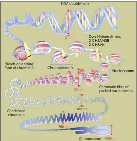
Chromatin state and epigenetic regulation... 32
7. 7.1. Chromatin organization and structure ... 33
7.1.1. Heterochromatin vs. Euchromatin ... 33
7.2. Nucleosome structure and accessibility ... 34
7.3. Histones ... 36
7.4. Histone modifications and biological effects ... 37
7.4.1. Histone H3 methylation status ... 38
Chromatin remodelling ... 39
8. 8.1. Heterochromatin protein 1 (HP1) ... 40
8.2. Nucleosome / chromatin remodelling and Deacetylase complex (NuRD) ... 42
8.3. Histone deacetylase enzymes (HDACs) ... 43
8.3.1. Class I HDAC ... 44
8.3.2. Class II HDAC ... 44
Epigenetic remodelling and Viruses... 45
9. Aims ... 47
10. CHAPTER II ... 49
Host DNA damage response facilitates African swine fever virus infection ... 49
Introduction ... 52
1. Material and Methods ... 53
2. 2.1. Cell Lines and Cultures ... 53
2.2. Virus and Infections ... 53
2.3. Protein Extraction and Western Blotting analysis... 54
2.4. Antibodies for immunoblotting and immunofluorescent analysis ... 54
2.5. Immunofluorescence Microscopy Analysis ... 55
Results ... 55
3. 3.1. ASFV infection activates key DNA damage response markers ... 55
3.2. ASFV specifically elicits the ATR-mediated DNA damage signalling ... 57
3.3. Inhibition of ATR activity disrupts ASFV protein synthesis ... 58
3.4. ATR kinase activity enhances ASFV infection ... 59
Discussion ... 62 4.
ix
CHAPTER III... 65
Early intranuclear replication of African swine fever virus genome modifies the landscape of the host cell nucleus ... 65
Introduction ... 68
1. Material and methods ... 70
2. 2.1. Swine monocyte-derived macrophages and Vero cell cultures ... 70
2.2. Viral isolates and infections ... 70
2.3. Microscopy analysis ... 71
2.3.1. G2/M Vero cell synchronization... 71
2.3.2. Indirect immunofluorescence for viral DNA labelling in swine MDMs and synchronized Vero cells... 71
2.3.3. DDR factors and nuclear domains immunolabelling ... 72
2.3.4. Image acquisition and processing ... 72
2.4. Western blotting analysis... 72
2.5. Antibodies ... 73
Results ... 73
3. 3.1. ASFV replicates its DNA in the host cell nucleus ... 73
3.2. ASFV/L60 isolate specifically activates the ATR pathway... 76
3.3. ASFV/L60 isolate disrupts the subnuclear organization ... 77
Discussion ... 79
4. CHAPTER IV ... 83
Alterations of nuclear architecture and epigenetic signatures during African swine fever virus infection ... 83
Introduction ... 86
1. Materials and Methods ... 87
2. 2.1. Vero cell culture and Lentiviral infection of shRNA ... 87
2.2. Virus and infections ... 88
2.3. Antibodies ... 88
2.4. Immunofluorescence studies ... 89
2.4.1. Microscopy and image processing ... 90
2.4.2. Radial analysis of site images ... 90
2.5. Western blotting analysis... 90
Results ... 91
3. 3.1. ASFV disrupts host subnuclear domains ... 91
3.2. ATR-related factors accumulate nearby PML-NBs during ASFV infection ... 93
x
3.4. ASFV modifies host chromatin epigenetic state ... 97
3.5. Subnuclear domains and ATR loci accumulate nearby heterochromatic regions during ASFV infection ... 100
Discussion ... 103
4. CHAPTER V ... 107
General discussion and conclusions ... 109
Future perspectives ... 117
xi
FIGURE INDEX
Figure 1 - Asfivirus virion ... 4
Figure 2 - ASFV DNA of infected swine macrophage detected by pulsed-field electrophoresis. ... 5
Figure 3 – Recent proposed model for virus entry and egress ... 7
Figure 4 – Overview model of the cellular DNA damage and consequences over cellular processes. 12 Figure 5 - Schematic illustration showing DNA lesion sensing, further stimulation of the signalling cascade. ...13
Figure 6 – Schematic representation of specific ATR inhibition resulting in the death of cancer cells. 18 Figure 7 – Schematic representation of DNA damage response activation upon virus replication and specific viral proteins interaction. ...20
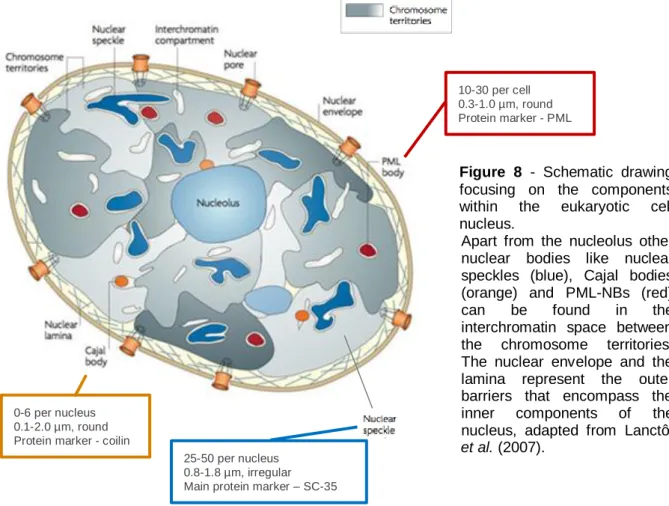
Figure 8 - Schematic drawing focusing on the components within the eukaryotic cell nucleus. ...23
Figure 9 – PML-NBs revised functions, from cell cycle regulation to antiviral activity. ...26
Figure 10 – Nucleosome, Chromatosome and Chromatin organization. ...35
Figure 11 – Histone modifications, chromatin structure and gene regulation modulation...38
Figure 12 - HP1 and the interacting partners responsible for chromatin status regulation. ...42
Figure 13 - ASFV activates DDR key sensors. ...56
Figure 14 - ɣH2AX and pRPA32 relocalize in response to ASFV infection. ...56
Figure 15 – Steady state levels of ATM and DNA-PKcs related factors. ...57
Figure 16 - ASFV infection elicits ATR pathway activation...58
Figure 17 - ASFV alters nuclear localization of ATR kinase phosphorylated form. ...58
Figure 18 - ATR inhibition by caffeine and wortmannin disrupts ASFV protein synthesis. ...59
Figure 19 - ASFV protein synthesis is modulated in ATR kinase-dead cells. ...60
Figure 20 - ASFV-p72 variable expression in ATR-inducible cells. ...61
Figure 21 - ATR-inducible cells displayed differential cytopathic effect. ...61
Figure 22 - ASFV progeny is reduced in ATR-kd cells. ...61
Figure 23 – ASFV genomes replicate in the host cell nucleus during the early phase of infection. ...75
Figure 24 – ASFV infection promotes activation of the ATR pathway in swine MDMs. ...76
Figure 25 – ASFV induced pRPA32 and pATR redistributions in swine MDMs. ...77
Figure 26 - ASFV-infected MDMs display subnuclear domains disruption. ...78
Figure 27 - ASFV induces the reorganization of subnuclear domains. ...92
Figure 28 - Subnuclear domains major constituents’ expression levels during ASFV infection. ...93
Figure 29 – PML-NBs and DDR factors juxtapose during ASFV infection. ...94
Figure 30 - PML has a proviral role in ASFV infection. ...95
Figure 31 – PML knockdown cells display aberrant ASFV factories. ...96
Figure 32 - ASFV progeny is reduced in Vero-shRNA-PML kd cells. ...97
Figure 33 - ASFV modifies the host chromatin state...99
Figure 34 – ASFV leads to juxtaposition of host heterochromatic regions, disrupted subnuclear domains and pATR foci. ...102
Figure 35 – Radial intensity profile analysis. ...102
xii
Figure 37 – Schematic representation of cellular compartments used by viruses for DNA replication, with an hypothetic ASFV DNA intranuclear replication scenario. ... 112 Figure 38 – ASFV DNA synthesised inside the nucleus of swine MDMs diffuses into viral cytoplasmic
factories. ... 113
TABLE INDEX
Table 1 - DDR mechanisms activated/inhibited by different viruses ... 21 Table 2 - Different classes of modifications identified on cellular histones... 38
xiii
ABBREVIATIONS
Ab Antibody
ASF African swine fever
ASFV African swine fever virus
ATM Ataxia telangiectasia mutated
ATR Ataxia telangiectasia mutated and Rad3 related
ATR kd ATR kinase dead
ATR wt ATR wild type
BrdU 3’-bromo-deoxyuridine
DAPI 4',6-diamidino-2-phenylindole
DDR DNA damage response
DNA /ADN (Pt) Desoxiribonucleic acid/ Ácido desoxirribonucleico (Pt) DNA-PKcs DNA-dependent protein kinase catalytic subunits
dNTP Deoxynucleotide
DSBs Double strand (DNA) breaks
HDACs Histone deacetylases
HP1 Heterochromatin Protein 1
HR Homologous Recombination
H2AX Histone H2A, member X
H3K9me (3) Histone H3 (tri)methylated at lysine 9 H3K27me3 Histone H3 trimethylated at lysine 27
IIF Indirect immunofluorescence
kbp kilo base-pair
kDa kilo Dalton
MDa Mega Dalton
MDMs Monocyte-derived macrophages
NFkB Nuclear Factor kappa-light-chain-enhancer of activated B cells
NHEJ Non-Homologous End-Joining
OIE Office International des Épizooties – World Animal Organization
PML Promyelocytic leukaemia protein
PML k/d PML knockdown
PML-NBs Promyelocytic leukaemia nuclear bodies
PI3K Phosphainositide-3-Kinase
p53 Tumour suppressor p53
SDS-PAGE SDS-polyacrylamide gel electrophoresis
ssDNA Single strand DNA
TNF Tumour necrosis factor
VP Viral protein (ASFV)
53BP1 Tumour suppressor p53-binding protein 1
1
CHAPTER I
3
INTRODUCTION
THESIS OUTLINE AND OVERVIEW
When infecting a host cell, viruses face challenges that must be overcome in order to successfully replicate. For example, viruses must avoid or subvert antiviral responses either by degrading or mislocalizing cellular factors that have been activated by its intrusion.
This study primarily concerns an investigation over African swine fever virus (ASFV) interactions with the host-cell nucleus, with particular emphasis on the DNA damage response (DDR) modulation, the viral DNA intranuclear replication, the subnuclear domains disruption and the nuclear architecture rearrangements. For this reason, the following sections in this Chapter will provide a detailed overview of the related topics mentioned above. Initially, the basic features of ASFV are revised, followed by its pathological outcome in swine organism. Attention then moves to the various host facets and mechanisms as well as the various stimuli described to possibly trigger the host nucleus remodelling and its biological importance, as: DNA damage response activation, subnuclear domains disruption and nuclear environment rearrangements derived from viral replication and infection.
The major aims of the present thesis are stated at the end of Chapter I, while research outcomes of this work that have been published, or are under review for publication, in international peer-reviewed journals represent Chapters II-IV. Major conclusions and future directions of the work will be entailed in Chapter V.
LITERATURE REVIEW
African swine fever virus
1.
1.1. Asfivirus - classification and morphology
African swine fever virus (ASFV) is the sole member of Asfarviridae family (Dixon et al., 2004) and most interestingly, the only arthropod-borne DNA virus known to this date.
ASFV genome, a linear double-stranded DNA molecule (between 170-190 kbp) has a guanine/cytosine content of approximately 39%, comprising several genes involved in nucleotide metabolism, transcription, replication, repair, immune evasion, and modulation of host cell apoptosis (Dixon, Chapman, Netherton & Upton, 2012). This viral genome also presents a genomic arrange closed by 37 nucleotide-long hairpin loop structures composed, almost entirely, of incompletely paired A and T residues, terminal cross-links and inverted terminal repeats (Gonzalez, Talavera, Almendral & Viñuela, 1986; Vega, González, Blasco, Calvo & Viñuela, 1994).
4
The Asfivirus is classified as a large enveloped, complex icosahedral virus since the virions have a complex multi-layered structure composed by a 30 nm nucleoid - that consists in the nucleoprotein structure, composed by the viral genome and different enzymes required for replication ultimately surrounded by an 80 nm core shell, a first lipid layer (inner envelope) and a 170-190 nm icosahedral capsid – Figure 1. The outline capsid is composed by capsomers that measure 13 nm in diameter and there are 1892-2172 of these per capsid, as reviewed by Salas & Andrés (2013).
Figure 1 - Asfivirus virion1
Recently, ASFV has been classified as belonging to the Nucleo-Cytoplasmic Large DNA Virus superfamily (NCLDV), a group composed, as the name indicates, of large DNA viruses whose replicative cycle may start in the nucleus and is further performed in the host cell cytoplasm (King, Lefkowitz, Adams & Carstens, 2011). This superfamily is constituted by Mimivirus and Mamavirus (Mimiviridae family), Marseillevirus and Lausannevirus (Marseilleviridae family), some members of the Ascoviridae family, Vaccinia virus (Poxviridae family), Iridoviruses, Phycodnaviruses and Asfivirus. The phylogenetic analysis on several NCLDV members revealed a common ancestral virus, presumably encoding in its genome conserved proteins, related to key-life processes, like DNA polymerases, DNA helicases and ATPase pumps for DNA packaging, Topoisomerase II, RNA polymerase, and that may explain these viruses relative autonomy towards host cells (Iyer, Balaji, Koonin & Aravind, 2006; Yutin & Koonin, 2009, 2012; Colson, de Lamballerie, Fournous & Raoult, 2012).
Even though ASFV genome fragments have been found in nuclear extracts, only its cytoplasmic DNA replication at a later phase of infection has been explored (Garcia-Beato, Salas, Viñuela & Salas, 1992; Rojo, García-Beato, Viñuela, Salas & Salas, 1999). Of utmost importance, enucleated cells have proved to impair ASFV-infection, strongly indicating that cellular nuclear factors are most probably indispensable in the early stages of viral DNA synthesis (Ortin & Vińuela, 1977; Tabares & Sánchez Botija, 1979; Tabarés, 1987). This fact is also related to ASFV full length genomes that are synthesized in the cytoplasmic factories, possibly from replicative intermediate forms head-to-head or tail-to-tail genomic concatemer
1
5
structures initially formed within the nucleus – Figure 2 (Gonzalez et al., 1986; Caeiro, Meireles, Ribeiro & Costa, 1990; Brookes, Dixon & Parkhouse, 1996; Rojo et al., 1999), and latter packaged into viral particles as described for Poxviruses structural assembly (e.g. Vaccinia virus) (Traktman, 1996).
Figure 2 - ASFV DNA of infected swine macrophage detected by pulsed-field electrophoresis. Adapted from Rojo et al. (1999).
From all the particularities that make ASFV a unique virus, its encoded DNA Pol X (Oliveros et al., 1997; García-Escudero, García-Díaz, Salas, Blanco & Salas, 2003; Lamarche, Kumar, & Tsai, 2006; Sampoli Benítez, Arora, Balistreri & Schlick, 2008) and apurinic/apyrimidinic (AP) endonuclease may better explain its genotype variety and its efficient replication success (Redrejo-Rodríguez, Ishchenko, Saparbaev, Salas & Salas, 2009).
After internalization, the virus instantly initiates gene expression, using enzymes and factors packaged within the virion core, as reviewed by Dixon et al. (2012). Viral gene transcription does not require the host RNA polymerase activity since the virion contains a RNA polymerase and specific virus-encoded transcription factors (Baxter, Wilkinson, Turner & Dixon, 1996). ASFV genes present a strongly regulated, time-dependent expression, divided in four classes: early, immediate-early, intermediate and late expressing (Netherton & Wileman, 2013). It has been shown that even though early genes expression can continue throughout infection, immediate-early expressing genes are silenced before the onset of virus DNA replication (Almazán et al., 1992); while genes classified as intermediate expressing are detected from 4 to 6 hours postinfection (hpi), concurring with the maximum expression of early genes (Rodríguez & Salas, 2013). This programmed gene expression is similar to the cascade model described for a few Poxviruses, with some factors required for transcription are synthesised during the previous temporal stage (Traktman, 1996; Moss, 2013). Yet, and curiously, the host translation machinery is still modified during ASFV infection (Castelló et al., 2009; Sánchez, Quintas, Nogal, Castelló & Revilla, 2013).
The genome length variation between different ASFV isolates is most probably due to DNA sequences deletion/addition in regions located in the genome ends, and may explain the virulence and pathogenicity of these viral isolates (Dixon, Bristow, Wilkinson & Sumption, 1990; Tulman & Rock, 2001; Chapman, Tcherepanov, Upton & Dixon, 2008; Michaud,
Putative concatemeric forms of ASFV genome
6
Randriamparany & Albina, 2013). Until now, twenty-two different genotypes have been identified based on sequencing of the C-terminal end of VP72 (Bastos et al., 2003; Lubisi, Bastos, Dwarka & Vosloo, 2005; Michaud et al., 2013), which combined to other viral protein genomic sequencing are thought to be useful for molecular epidemiology studies and viral subgroups determination (Lubisi et al., 2005; Nix, Gallardo, Hutchings, Blanco & Dixon, 2006; Gallardo et al., 2009; Chapman et al., 2011; Portugal et al., 2015).
The complete genome sequences of several ASFV isolates have been determined and are available at GenBank2, with preliminary annotations on the website www.virology.ca. These genomes are predicted to encode between 151 and 167 open reading frames (ORFs) which are read from both DNA strands across the length of the genome (Yáñez et al., 1995). The number of these ORFs has also been shown to depend on the virus isolate with only 109 ORFs being conserved among the different viral isolates, and encoding for proteins either involved in virus replication, virus assembly or host immune response interference (Chapman et al., 2008; de Villiers et al., 2010).
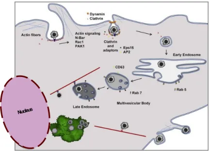
During the late phase of infection, the cellular microtubule network is required for the cytoplasmic viral factories maintenance that is organised in vimentin cages. These perinuclear viral factories resemble aggresomes, which are formed in response to misfolded proteins or even as antiviral defence (Wileman, 2007). Additionally, and following mature virus particles assembly, cellular microtubules also serve as transport interface from the viral factories to the cell surface, conveying the full infectious viral particles egress in a budding form (Jouvenet, Monaghan, Way & Wileman, 2004; Netherton et al., 2006). This budding process through the cellular plasma membrane capacitates virions to acquire an external membrane (Carrascosa et al., 1984; Valdeira, Bernardes, Cruz & Geraldes, 1998; Cuesta-Geijo et al., 2012; Sánchez et al., 2012; Alonso et al., 2013). Lastly, the ASFV coating strongly supports the notion of host defence evasion by its infection, especially given the fact that ASFV infects swine monocyte-macrophage lineage cells (Rouiller, Brookes, Hyatt, Windsor & Wileman, 1998), and also because of the viral proteins that can be found in the cellular membrane (Camacho & Viñuela, 1991). Moreover, recent work showed that endoplasmic reticulum and an intact host cholesterol biosynthesis are required for an effective virus morphogenesis and early cytoskeleton modulation (Quetglas et al., 2012). As previously mentioned, ASFV primarily infects cells belonging to the monocyte-macrophage lineage, after endosomal internalization (Figure 3), thus promoting the easy access to host factors (Hernaez & Alonso, 2010; Alonso et al., 2013), and subversion of factors that allow the viral infection (Gómez del Moral et al., 1999), particularly through a tight regulation of cellular apoptosis (Revilla et al., 1997; Nogal et al., 2001; Granja et al., 2004; Hernáez et al., 2004; Hurtado et al., 2004).
2
7
Figure 3 – Recent proposed model for virus entry and egress Adapted from Alonso et al. (2013).
Still, in later phases of infection in host animals, ASFV is also recognized to replicate in other cell types, like endothelial cells, though efficient replication in mononuclear-phagocytic cells appears the most crucial factor for pathogenesis in domestic swine (Basta, Gerber, Schaub, Summerfield & McCullough, 2010; Blome, Gabriel & Beer, 2013; Galindo-Cardiel et al., 2013; Gómez-Villamandos, Bautista, Sánchez-Cordón & Carrasco, 2013). The mediated cytokine expressions of the immune response are subverted, varying with the specific viral isolate (Martins & Leitão, 1994; Gil et al., 2003; Portugal, Leitão & Martins, 2009), along with the inhibition of Toll-like receptors by ASFV genes expression (de Oliveira et al., 2011; Correia, Ventura & Parkhouse, 2013).
1.2. ASFV and hosts/vectors interactions
ASFV is transmitted by direct contact among pigs, pig meat and other contaminated materials (e.g. fomites spread by people and transportation), entering the body via the tonsils or dorsal pharyngeal mucosa, heading to the mandibular or retropharyngeal lymph nodes, from where the virus spreads through viraemia (Sánchez-Vizcaíno et al., 2009).
The virus can also be transmitted by soft ticks, Ornithodorus moubata complex – prevalent in Africa and Ornithodorus erraticus (present in the Iberian Peninsula) where it can persist for long periods of time (Basto et al., 2006; Sánchez-Vizcaíno et al., 2009; Boinas, Wilson, Hutchings, Martins & Dixon, 2011). These ticks play an important role in the transmission of the disease by feeding on wild suids, therefore acting as vectors in the sylvatic cycle (Jori & Bastos, 2009; Sánchez-Vizcaíno et al., 2009; Burrage, 2013). In addition, stable flies (Stomoxys spp.) have also been identified as mechanical vectors, maintaining ASFV for at
8
least 48 hours (Mellor, Kitching & Wilkinson, 1987), reflecting the possibility of other insects/blood-sucking ectoparasites to play a role in ASFV transmission.
Foremost importantly, both the African bush pig (Potamochoerus porcus) and the warthog (Phacochoerus aethiopicus) are considered to be natural vertebrate reservoirs of ASFV (Costard, Mur, Lubroth, Sanchez-Vizcaino & Pfeiffer, 2013) since the viraemia persists in these hosts, even asymptomatically, for months or years, reflecting the long-term host-pathogen co-evolution, corroborated by the existence of many ASFV genes that evolved to escape the full host immune response presenting its infection as non-pathogenic (Sánchez-Vizcaíno et al., 2009).
In contrast, the acute disease in domestic pigs is characterized by massive apoptosis of lymphocytes and an haemorrhagic pathology with extensive vascular damage, probably due to molecules released from the infected macrophages, although infected endothelial cells may contribute to the pathogenesis (Oura, Powell & Parkhouse, 1998; Blome et al., 2013).The extent of lymphocyte apoptosis correlates with the level of ASFV replication and with the virulence of the isolate (Portugal et al., 2009; Galindo-Cardiel et al., 2013).
Alarmingly, recent studies have also demonstrated the presence of viral particles in the air of infected swine pens, and that the virus can survive outside the host and exposed to environmental conditions for a few days (Ferreira, 2013).
1.2.1. Modulation of host defence response
Large DNA viruses encode many proteins involved in the evasion of host immune responses (Iyer et al., 2006). As ASFV replicates in monocyte-derived macrophages (MDMs) it interferes with both the initial innate and later acquired immune responses by modulating macrophage immunoregulatory proteins and, lastly, the macrophage functions (Martins, Scholl, Mebus, Fisch & Lawman, 1987). Indeed, one of the major strategies used by ASFV, is the manipulation of different cellular signalling pathways that leads to the regulation of cytokines transcription (Dixon et al., 2004; Correia et al., 2013), or to the inhibition of apoptosis (Revilla et al., 1997; Nogal et al., 2001). One of the first evasion molecules described in ASFV was the A238L protein (Powell, Dixon, & Parkhouse, 1996), which displays dual functions by inhibiting both NFкB and TNFα activities (Gil et al., 2003; Le Negrate, 2012; Sánchez et al., 2012).
1.3. African swine fever – nosological entity and history
African swine fever (ASF) is mostly considered an acute, highly contagious and majorly fatal disease of domestic swine (Sus scrofa). First described by Montgomery, as an ignored pathological identity of swine in Kenya (Montgomery, 1921), the disease is responsible for a variety of clinical signs ranging from the widespread haemorrhages with the involvement of
9
the lymph-reticuloendothelial cells (typical acute form ranging 100% mortal cases within 6 to 13 days postinfection), to the loss of physical condition and respiratory disorders in sub-acute forms. Surviving animals chronically present emaciation, stunted growth and necrotic lesions and are virus carriers for periods of at least 6 months, presenting elevated morbidity rates (Sánchez-Vizcaíno et al., 2009). Utmost importantly, infection caused by highly virulent isolates is characterized by few or non-detectable antibodies, resulting in the absence of a neutralizing immune response, which has ultimately stalled the development of a conventional vaccine.
Since it was first reported, ASF remained confined to Africa until it was introduced in Portugal in 1957, causing a hyper acute form of disease with a 100% mortality rate. Despite the prompt eradication, a new outbreak succeeded in 1960, spreading into Spain. In this case, the epidemic range supported the endemicity status to the Iberian Peninsula until mid-1990s. Several ASF outbreaks were also reported in other European countries until the 1990s, and although all the involved countries managed to eradicate the disease, the Italian island of Sardinia maintains its status of endemicity since 1982 (Mannelli et al., 1997; Mur, Martínez-López & Sánchez-Vizcaíno, 2012; Costard et al., 2013). Although other parts of the globe have been previously affected, the 2007 outbreak in Georgia (Rowlands et al., 2008) has gained threatening proportions, reminding the precarious free-disease state reality (reviewed in Sánchez-Vizcaíno, Mur, Gomez-Villamandos & Carrasco, 2015). The later ASFV isolate to be introduced has been found to be related to Eastern Africa isolates, and has spread to other countries of the Transcaucasian region and the Russian Federation (Gogin, Gerasimov, Malogolovkin & Kolbasov, 2013; Oganesyan et al., 2013; Malogolovkin et al., 2015; Sánchez-Vizcaíno et al., 2015). In 2015, several outbreaks were declared in Estonia, Latvia, Lithuania and Poland (Sánchez-Vizcaíno et al., 2015), and are believed to be disseminated through the “sylvatic cycle” by European wild boars, as previously characterized (Costard et al., 2013).
ASF infection outcome depends on the virus isolate virulence, host susceptibility, presence of tick vectors, and the probable interaction amongst host suids and vectors. Therefore, three main cycles or epidemiological scenarios can be distinguished in general: the “sylvatic cycle”, the “intermediate enzootic cycle”, and the “domestic cycle” (Sánchez-Vizcaíno et al., 2009).
1.3.1. Legal trade implications, control strategies, diagnosis and epidemiology
African swine fever (ASF) can be accounted for extensive financial hazards to the pig industry with severe sanitary and socio-economic consequences, since the absence of a vaccine or an effective treatment makes it an expensive disease to eradicate (Sánchez-Vizcaíno et al., 2009). Its control is based on rapid laboratory diagnosis and the enforcement of strict hygio-sanitary measures with preventive culling of all infected and susceptible animals, movement restrictions and notification (FAO, 2000; Wieland, Dhollander, Salman &
10
Koenen, 2011; OIE, 2012; EFSA, 2014). These facts allied to an increasing global travelling and the international economic scenario compelled OIE - World Animal Health Organization, to list ASF as an obligatory notifiable disease (Vizcaíno et al., 2009; Sánchez-Vizcaíno, Mur & Martínez-López, 2013; Costard et al., 2013).
To further complicate this hazardous transmissible disease, its field diagnosis still reflects difficulties due to the pathogenesis similarity to other swine diseases and also for the varied array of signs caused by different isolates. All this aggravated by the necessity to transport infectious samples to certified labs posing additional threat for the disease spreading if proper care is not taken (Costard et al., 2009; Ferreira, 2013). Earlier, it would take weeks to check the outbreak disease status since most of the official reference laboratories are mostly distant and very difficult to access, especially in sub-Saharan countries, thus hampering the critical sanitary decisions to restrain the outbreak spread (FAO, 2000; Sánchez-Vizcaíno et al., 2009; de León, Bustos & Carrascosa, 2013). Fortunately, local Animal Health Services/Veterinary authorities have grown aware and have diagnostic tools that provide an accurate risk assessment and institute effective control measures (Mur et al., 2013), as other rapid diagnostic techniques are continuing to be developed (Oura, Edwards & Batten, 2013). Nowadays, virus genotyping greatly contributes to identify the origin, monitor sources and risks and, better manage the outbreaks spread, adding to an enhanced surveillance capacity of pig health in buffer areas (e.g. swine farming, airport surroundings). Additionally, the molecular classification is currently being used for epidemiological studies and prediction models, assessing spreading possibilities as: serological survey of adjacent zones, wild suids and tick involvement, sentinel animals to be used for ASFV detection, and control confirmation in outbreak areas (Costard et al., 2009, 2013; de Villiers et al., 2010; Mur, Martínez-López & Sánchez-Vizcaíno, 2012; Mur et al., 2013; Sánchez-Vizcaíno & Arias, 2012; Sánchez-Vizcaíno, Mur & Martínez-López, 2012; Sánchez-Vizcaíno et al., 2013; Oganesyan et al., 2013), and further contributing for the virus classification (Michaud et al., 2013).
The devastating impact of ASF on both, epidemic and endemic areas is well known, accounting for major economic losses, mainly due of costs for eradication and surveillance, but also because the latter effects on pork production and trade restrictions. These total costs are most apparent in countries with industrialized pig production; although the impact inflicted on poorer pig producers (especially in developing countries) can often be the reason why animal production cannot re-start, denying a better subsistence and lifestyles (Costard et al., 2009).
Importing activities of either pig products or live suids are considered as the major source for disease introduction, especially the illegal imports and associated transit (Mur et al., 2012; Costard et al., 2013). Considering that European free borders and easy transportation are critical in the introduction and spreading of this disease to all the community countries, for all
11
this should be properly appraised by the previously developed risk models, not forgetting the fact that competent tick vectors do not exist in all regions, epidemiological investigations should also assess the abundance of soft ticks (Mur et al., 2012; Costard et al., 2013).
Recent projects entail the development of deletion mutants where essential viral genes are to be deleted hence providing base for Defective Infectious Single-Cycle (DISC) viral particles and its possible use as vaccines. Under the threat posed by the outbreaks in Europe’s outskirts, research has been intensified, namely under the 7th Framework Programme of the EU.
Cellular mechanisms and DNA Integrity
2.
Since the early arguments on cell theory ‘Omnis cellula et cellula’ ("All cells [are] from cells") defended by Rudolf Virshow in 1958, notions about the genetic information integrity and maintenance have been addressed (Hakem, 2008; Schultz, 2008). At the same time, cellular stress has been defined as a variety of processes that are triggered by an acute or chronic shift from the usual cellular conditions and homeostasis to counteract the insult and repair the damage and, to eventually protect the cell or organism (Fulda, Gorman, Hori & Samali, 2010). This definition indicates that there are many conditions that alter the cellular environment and activate a plethora of responses required for cell adaptation and recovery. It is known that mutations can interfere with the cell cycle and may cause severe disorders (e.g. metaplasia), whereas cellular surveillance mechanism are responsible for maintaining DNA integrity and repair or, ultimately, promoting an apoptotic fate of the injured cell (Ciccia & Elledge, 2010).
A great number of factors can cause DNA damage in cells, as ionizing radiation, UV-light, oxidative stress (e.g. reactive oxygen species) and alkylating substances, thus leading to different forms of genomic injuries (Stokes & Comb, 2011). As previously described, UV-light can induce pyrimidine dimers formation, whereas genotoxic substances as Doxorubicin can lead to histone eviction and inhibit DNA biosynthesis; and, ionizing radiation can lead to the formation of single and double-strand breaks (Sancar, Lindsey-Boltz, Unsal-Kaçmaz & Linn, 2004; Jackson & Bartek, 2009). But not only external factors are responsible for DNA injuries, since cell-cycle replication stress can itself originate strand-breaks, base-pair mismatches and other DNA abnormalities (Andreassen, Ho & D’Andrea, 2006; Gasser & Raulet, 2006; Peng, Yamamoto, Goldberg & Maller, 2010).
In order to detect and circumvent these various DNA damages, eukaryotic cells have developed numerous strategies that enable the recovery of genomic integrity or, in those cases beyond repair, the proceeding to programmed cell death (d’Adda Di Fagagna, 2008; Jackson & Bartek, 2009; Freeman & Monteiro, 2010). These are referred as DNA damage response (DDR) mechanisms.
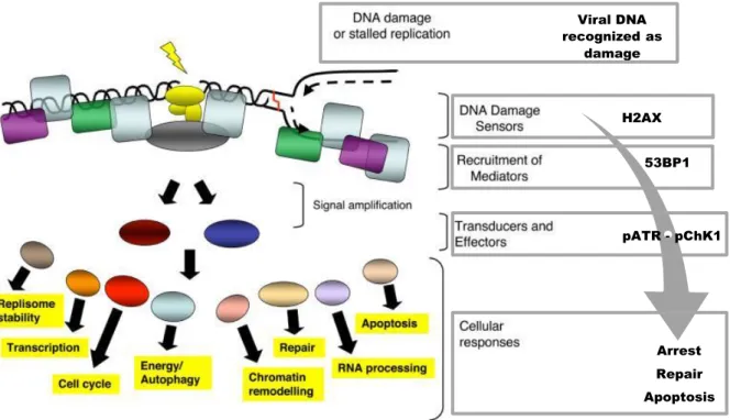
12 Viral DNA recognized as damage H2AX 53BP1 pATR - pChK1 Arrest Repair Apoptosis
2.1. DNA damage response (DDR) activation
Cellular DDR mechanisms protect and preserve the integrity of the genome, comprising pathways which are activated in response to different forms of DNA damage and involve a number of proteins that participate in both DNA repair and cell cycle checkpoint, commanding progression or apoptosis (Hoeijmakers, 2001; Andreassen et al., 2006; Ciccia & Elledge, 2010). As for example, repair of double-strand DNA breaks (DSBs) starts with the activation of intricated cellular DDR downstream events, which includes the sensing of the DNA damage (DNA-damage binding proteins), subsequently amplifying and transmitting a damage signal to transducers (proximal/distal protein kinases), in order to generate a myriad of cellular responses (Figure 4). DDR pathways are ubiquitous, in some ways interlinked by sharing sensors, mediators or other related-factors, and the majority of the involved proteins being highly conserved from yeast to humans (Finn, Lowndes & Grenon, 2011).
Figure 4 – Overview model of the cellular DNA damage and consequences over cellular processes. A possible viral-induced DDR activation represented at the right. Adapted from Jackson & Bartek (2009).
Single-strand breaks (SSBs) are differently tackled from DSBs, because cells are set to respond differently to these events during cell-cycle stages. For instance, in replicating cells, sensed DSBs trigger a slower cycle progression through “cell-cycle checkpoints”, most probably ensuring the DNA repair before DNA polymerase proceeds (Finn et al., 2011; Polo & Jackson, 2011). For this reason, the most dangerous DNA damages for the cell are DSBs. Due to the linear nature of eukaryotic chromosomes unrepaired DSBs that can lead to partial aneuploidy (by losing big parts of chromosomes), cell death often occurs, while misrejoined
13
DSBs can lead to potentially carcinogenic chromosome rearrangements (Wyman & Kanaar, 2006; Hanahan & Weinberg, 2011).
Such sensing-repair mechanisms are highly selective and present an immediate onset, specifically targeting DNA damages. If the DNA break cannot be repaired or the extent of the damage is too great, DDR signals promote cellular senescence or apoptosis. The downstream responses of the DDR cannot occur without the activation of damage sensors, which enhances the importance of these proteins for a robust DDR (Harper & Elledge, 2007; Freeman & Monteiro, 2010).
2.2. Homologous Recombination vs. Non-Homologous End Joining
In general most of the DNA damages can be repaired very efficiently, and about the molecular level of DSBs repair much is already known (reviewed in Polo & Jackson, 2011). DSBs repair is mostly carried out by two main pathways: Homologous Recombination and Non-Homologous End Joining mechanisms – Figure 5. The relative contribution of these mechanisms is cell-cycle regulated (reviewed in Branzei & Foiani, 2008; Ciccia & Elledge, 2010), and preferentially used upon different DNA injuries, appearing to be a critical factor in order to maintain genomic integrity (reviewed in Freeman & Monteiro, 2010; Sirbu & Cortez, 2013).
Figure 5 - Schematic illustration showing DNA lesion sensing, further stimulation of the signalling cascade.
Hence activating transducers and effectors, the later are responsible to trigger cell cycle checkpoints. This cascade of events insures time for DNA repair, either by coordinating cellular activities to repair DNA by Homologous recombination (HR) or Non-homologous end-joining (NHEJ), adapted from Mullenders, Atkinson, Paretzke, Sabatier, & Bouffler (2009).
Homologous Recombination (HR) is an error proof mechanism that uses a sister chromatid as a repairing template to achieve very faithful restoring events. Due to this unique feature, it is the predominant process of DSBs repair during the S and G2 phases of the cell cycle (Bartek, Lukas, & Lukas, 2004; Stiff, Cerosaletti, Concannon, O’Driscoll, & Jeggo, 2008; Willis & Rhind, 2009). This mechanism requires the initial enrolment of Mre11-Rad50-Nbs1 proteins (MRN) complex and is carried out by the Rad family members of DNA repair
14
proteins (Carson et al., 2003). Also importantly, HR-dependent DSBs repair require the processing of the present dsDNA ends, creating extended 3’ ssDNA tails (further coated with Replication Protein A - RPA) and trimming the 5’ ends of the DSBs (Andreassen et al., 2006; Flynn & Zou, 2011; Liu et al., 2012).
The serine-threonine kinases Ataxia telangiectasia mutated (ATM), Ataxia telangiectasia mutated and Rad3-related (ATR), and DNA protein kinase catalytic subunit (DNA-PKcs) are differentially activated in response to distinct types of damage (reviewed in Yang, Yu, Hamrick & P.J., 2003; Falck, Coates & Jackson, 2005; Tomimatsu, Mukherjee & Burma, 2009; Liu et al., 2012). These proteins are members of the phosphatidylinositide 3-kinase (PI3Ks) family, as reviewed by Abraham (2001) and Yang et al. (2004), and relay the message of the damaged DNA by phosphorylating downstream effectors at serine or threonine residues that are followed by a glutamine - SQ/TQ motifs (reviewed in Freeman & Monteiro, 2010). These PI3Ks enzymes share downstream phosphorylation targets, eliciting some redundancy and compensation during DNA damage signalling. Recently, some groups have reported large-scale screens to identify previously unknown ATM and ATR targets upon DNA damage (Smolka, Albuquerque, Chen & Zhou, 2007; Chen, Albuquerque, Liang, Suhandynata & Zhou, 2010), since both pathways are the major HR repair mechanisms. Individually, ATM and ATR pathways lead to cell cycle arrest mediated through the phosphorylation of the Chk2 and Chk1 kinases, respectively (Reinhardt & Yaffe, 2009), which mark the damage sites for further recruitment of proteins, cooperating with the phosphorylation signal of histone ɣH2AX variant (Dickey et al., 2009; Fragkos, Jurvansuu & Beard, 2009; Yuan, Adamski & Chen, 2010). Interestingly, ATM and ATR pathways can also activate each other, and functionally overlap, resulting in an extended response of dual kinase activation allowing more time for repair, as reviewed by Maréchal & Zou (2013). As for example, after replication stress, the active ATR kinase activity can lead to ATM activation by inducing ATM phosphorylation (Ser 1981), which is also the site of ATM auto-phosphorylation (Dodson, Shi, & Tibbetts, 2004; So, Davis, & Chen, 2009).
By contrast, the Non-homologous end-joining (NHEJ) is considered an error-prone DNA repair process, since it performs the gap filling without homologous regions whilst overlapping single stranded DNA are removed at the extremity of the breaks and the resulting blunt DNA ends are just fused together. DNA-PK is a protein complex which interacts with Ku70 and Ku80 protein subunits, and that recognizes DNA breaks, later on recruiting and binding the catalytic subunit DNA-PKcs, forming the DNA-PK holoenzyme complex, which in turn recruits DNA ligase finalizing the NHEJ repair (reviewed in d’Adda Di Fagagna, 2008; Ciccia & Elledge, 2010; Lieber, 2010; Wang & Lees-Miller, 2013). NHEJ is the major pathway for repairing non-replication-associated breaks and occurs predominantly in G1 phase of the cell cycle (Sancar et al., 2004; FitzGerald et al., 2011). Critically, at sites
15
of multiple DSBs this can lead to misrejoined DSBs and thus to chromosome translocations (Jackson & Bartek, 2009).
2.3. Sentinel factors – H2AX, RPA32 and p53
2.3.1. Histone H2A.X variant (H2AX)
Phosphorylation of the cellular H2AX is one of the earliest events of the activated DNA damage cascade and is crucial for the efficient recruitment of DNA damage signalling and repair proteins to the sites of damage (Dickey et al., 2009; Fragkos et al., 2009; Solier, Sordet, Kohn & Pommier, 2009). The main role of H2AX phosphorylation is to mediate successful DNA repair, as reviewed by Yuan et al. (2010).
Upon recognition of DNA damage by the cell, H2AX is phosphorylated on its extended C-terminal tail over a distance of one to two megabases surrounding the DNA break by the phosphatidylinositol 3-kinase-related kinases ATM, ATR, or DNA-PK (Tomimatsu et al., 2009; Ward, Minn, Jorda & Chen, 2003; J Yang et al., 2003; Jun Yang et al., 2004). In damaged cells, phosphorylated H2AX can be visualized within the cell nucleus as distinct foci, termed ɣH2AX foci which appear prior to the generation of foci containing damage repair molecules such as the MRN and RPA complexes (Olson, Nievera, Lee, Chen, & Wu, 2007; Freeman & Monteiro, 2010; Liu et al., 2012).
The histone H2AX has been conserved throughout evolution as a separate species from the rest of the H2A histones (Macadangdang et al., 2014). In contrast to the majority of cellular histones, H2AX synthesis can be upregulated in response to DNA damage and sensitize cells to undergo apoptosis (Liu, Parry, Chin, Duensing & Duensing, 2008; Gagou, Zuazua-Villar & Meuth, 2010).
Incubation of cells with drugs that inhibit DNA replication (e.g. Hydroxyurea), generates multiple stalled replication forks, while H2AX synthesis is not inhibited instead leading to an accumulation of soluble H2AX within the cell, causing chromatin aggregation which leads to the increase of cellular apoptosis (Campos & Reinberg, 2009; Solier et al., 2009; Kobayashi, Kato, Ota, Ohba & Komatsu, 2010). In contrast, apoptotic mechanisms are not increased upon over-expression of H2AX in cells (Liu et al., 2008), since many studies have demonstrated that ɣH2AX acts as a scaffolding protein to which a number of DNA repair factors can dock to facilitate repair of the damaged DNA (Ward et al., 2003; Solier et al., 2009; Boichuk, Hu, Makielski, Pandolfi & Gjoerup, 2011; Polo & Jackson, 2011). Although initially thought to be associated only with double-stranded DNA breaks, ɣH2AX foci have been shown to be enriched for proteins involved in both Homologous recombination pathways and also Non-homologous end joining, such as NBS1, RPA32 and p53 (reviewed in d’Adda Di Fagagna, 2008; Soutoglou, 2008; Jackson & Bartek, 2009; Freeman & Monteiro, 2010; Polo & Jackson, 2011). Overall, it appears that histone H2AX may have
16
multiple functions in maintaining genomic stability involving both DNA damage responses and/or the induction of cell death (Fragkos et al., 2009; Yuan et al., 2010).
2.3.2. Replication protein A (RPA)
RPA is a key component of cellular DNA metabolism comprising DNA replication, recombination, and repair as well as DDR signalling. It is closely involved in replication during S phase, in HR-based repair mechanisms during both S and G2 phases and in checkpoint signalling (Borgstahl et al., 2014). It is composed of an heterotrimeric complex consisting in 70 kDa, 32 kDa, and 14 kDa subunits, being the single-stranded DNA (ssDNA) binding protein required for multiple aspects of DNA metabolism. Actually, RPA is involved in initiating and elongating DNA replication, and in the binding of ssDNA structures that arise as a result of DNA repair, including the nucleotide excision, the base excision, and the double-stranded breaks (DSBs) repair mechanisms, as reviewed by Zou, Liu, Wu, & Shell (2006). RPA-bound ssDNA has been shown to be necessary for ATR activation and for the localization of various DNA damage sites (Zou & Elledge, 2003; Olson et al., 2007; Flynn & Zou, 2011; Liu et al., 2012).
The latest studies reveal that RPA exhaustion leads to augmented fork breakage in replication sites, while the excess of RPA molecules delays fork breakages, even without functional loads of ATR. These evidences could explain that when all RPA becomes sequestered, every active replicon generates unprotected ssDNA, which is rapidly converted to DSBs, ultimately resulting in ATR incapacity to restore all firing points (Toledo et al., 2013).
As a component of the conserved repair mechanisms, RPA also interacts with the promyelocytic leukaemia (PML) protein, providing a functional connection between the homologous recombination-mediated repair machinery and PML subnuclear domains, which usually colocalize with the RPA foci (Boichuk et al., 2011).
2.3.3. Tumour suppressor p53
The tumour suppressor p53 is a central protein in mammalian cell stress response. Due to its ability to integrate many different signals controlling cell life and death, p53 has been named the “guardian of the genome” and plays a crucial role in maintaining genomic stability in somatic cells (reviewed in Levine & Oren, 2009; Vousden & Prives, 2009). Under normal physiological conditions p53 remains inactive and unstable, but is rapidly stabilized and activated by upstream kinases in response to genotoxic and oncogenic stresses, leading to cell cycle arrest, apoptosis or senescence (Chao et al., 2000; Aparicio & Eaves, 2009; Zhao & Xu, 2010). These functions of p53 can protect the genome from accumulating genetic mutations, by allowing the time for faithful repair of DNA damage or by eliminating cells with excessive DNA damage (Maclaine & Hupp, 2009). This protein functions mainly as a
DNA-17
binding, sequence-specific transcription factor that activates the expression of multiple genes which in turn mediate p53 functions in cell cycle regulation (reviewed in Bannister & Kouzarides, 2005; Jackson & Bartek, 2009; Kondo, 2009; Pal et al., 2010; Hanahan & Weinberg, 2011; Palomera-Sanchez & Zurita, 2011).
Upon DNA damage, p53 is also known to be recruited to subnuclear domains, as reviewed by Hofmann & Will (2003), more specifically to promyelocytic leukaemia nuclear bodies (PML-NBs) where it is post-translationally modified and activated to perform pro-apoptotic functions (Guo et al., 2000; De Stanchina et al., 2004).
Ataxia Telangiectasia Mutated and Rad3-related (ATR) 3.
The ATR pathway can be activated in response to a wide range of exogenous and endogenous stresses, the latter characterized as possible base or nucleotide excision, double-strand DNA breaks (DSBs), or single-strand DNA breaks, although the major functions appear to be responding to DNA damage and replication stress, particularly stabilizing stalled replication forks, either promoting their restart in order to prevent the generation of DSBs or activating checkpoints that hamper the entry into mitosis in the presence of unreplicated DNA (Smits, Warmerdam, Martin, & Freire, 2010; Nam & Cortez, 2011; Toledo et al., 2013; Mazouzi, Velimezi, & Loizou, 2014). DNA damage caused by stalled replication forks is characterized by long stretches of ssDNA and ssDNA/dsDNA junctions occurring due replication stress, and have emerged as potent ATR activators as reviewed by Zou (2007); Cimprich & Cortez (2008) and Jones & Petermann (2012).
It is established that ATR activation majorly occurs with the recognition of RPA-coated ssDNA molecules (reviewed in Andreassen et al., 2006; Flynn & Zou, 2011), also requiring the association with the ATR-interacting protein (ATRIP), that is bound to ssDNA via interaction
to RPA (Xu & Leffak, 2010; L. Zou & Elledge, 2003) and p53-binding protein (53BP1) (Martínez, Flores & Blasco, 2012). ATR pathway is also activated by the phosphorylation of its homologous enzyme (ATR) and also by the phosphorylation of its substrates, like checkpoint kinase 1 (Chk1) and the 32 kDa RPA subunit (RPA32) (Liu et al., 2000; Casper, Nghiem, Arlt & Glover, 2002; Durkin, Arlt, Howlett & Glover, 2006; Rodriguez & Meuth, 2006; Mordes & Cortez, 2008; Rozenzhak et al., 2010; Smits et al., 2010).
Mutations in ATR are rare, as complete loss of its function is embryonic lethal as both ATR and ChK1 are essential for mammalian cells development (Brown & Baltimore, 2000; Nghiem, Park, Kim, Vaziri & Schreiber, 2001; Nghiem, Park, Kim Ys, Desai & Schreiber, 2002). In their absence, stalled forks persist while additional replication origins are fired, leading to dNTP pools exhaustion and a consecutive accumulation of non-progressive forks. Whenever ssDNA amount exceeds RPA availability, replication forks collapse thus inducing the generation of DNA DSBs (Toledo et al., 2013). However, ATR functions have been
18
studied in ATR kinase-dead human cell lines (Nghiem et al., 2001), derived from patients with Seckel syndrome, an hypomorphic mutation, originating a splicing defect in ATR mRNA transduced into lower protein levels (reviewed in O’Driscoll, Gennery, Seidel, Concannon & Jeggo, 2004), and also cells with conditional disruption of ATR (Brown & Baltimore, 2003). These and other systems reveal the importance of ATR within normal DNA replication, and in G1/S and G2/M checkpoints. Without ATR expression cells accumulate damage during S phase that likely contribute to lethal genomic instability (Shechter, Costanzo & Gautier, 2004). Intriguingly, recent reports suggest that ATM and ATR activate each other in mammalian cells, and do not simply exist as parallel pathways (Shengqin Liu et al., 2012; Shizhou Liu et al., 2011; Smits et al., 2010; L. Zou & Elledge, 2003; Y. Zou et al., 2006). For example, ATR activation occurs in S or G2 phase cells in response to DSBs, and requires ATM and DNA processing (Jazayeri et al., 2006). Another study suggests that ATR mediates ATM activation in response to UV and Hydroxyurea insults, as reviewed by Stiff et al. (2006) and Stokes & Comb (2011).
3.1. Inhibitors and pharmacological interference of the ATR pathway
Accumulating evidences suggest that targeting ATR can selectively sensitize mutated/cancer cells but not normal cells to DNA damage. Furthermore, in hypoxic conditions, ATR blockage results in overloading replication stress and DNA damage response causing cell death (d’Adda Di Fagagna, 2008). Despite the attractiveness of ATR inhibition in the treatment of cancer, specific ATR inhibitors have remained elusive. In the last two years however, selective ATR inhibitors suitable for in vitro and, most recently, in vivo studies have been identified, as reviewed by Fokas et al. (2014), and summarized in Figure 6.
Figure 6 – Schematic representation of specific ATR inhibition resulting in the death of cancer cells. The use of caffeine and wortmannin, at specific concentrations can inhibit ATR-dependent mechanisms. Adapted from Fokas et al (2014).
Specific concentrations of ATR inhibitors Specific concentrations of ATR inhibitors