online | memorias.ioc.fiocruz.br
Flaviviruses are arthropod-borne viruses that may cause severe acute infectious diseases, such as dengue fever and yellow fever (YF). Dengue fever is the most im-portant arthropod-borne emerging viral disease in tropi-cal countries due to high morbidity and increased risk of mortality (Gubler 2002). The YF17DD vaccine induces long-lasting immunity, similar to wild viruses (Monath 2001, Halstead 2007). For dengue fever, a chimeric YF/ dengue vaccine (ChimeriVaxTM technology) is currently
being tested in clinical trials (Guy et al. 2010). Both den-gue and YF wild viruses can induce a broad spectrum of clinical manifestations from asymptomatic to severe clinical features, the latter of which is characterised by haemorrhaic manifestations and shock syndrome, which are associated with vascular permeability and leakage. Cytokines play a key role in the generation of these physiopathological processes (Geisbert & Jahrling 2004). Moreover, the occurrence of rare fatal cases and adverse clinical manifestations has been associated with the YF vaccine (Vasconcelos et al. 2001, Silva et al. 2010).
Several cytokines have been associated with disease severity in patients. For dengue fever, tumor necrosis factor (TNF)-α (Hober et al. 1993, Braga et al. 2001), interleukin (IL)-10 (Green et al. 1999, Azeredo et al. 2001), IL-6 (Nguyen et al. 2004), macrophage inhibitory factor (Chen et al. 2006, Assuncao-Miranda et al. 2010)
and interferon (IFN)-γ (Bozza et al. 2008), among oth -ers (Srikiatkhachorn & Green 2010), have been reported. Chemokines also have an important role in pathogenesis. For example, IP-10/CXCL-10 was found to be elevated in dengue haemorrhagic fever (DHF) (Fink et al. 2007) and
MCP-1/CCL-2 (Lee et al. 2006), while MIP-1β/CCL-4 is associated with a good prognosis (Bozza et al. 2008). El
-evated serum levels of IL-6, IL-8, TNF-α, MCP-1, IL-1
receptor agonist (Ra) and IL-10 were observed in fatal YF cases when compared with non-fatal cases (Bae et
al. 2008). Finally, in vaccinated individuals, high levels of IP-10, IL-1β, TNF-α, IFN-γ and IL-10 were detected
(Querec et al. 2009, Silva et al. 2011).
Monocytes, macrophages and dendritic cells (DCs) are the main targets for viruses involved in vascular permeability induction (Schnittler & Feldmann 2003, Clyde et al. 2006). Mononuclear phagocytes become ac-tivated by virus, which induces synthesis and release of
cytokines (e.g., TNF-α, IL-6, IL-1β, IFN-α/β and IL-10) and chemokines (e.g., IL-8, MIP-1α and MCP-1). These
factors produced by in vitroinfected cellsare known to induce alterations in the endothelium, which leads to the imbalance of fluid between the intra and
extra-vascu-Financial support: IOC/Bio-Manguinhos/FIOCRUZ, DECICT/CNPq, FAPERJ, ICGEB
+Corresponding author: [email protected] Received 7 February 2011
Accepted 8 June 2011
Dengue-2 and yellow fever 17DD viruses infect human dendritic cells,
resulting in an induction of activation markers, cytokines and
chemokines and secretion of different TNF-
α
and IFN-
α
profiles
Mariana Gandini1, Sonia Regina Nogueira Ignacio Reis1, Amanda Torrentes-Carvalho1, Elzinandes Leal Azeredo1, Marcos da Silva Freire2, Ricardo Galler2, Claire Fernandes Kubelka1/+
1Laboratório de Imunologia Viral, Instituto Oswaldo Cruz
2Departamento de Desenvolvimento Tecnológico, Instituto de Tecnologia em Imunobiológicos-Fiocruz,
Av. Brasil 4365, 21045-900 Rio de Janeiro, RJ, Brasil
Flaviviruses cause severe acute febrile and haemorrhagic infections, including dengue and yellow fever and the pathogenesis of these infections is caused by an exacerbated immune response. Dendritic cells (DCs) are targets for dengue virus (DENV) and yellow fever virus (YF) replication and are the first cell population to interact with these viruses during a natural infection, which leads to an induction of protective immunity in humans. We studied the infectivity of DENV2 (strain 16681), a YF vaccine (YF17DD) and a chimeric YF17D/DENV2 vaccine in monocyte-derived DCs in vitro with regard to cell maturation, activation and cytokine production. Higher viral antigen posi-tive cell frequencies were observed for DENV2 when compared with both vaccine viruses. Flavivirus-infected cul-tures exhibited dendritic cell activation and maturation molecules. CD38 expression on DCs was enhanced for both DENV2 and YF17DD, whereas OX40L expression was decreased as compared to mock-stimulated cells, suggesting that a T helper 1 profile is favoured. Tumor necrosis factor (TNF)-α production in cell cultures was significantly higher in DENV2-infected cultures than in cultures infected with YF17DD or YF17D/DENV. In contrast, the vaccines induced higher IFN-α levels than DENV2. The differential cytokine production indicates that DENV2 results in TNF induction, which discriminates it from vaccine viruses that preferentially stimulate interferon expression. These dif-ferential response profiles may influence the pathogenic infection outcome.
lar areas of tissues and ultimately culminates in shock (Srikiatkhachorn 2009). Coagulation features that oc-cur during dengue fever may be linked to mononuclear phagocyte and endothelial cell activation because pro-inflammatory cytokines interfere with the activation of coagulation factors (Suharti et al. 2002).
After virus infection, DCs become activated and ma-ture. During maturation processes, DCs usually
upregu-late co-stimulatory molecules, such as CD80, CD86, CD83, CD40, CD38 and OX40L (Quah & O’Neill 2005).
These molecules may be involved in the polarisation of T helper (Th) cells into either Th-1 or Th-2 subsets.
CD38, IFN-γ and IL-12p70 are associated with Th-1 re -sponses (Frasca et al. 2006). On the other hand, OX40L expression on DCs contributes to Th-2 polarisation (De-lespesse et al. 1999). Mature DCs are able to activate T lymphocytes and drive the type of the immune response (Lanzavecchia & Sallusto 2004). It is believed that T cell activation in patients with severe dengue leads to T cell responses with less avidity for serotype-specific responses than mild dengue, resulting in a pathologic cytokine storm with limited antiviral responses (Du-angchinda et al. 2010).
The aim of this investigation was to study the in-nate immune response induced in vitro by distinct fla-viviruses. We selected a pathogenic strain of dengue
vi-rus (DENV) 2 (16681) originated from a DHF SE Asian
case and the Brazilian YF17DD, which is known for its protective properties with minimal deleterious effects. A
chimeric YF17D/DENV2 vaccine virus (Caufour et al.
2001) was assayed as well. We report for the first time that flaviviruses induce the expression of the DC
matura-tion marker CD38 and downregulate OX40L, suggesting
that a Th-1 response has been generated. Additionally,
differential TNF-α and IFN-α expression by DCs in
-fected with DENV and vaccine YF17DD or 17D/DENV2
viruses are discussed, with regard to their possible influ-ence on pathogenicity and virus clearance.
MATERIALS AND METhoDS
Virus strain, cell cultures, virus stock preparation and titration - DENV2 (strain Thailand/16681/1984) was
provided by Dr SB Halstead (Naval Medical Research Centre, USA). YF strain 17DD is the live attenuated vi-rus used in the YF vaccine manufactured by Bio-Man-guinhos, Oswaldo Cruz Foundation, Rio de Janeiro (RJ), Brazil (Post et al. 1991). For virus stock preparation, Vero
cells (CCL 81, ATCC) were maintained in 199 medium with Earle’s salts buffered with sodium bicarbonate and supplemented with 5% foetal bovine serum (FCS) and
antibiotics (Gibco, Invitrogen). Viral stock was pre-pared by flavivirus infection of a Vero cell monolayer
in T175 flasks. When cytopathic effects were observed
seven days later, the supernatant was harvested and
sup-plemented with 30% FCS and aliquots were frozen at
-70ºC. Virus stock was titrated by serial dilution cultures in microtitre plates and detected by immunofluorescence as previously described (Miagostovich et al. 1993). An uninfected flask was also maintained and the supernatant was collected to be used as a mock inoculum. A mock infection was included in each experiment.
A set of experiments was performed using the
chi-meric virus YF/DENV2 strain 44/3. This virus contains the pre-M and E genes from DENV2 strain New Guinea C, with the carboxyl end of protein E (E261-E495 amino
acids) that belongs to the Brazilian strain 44/2 (Caufour et al. 2001). This chimeric virus elicits a protective
im-mune response against DENV challenge both in mice
and Rhesus monkeys, which includes neutralising
anti-bodies (Caufour et al. 2001, Galler et al. 2005).
Virus titre was calculated as 50% tissue culture in -fectious dose (TCID50/mL). However, when the virus was compared with the chimeric virus 44/3 (Fig. 1), all
three viruses were titrated by the plaque-forming assay as described before (Galler et al. 2005).
Human monocyte-derived DCs (MoDCs) - Human peripheral blood was obtained from University Hospi-tal Clementino Fraga Filho, Federal University of Rio de Janeiro (UFRJ), RJ. Peripheral blood mononuclear cells
(PBMCs) were isolated from healthy donors’ buffy coats
by centrifugation on a density gradient (400 g for 30 min
in Ficoll-Paque Plus, Amersham Biosciences Corp), as
described previously (Reis et al. 2007). Cells were re-suspended at 106 cells/mL in RPMI 1640 supplemented
with 200 mM glutamine, 100 U/mL penicillin and 10 mg/mL streptomycin (Gibco, Invitrogen) and incubated
at 37ºC under a humid atmosphere with 5% CO2. The
cells where then allowed to adhere to uncoated
polysty-rene flasks (150 cm3) for 90 min to enrich for monocytes.
Non-adherent cells were gently removed by washing and the adherent cells were detached by mechanical cell har-vesting with cell scrapers in cold cell culture medium. Cell viability was verified in the cultures by Trypan blue
exclusion and was ≥ 95%. Enriched monocytes were
suspended in RPMI 1640 medium supplemented with
10% FCS, 500 U/mL human rGM-CSF and 500 U/mL
rIL-4 (Peprotech) and seeded at 1 x 10⁶ cells/mL on
24-well plates (Nunc, Thermo Fisher Scientific). Cytokines were added every other day for six days. The appropriate phenotype of DC differentiation was confirmed before each experiment by flow cytometry.
Infection of human MoDCs by flaviviruses - After a six-day cell differentiation period, MoDCs were
incu-bated with YF17DD, a dose equivalent to that used for DENV2 (2.5 x 10⁵ TCID
50/mL), a dose five times more
concentrated or cell culture supernatant as mock
treat-ment diluted inoculum (500 μL). This compensation
for the infectivity of different flavivirus has also been previously reported (Youn et al. 2010). For experiments
comparing the YF17DD, DENV2 and YF17D/DENV2
viruses, the multiplicity of infection (MOI) was 4 (4x10⁶ plaque-forming unit/106 cells).
After three-hour incubation for adsorption at 37ºC
Detection of viral antigen and specific co-stimulatory markers by flow cytometry in human MoDCs - Cells were harvested by vigorous pipetting using cold wash buffer
[phosphate-buffered saline (PBS) pH 7.4, 1% bovine se-bovine se-rum albumin (BSA) (Sigma) and 0.1% sodium azide] and were resuspended at 1 x 10⁶ cells/microtube. The cells
were then centrifuged (260 g, 7 min) and washed once more with wash buffer. Single or double cell labelling of infected and uninfected cultures was performed. For
surface labelling, the cells were blocked with 5%
heat-inactivated human plasma in wash buffer for 30 min at 4ºC and further incubated for 30 min with anti-human
CD1a-PE (Iotest Immunotech, Beckman Coulter Inc,
Brea, CA, USA), DC-specific intercellular adhesion molecule-3-grabbing non-integrin (DC-SIGN)-FITC
(R&D Systems), CD14-PE (DakoCytomation), HLA-DR,DP,DQ-FITC (Dako Cytomation), CD80-FITC (Iotest Immunotech), CD38-FITC (Caltag, Invitrogen) or OX40L-PE (BD Biosciences Pharmingen). Matching iso -type antibodies were used as negative controls. The cells
were washed and subsequently fixed with 2% parafor -maldehyde. Intracellular staining was performed accord-ing to previously described methods (Neves-Souza et al.
2005), with slight modifications. Briefly, the cells were fixed with cold 2% paraformaldehyde in PBS for 20 min
and, after centrifugation, the membranes were
permeabi-lised with 0.15% saponin in wash buffer. The cells were blocked with 5% heat-inactivated human plasma in wash
buffer for 30 min at 4ºC. Primary monoclonal antibody incubation was performed with anti-Dengue Complex (Chemicon, Millipore, Billerica, MA, USA), anti-Yellow Fever (Biogenesis, AbD Serotec) or an isotype-matched
antibody diluted in wash buffer containing 0.15% sapon -in for 60 m-in at 4ºC. The cells were washed once with wash buffer and further incubated with secondary
anti-mouse labelled with Alexa Fluor-488 (Molecular Probes, Invitrogen) for 30 min at 4ºC. The cells were acquired
(10,000 events for DC-SIGN positive gate) on a FACS-Calibur flow cytometer (BD Biosciences). Analysis was performed using FlowJo (TreeStar Inc, Ashland, OR, USA) or Summit (Dako Automation).
Enzyme-linked immunosorbent assay (ELISA) for TNF-α - ELISA plates (MaxiSorp, Nunc) were coated overnight at room temperature (RT) with 1 μg/mL at 100 μL/well of rabbit anti-human TNF-α antibody pro
-vided in the Human TNF-α ELISA Development Kit (Peprotech). The plates were washed with 0.05% Tween 20 (Sigma) in PBS and were incubated with 1% BSA in PBS at 250 μL/well for 90 min at RT. The samples and standards were added in duplicate at 100 μL/well and the plates were incubated overnight at 4ºC. Subsequent in
-cubation with 0.25 μg/mL at 100 μL/well of biotinylated detection antibody for TNF-α was performed for 120
min at RT. This procedure was followed by a 30-min incubation with streptavidin-peroxidase (Streptavidin,
Zymed, Invitrogen) at 1:2,000 dilution with 100 μL/well.
Tetramethylbenzidine (KPLInc) was added and the
plates were read at 630 nm after 15-min incubation. Cytokine detection in cell culture supernatant by mul-tiplex microbead immunoassay - A multiplex biometric immunoassay, which contains fluorescent microspheres conjugated with a monoclonal antibody specific for a tar-get protein, was employed for cytokine and chemokine
measurement according to the manufacturer’s instruc -tions (Upstate, Millipore) as described previously (Reis et al. 2007). Measured cytokines including the
follow-ing: IFN-α, IL-6, IL-1Ra, IL-10, MCP-1/CCL-2, MIP-1β/ CCL-4, IP-10/CXCL-10 and RANTES/CCL-5. Briefly, cell culture supernatants (50 μL) were incubated over -night at 4ºC with pooled antibody-coupled beads in bot-tom filter plates (Millipore). The complexes were washed and incubated with pooled biotinylated detection antibod-ies for 90 min at RT. Then, streptavidin-phycoerythrin was added for 30 min at RT prior to assessing cytokine
concentration titres. A broad range (1.95-8,000 pg/mL) of
concentrated human recombinant cytokines, provided by the vendor, was used to establish standard curves to max-imise assay sensitivity and dynamic range. Cytokine lev-els were determined with a multiplex array reader from Luminex Instrumentation System (Bio-Plex Workstation from Bio-Rad Laboratories). The analyte concentration
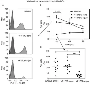
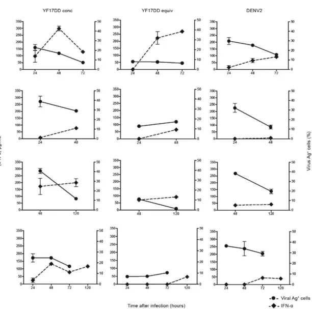
Fig. 1: monocyte-derived dendritic cells (MoDCs) infection with
dengue virus 2 (DENV2) infection and yellow fever (YF) 17DD. MoDCs were infected with DENV2 or YF17DD at equivalent mul
-tiplicity of infections (MOIs) [2.5 x 10⁵ 50% tissue culture infectious
dose (TCID)/mL] or YF17DD at a higher MOI (1.25 x 10⁶ TCID
50/
mL) or mock-infected. Cells were labelled with DENV or
anti-YF monoclonal antibodies using triplicates for each peripheral blood mononuclear cells (PBMC) donor. A: flow cytometry patterns for viral antigens on gated MoDCs 24 h after in vitro infection.
Histo-grams demonstrate cell distribution by Alexa Fluor-488 fluorescence
intensity (FL1-H: level of fluorescence). Cells were cultured with
mock (dark grey) or DENV2 or YF17DD (light grey) at concentrated (conc) or equivalent (equiv) doses; B: viral antigen detection kinetics
on infected MoDCs (data from four PBMC donors) [hpi: hours
post-infection; X axis: time after infection (hours); Y axis: percentage of
viral antigen positive cells (Ag+)]; C: intracellular viral antigen detec
-tion 24 h after infec-tion representing 11 cell donors (horizontal lines:
distribution mean; X axis: viral inoculum; Y axis: percentage of viral
Ag+ cells. Asterisks mean: p< 0.0001 in one-way analysis of variance
was calculated by the software provided by the manu-facturer (Bio-Plex Manager Software), which provided
a regression analysis to derive the equation for cytokine
concentration prediction in cell culture samples.
Ethics - Procedures performed in this work were
ap-proved by the Ethical Committee of the Fiocruz, Bra -zilian Health Ministry (recognised by the Bra-zilian
National Ethics Committee) (111/00 and CAAE-0064
.0.011.000-07).
Statistical analyses - Data were first tested for nor-mality with Prism version 4.0 for Windows (GraphPad Software). Flow cytometry data exhibited normal distri-butions, while normality was not detected for cytokine and chemokine production. To determine whether there were significant differences in viral antigen expression, data values were subjected to one-wayanalysis of vari-ance followed by Tukey’s multiple comparison test. Data from cytokine and chemokine assessments were submit-ted to a Wilcoxon signed rank test.
RESuLTS
DENV2 and YF17DD infection kinetics in human MoDCs - Considering that DCs have been described as
targets for DENV and YF vaccines (Barba-Spaeth et al. 2005, Neves-Souza et al. 2005) we investigated these
host-virus interactions in detail. MoDCs that originated from healthy human PBMC donors exhibited
charac-teristic downregulation of CD14 (1.8 ± 0.9%) and up
-regulation of DC-SIGN (75 ± 5%) and CD1a (54 ± 6%). These cells expressed low levels of CD80 and less
HLA-DR,P,Q than MoDCs that were stimulated with bacterial
lipopolysaccharides and human IFN-γ (data not shown).
The MoDCs exhibit characteristics of immature DCs, which have the ability to perform endocytosis and cap-ture antigens and are more susceptible to virus infection (Steinman & Nussenzweig 2002).
MoDC cultures were incubated with YF17DD
vac-cine virus at a dose either equivalent to that used for DENV2 (2.5 x 10⁵ TCID
50/mL) or with a dose five times
more concentrated. Fig. 2A shows histograms obtained by flow cytometry analysis after intracellular viral an-tigen labelling, representing data from cells incubated with the three different inocula at 24 h after infection. Fig. 1B shows percentages of viral antigen-positive cell (Ag+) rates from 24-72 h post-infection (hpi). Both
vi-ruses were able to infect MoDCs, but higher inoculum
doses of YF17DD were required to generate similar frequencies of infected cells compared with DENV2.
The peak infection rate was detected at 24 hpi, although
significant percentages were still detected at 48-72 hpi.
Fig. 2C shows viral antigen detection in MoDCs origi-nated from 11 different PBMC donors, confirming that
DENV2 is significantly more infectious than YF17DD
when the same MOI is used (p < 0.0001). Cells cultured with mock or heat-inactivated inocula contained less
than 0.5% positive labelled cells PBMC donors in all
assays (data not shown).
Except when mentioned, the assays described be -low were performed using YF17DD at the higher MOI, which induced similar rates of cell infection when
com-pared to DENV2.
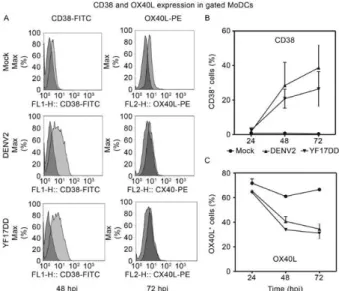
CD38 and OX40L activation/maturation markers are regulated during MoDCs infection with DENV2 and YF17DD - DCs interact with pathogens or exogenous molecules and undergo activation and maturation pro-cesses, resulting in reduced antigen processing capacity and increased expression of co-stimulatory and adhesion molecules. These molecules may induce tissue migration, antigen-specific immunological responses and Th
polari-sation (Quah & O’Neill 2005, Wallet et al. 2005). CD38
is involved in chemotaxis and calcium mobilisation and binds to CD31 on endothelial cells, facilitating DC migra-tion through endothelium (Frasca et al. 2006). It may par-ticipate in the Th-1 response, which involves lymphocytes
producing IL-12 and IFN-α. On the other hand, OX40L
expression on DCs (Ohshima et al. 1997) may contribute to the Th-2 polarisation by enhancing IL-4 and IL-13
in-duction and suppressing IFN-α after it binds to OX40 on T cells (Ohshima et al. 1998, Delespesse et al. 1999).
MoDCs were cultured with DENV2, YF17DD or mock supernatants. Fig. 3A shows that CD38 was weakly
expressed on infected cells but increased when fluores-cence intensity virus was present. DC activation occurs
between 48-72 hpi (Fig. 3B). MoDCs express OX40L ear
-ly in infection, but this molecule was downregulated at 48
and 72 hpi (Fig. 3C). This shift is shown in the histograms from flow cytometry data (Fig. 3A), whereas the mock-infected cells remain unchanged and express OX40L.
Fig. 2: CD38 and OX40L maturation markers expression during dengue virus 2 (DENV2) and yellow fever (YF) 17DD
monocyte-derived dendritic cells (MoDCs) infection. MoDCs were infected
with DENV2 [2.5 x 10⁵ 50% tissue culture infectious dose (TCID)/
mL] or YF17DD (1.25 x 10⁶ TCID
50/mL) or mock-infected. Cells were
labelled with anti-CD38-FITC and anti-OX40L-PE monoclonal anti -bodies. A:flow cytometry patterns on gated MoDCs for CD38 at 48 h (left panel) and OX40L at 72 h (right panel) after in vitro
incuba-tion with mock, DENV2 or YF17DD. Overlapping histograms from labelled cells (light gray for CD38 or OX40L) and isotype control
labelling (dark gray). FL1-H: level of fluorescence; hpi: hours post-infection; B:CD38 or OX40L (C) expression on MoDCs incubated
with DENV2, YF17DD or mock. Data were calculated by statistically
The ratio of percentages of CD38 and OX40L-ex -pressing cells was calculated for each PBMC donor. In
the presence of DENV2, there was an increase from 0.49 ± 0.01 at 24 hpi to 1.90 ± 0.34 at 48 hpi. A similar pattern
was observed for the YF17DD-infected cultures, with a
ratio of 0.50 ± 0.15 at 24 hpi and one of 1.55 ± 0.03 at 72
hpi. For mock-infected cells this ratio remained below
0.5 (data from 2 cell donors). We therefore observed a change in the CD38 and OX40L balance during infection
by both flaviviruses.
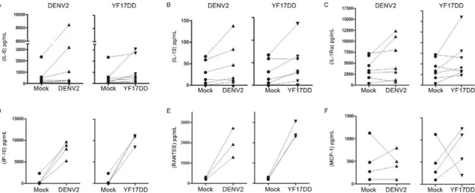
Several cytokines and chemokines are present in supernatants of virus-infected MoDCs - Cytokines and chemokines play important roles in dengue
physiopathol-ogy (Bozza et al. 2008, Noisakran & Perng 2008). Since
DCs are one of the main sources for these molecules early in viral infection, we investigated the production
of several of them after MoDC infection by DENV2 and
YF17DD from 24-120 hpi. We searched for the presence of inflammatory cytokines, chemokines and antiviral molecules that could drive T cell polarisation, either in-ducing vascular permeability or controlling infection.
We detected TNF-α, IFN-α, IL-6, IL-1Ra, IL-10, MIP-1β/CCL-4, MCP-1/CCL-2, IP-10/CXCL-10 and RANT
-ES/CCL-5 in cell culture supernatants. A mock-infected
MoDc culture supernatant was also assayed.
Cytokines were detected after infection by both fla-viviruses. IL-6 and IL-10 (Fig. 4A, B) were detected in cultured PBMC from seven donors at significant levels
(p < 0.5, in a Wilcoxon signed rank test). These factors
are known to modulate the immune response (Sabatte et al. 2007). IL-1Ra, the antagonist of the
pro-inflamma-tory cytokine IL-1β and chemotactic factor IP-10 were
also increased in most of the donors tested (Fig. 4C, D), although with borderline significance (0.0624 < p
< 0.0782; in a Wilcoxon test). RANTES and MCP-1 are
Fig. 3: multiple cytokine and chemokine induction by dengue virus 2 (DENV2) or yellow fever (YF) 17DD after monocyte-derived dendritic
cells (MoDCs)infection.MoDCs were infected with DENV2 [2.5 x 10⁵ 50% tissue culture infectious dose (TCID)/mL], YF17DD (1.25 x 10⁶
TCID50/mL) or mock-infected. Supernatants were collected 48 h after infection and analyzed by immunofluorescent multiplex-bead assay. A-F:
lines represent paired production for each donor of interleukin (IL)-6 (A), IL-10 (B), IL-1 receptor agonist (Ra) (C), CXCL-10/IP-10 (D), CCL-5/ RANTES (E)and CCL-2/MCP-1 (F)induced after mock virus infection. Three-seven peripheral blood mononuclear cells donors were used.
both chemokines related to severity of infection (Lee et
al. 2006, Pulendran et al. 2008) and were only slightly in
-creased after infection (Fig. 4E, F). However, low donor
numbers did not allow us to perform reliable statistical
analysis between the two viruses. MIP-1β/CCL-4, which
is correlated with non-severe disease, was detected in mock cultures from seven PBMC donors and, appar-ently, its production was not altered after infection with either virus (data not shown). In vivo, other cells may be responsible for its synthesis and release.
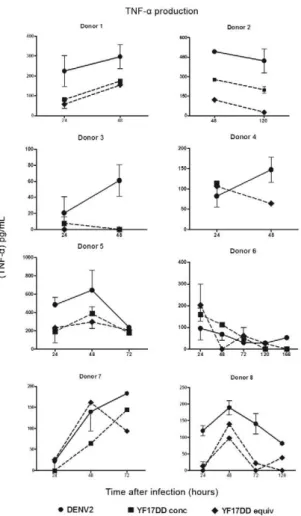
Significant TNF-α amounts were already detected at
24 hpi in the presence of either virus when compared
to the mock culture (data not shown), with a peak at 48
hpi and a decline thereafter in most PBMC donors
test-ed (Fig. 5). DENV2 inductest-ed the highest TNF-α levels (255 ± 75 pg/mL), which were significantly higher when compared with those produced by YF17DD (121 ± 31 pg/ mL) in eight different PBMC donors assayed at 48 hpi (p = 0.0156 in the Wilcoxon test).
In Fig. 6, IFN-α is plotted against viral Ag+ cells
dur-ing the course of infection in four different PBMC
do-nors. At 24 hpi, IFN-α is already detected in YF17DD-infected MoDCs and at 48 hpi in DENV2-YF17DD-infected MoDCs. Virus load decreases as the IFN-α levels in -crease. MoDCs from seven donors were assayed
side-by-side at 48 h with both viruses and those infected with YF17DD produced significantly higher IFN-α levels (393 ± 139 pg/mL) than those infected with DENV2 (72 ± 29 pg/mL; p = 0.0156 in a Wilcoxon test).
When the ratio of TNF-α and IFN-α levels for each
cell donor is calculated, significantly higher indexes
were obtained for DENV2 (13 ± 6) than for YF17DD (1.1 ± 0.3; p = 0.0156 in a Wilcoxon test).
As mentioned, data shown in Fig. 4B were generated
with higher doses of YF17DD than DENV2 inoculum
YF17DD was inoculated at DENV2 equivalent MOIs (thus resulting in lower infection rates) (Fig. 2), IFN-α
levels were 2-10 times higher in YF17DD cultures than
those in DENV2. Therefore, the TNF-α vs. IFN-α cy -tokine imbalance has a different pattern in the two flavi-virus studied irrespective of the MOI used.
YF17D/DENV2 vaccine virus infection compared with DENV2 or YF17DD infections - To investigate whether a chimeric dengue target vaccine virus could induce similar responses to the YF17DD vaccine virus, we performed pilot experiments infecting MoDCs with
the YF17D/DENV2 vaccine virus. Cell infection rates
were lower for the chimeric virus compared with the
other two viruses (DENV2 16681 strain and YF17DD) (Fig. 6). IFN-α levels detected in experiments with this
chimeric virus were slightly higher than those produced
by DENV2, but these differences were not statisti -cally significant (Wilcoxon signed rank test). However,
TNF-α levels were significantly higher after DENV2 in
-fection when compared with YF17D/DENV2 in-fection
(p = 0.0313). Therefore, thechimeric vaccine virus ap-parently displayed a similar cytokine response profile to YF17DD, even though it had lower replication rates.
DISCuSSIoN
During either a natural infection or a vaccination, the entry site of flaviviruses into the vertebrate host is the dermis. Langerhans cells were characterised as
permis-sive cells for DENV in vivo replication (Wu et al. 2000).
Indeed, DCs have a crucial role in initiating host defence
mechanisms; through antigen presentation and cytokine
production, they can define the fate of the
immunologi-cal response (Blanco et al. 2008). Both DENV and YF
vaccine viruses are known to infect DCs and monocytes in vitro(Wu et al. 2000, Barba-Spaeth et al. 2005, Reis et al. 2007). We aimed to characterise the similarities and differences between flavivirus infections with distinct virulence patterns that elicit long lasting in vivo immu-nity (Monath 2001, Halstead 2007).
DC infections with the YF Brazilian vaccine strain
17DD and with YF17D/DENV2 (44/3) chimeric virus
were reported here for the first time. The YF17DD
vac-cine virus infects cells at inoculum doses equivalent to those used for DENV2 (16681 strain) but showed re -duced expression of viral antigens within DCs at all time
points tested when compared to DENV. Similar results
were described for the YF vaccine strain 204, which is
poorly infectious compared with DENV (Deauvieau et al. 2007). Other authors used higher MOIs for DENV
to obtain the same relative infection level as West Nile virus because these viruses replicate at different rates. They then studied NS1 antigenic expression by flow cy-tometry analysis (Youn et al. 2010).
DENV sequences from YF17D/DENV2 (44/3) stud
-ied here are from the NGC virus. Although our DENV2 originated from strain 16681, NGC and 16681 are very
similar with regard to their genome and they belong to the
same genotype - SE Asian strains - that characteristically
induce the most severe forms of dengue fever (Leitmeyer et al. 1999). We do not intend to compare the virus struc-tures but instead the virulence (pathogenic vs. vaccine).
Virulent YF wild-type or Asibi strains have not been reported to infect DCs, but a few studies compared these viruses with YF17DD during infection in Kupffer or
en-dothelial cells (Khaiboullina et al. 2005, Woodson et al.
2011). Both viruses infect these cells, but the resulting
cy-tokine production is quite different. The virulent YF strains
produce a much more intense pro-inflammatory cytokine
response (including TNF-α, IL-6, IL-8 and RANTES/ CCL5) than the YF vaccine, which produces more IL-10.
During infection by viruses, DCs may undergo sev-eral phenotypic changes to become activated and capa-ble of antigen presentation to T lymphocytes. Among these changes is the upregulation of surface markers and soluble molecules related to the polarisation of the T effector cell response. YF17DD immunisation leads to natural killer (NK) cell and monocyte activation
(Mar-tins et al. 2008). With respect to DENV and YF17D/
Fig. 4: tumor necrosis factor (TNF)-αinduction by dengue virus 2
(DENV2) or yellow fever (YF) 17DD after monocyte-derived dendritic cells (MoDCs). MoDCs were infected with DENV2 or YF17DD equiva
-lent (equiv) doses [2.5 x 10⁵ 50% tissue culture infectious dose (TCID)/
mL] and YF17DD at concentrated (conc) dose (1.25 x 10⁶ TCID
50/mL).
Cultures were incubated from 24-168 h. TNF-α cell culture superna -tant content was determined by enzyme-linked immunosorbent assay. Individual data for eight different peripheral blood mononuclear cells donors are shown. Mock-infected cells were included for each donor,
but no detectable TNF-α was recorded (data not shown). Mean and
DENV chimeric viruses, earlier reports showed that ac
-tivation molecules, such as CD80, CD86 and CD83, can
be upregulated after DC infection (Libraty et al. 2001, Deauvieau et al. 2007, Sun et al. 2009). Co-stimulation
molecules, such as CD38, are present in circulating
monocytes but are poorly expressed after their differen-tiationinto immature DCs. During the maturation
proc-ess, CD38 is re-expressed onDCs (Fedele et al. 2004). In the present study, we observed that flavivirus
infec-tion favours CD38 expression on DCs. This molecule is
involved in cytoplasmatic calcium release, chemotaxis
and IFN-γ production, indicating a role for CD38 in Th-1
polarisation (Frasca et al. 2006).
OX40L expression is a fundamental requirement
for optimal induction of primary and memory Th-2 re-sponses in vivo. It binds to OX40 on T lymphocytes and stimulates the appropriate expansion and/or survival of T cells, leading to IL-4 and IL-13 production (Jenkins
et al. 2007, Blazquez & Berin 2008). Here we observed that the immature DCs expressed OX40L in culture and that this expression was downregulated as infection progressed in both viral infections. These data are in-formative with respect to Th-1/Th-2 axis determination by surface markers on DCs after flavivirus infection.
CD38 upregulation and OX40L downregulation indicate
that both viruses favour a Th-1 type response during the
Fig. 5: viral antigen positive cells (Ag+) vs. interferon (IFN)-α production by monocyte-derived dendritic cells (MoDCs)after dengue virus 2
(DENV2) or yellow fever (YF) 17DD infection.MoDCs were infected with DENV2 or YF17DD equivalent (equi) doses [2.5 x 10⁵ 50% tissue
culture infectious dose (TCID)/mL] or YF17DD at concentrated (conc) dose (1.25 x 10⁶ TCID
50/mL). Cultures were incubated from 24-120 h. X
axis represents time after infection. Left Y axis, the percentage of viral Ag+ cells obtained after flow cytometry analysis. Right Y axis, the IFN-α
cell supernatant content determined by enzyme-linked immunosorbent assay. Individual data for four different peripheral blood mononuclear
cells donors are shown. Mock-infected cells were included for each donor, but no detectable IFN-α was recorded (data not shown). Mean and
early immune response, which likely plays a role in vi-rus clearance, although a Th-2 response may be present as well during infections by both viruses (Bozza et al.
2008, Querec et al. 2009).
Indeed, Th-1/Th-2 mixed cytokine patterns, in which an early Th-1 profile gives rise to a late Th-2-predomi-nant pattern, likely occur during the course of a dengue fever infection (Chaturvedi et al. 2000, Mustafa et al. 2001, Nguyen et al. 2004). The difference in timing of
IFN-γ peaks in plasma have influences the severity of
disease. On the other hand, reports of an association be-tween low T-bet mRNA expression and high IL-10 levels may indicate a Th-2 role in the pathogenesis of dengue
hemorrhagic fever (Chen et al. 2005). YF17DD vacci -nation leads to a mixed pro/anti-inflammatory cytokine profile, including TNF+ monocytes, IFN-γ+ NK cells and
IL-10+ cells (Silva et al. 2011).
Infection of DCs by DENV induces T cells to produce IL-4, IL-10 and IFN-γ, suggesting a mixed Th cytokine
pattern (Ho et al. 2004). DCs infected with YF17DD
stimulate CD8+ T cells from YF17DD-immunised or naive donors and induce IFN-γ and present antigen spe
-cifically to CD8+ and CD4+ T cells (Barba-Spaeth et al. 2005). In the present study, DC cultures infected with
flaviviruses were able to produce a panel of several cy-tokines and chemokines, which were similar in both vi-ral infections. It is likely that chemokines produced by
DCs after flavivirus infections, such as IP-10, RANTES
and MCP-1, might be signalling to monocytes, NK cells and Th-1 type lymphocytes, resulting in modulation of endothelial permeability, chemotaxis to the site of in-flammation, migration to inflammatory sites and T cell polarisation (Aliberti et al. 2000, Thomsen et al. 2003,
Nightingale et al. 2008). These cell subsets are crucial
for viral clearance and efficient immunological response generation, but we cannot exclude the possibility that an excessive inflammatory reaction may result in exacerba-tion of disease severity.
The most striking differences between the DENV2 and YF17DD viruses were found in TNF-α and IFN-α produc
-tion by DCs. TNF-α levels are significantly increased after
DENV2 infection in DC cultures, but only slightly altered in the presence of YF17DD and YF17D/DENV2. On the other hand, these viruses were able to elicit high IFN-α levels that not reached during DENV2 infection.
TNF-α is a pro-inflammatory cytokine that has of -ten been found in patients with acute dengue fever and high levels are associated with haemorrhagic
manifesta-tions (Hober et al. 1993, Kubelka et al. 1995, Braga et
al. 2001, Chakravarti & Kumaria 2006). Together with
IL-1β, TNF-α is known to increase acute phase protein
production and act in synergy with other factors to in-duce microbicidal activity during phagocytosis (Clark 2007). It also has the capacity to induce the upregulation of endothelial adhesion molecules, which in turn signal to chemotactic peptides and lipid mediators and facili-tate leukocyte recruitment, which may result in plasma leakage and hypovolemic shock. In our in vitro infection
model, we observed markedly higher TNF-α produc
-tion after infec-tion with pathogenic virus (DENV2) as
compared to the non-pathogenic viruses (YF17DD and
YF17D/DENV2), providing further evidence that this
cytokine plays a role in flavivirus immunopathogenesis.
Other authors have also observed TNF-α production in DC or monocyte cultures in the presence of DENV2
and some authors even associate it with the infected cell
(Hacker et al. 1998, Querec et al. 2006, Deauvieau et al. 2007, Reis et al. 2007, Ahmad et al. 2008, Nightingale et al. 2008, Levy et al. 2009). A moderate level of TNF-α
production may be beneficial to mature cells that may become good antigen presenters. This cytokine was de-tected in YF vaccinated individuals (Roers et al. 1994, Querec et al. 2006, Deauvieau et al. 2007, Levy et al. 2009, Silva et al. 2011).
IFNs are classically known as antiviral molecules and some reports confirm that flaviviruses are suscep-tible to these molecules (Roers et al. 1994, Diamond et
al. 2000). Indeed, these viruses stimulate IFN-α produc -tion (Libraty et al. 2001, Deauvieau et al. 2007, Palmer et
al. 2007, Querec et al. 2009), although IFN-α induction
was not compared between viruses in previous reports.
Earlier studies show that DENV is able to escape IFN
Fig. 6: viral antigen positive cells (Ag+), tumor necrosis factor (TNF)-αor interferon (IFN)-α production by monocyte-derived dendritic cells
(MoDCs)after yellow fever (YF) 17DD/dengue virus 2 (DENV2) infections are compared to DENV2 or YF17DD infection.MoDCs were infected
with YF17D/DENV2, DENV2 or YF17DD at multiplicity of infection 4 (4 x 10⁶plaque-forming unit/106 cells). Cultures were incubated from
one-seven days (X axis: time after infection; Y axis: percentage of viral Ag+ cells obtained after flow cytometry analysis). TNF-α or IFN-α cell su
-pernatant content are determined by enzyme-linked immunosorbent assay. Mean and standard error from duplicates were plotted. Mock-infected
cells were included for each donor, but no detectable TNF-α or IFN-α was recorded and viral Ag+ cells were present at < 2% (data not shown). Data
action by inhibiting steps of IFN type I activation path-ways through a decrease in the signal transducer and activator of transcription-2 expression (STAT-2) (Jones
et al. 2005). This effect is meditated by the viral non-structural protein 4. Recently, low levels of IFN-α/β pro
-duction by DENV2-infected MoDCs has been reported
as playing a role in the failure of DCs to prime T cells (Rodriguez-Madoz et al. 2010).
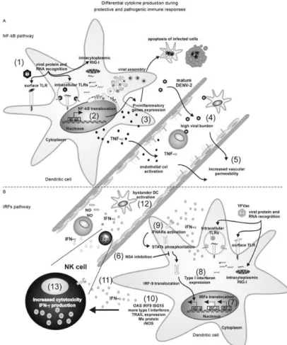
In Fig. 7, we outline a theoretical model for flavivi-rus interactions with DCs. Flaviviflavivi-rus infection may trig-ger differential signalling pathways that lead to an early clearance of virus. These pathways might include an IFN pathway that activates IFN- regulatory gene (IRF)-7 and
IFN-stimulated response element (ISRE), which in turn further induces high IFN-α and nitric oxide production.
Alternatively, the blockage of antiviral pathways, such
as STAT activation by non-structural viral proteins, may lead to higher virus load and intense stimulation of
inflammatory/pathologic mechanisms such as TNF-α. The IFN-α produced by target cells may act on IFN-α
receptors on bystander cells to protect them from further
infections. Furthermore, IFN-α activates natural killer
cells, which have cytotoxic and antiviral functions and produce IFNs that in turn can activate monocytes to ex-ert antiviral functions through nitric oxide production.
Here we observed that DCs infected with YF17DD
and YF17D/DENV2 produce more IFN-α than those infected with DENV2. These data suggest that the vac -cine viruses may have different virulent properties than
DENV2 and that they may have lost (or never acquired) the ability to inhibit the ISRE pathway that was hypoth
-esised to play a role in DENV infection in Fig. 7. YF Fig. 7: theoretical model for dendritic cell (DC)-flavivirus interaction. A: during target cell infection, dengue virus (DENV) is sensed/recog -nized by pattern recognition receptor (1), triggering preferentially pro-inflammatory genes such as for nuclear factor kappa B (NF-κB) (2), resulting in a high tumor necrosis factor (TNF)-αproduction (3) , while in Binterferon (IFN)-α is partially inhibited by DENV proteins, such
as NS4, through signal transducer and activator of transcription-2 expression (STAT) inhibition (4). Back to A, DENV infection outcome con
-sists in a high viral load (5) followed by an enhancement of pro-inflammatory gene activation and induction of plasma leakage (6). In B, yellow fever (YF) vaccine virus induces preferentially the IFN pathway activating IRF-7 and IFN-stimulated response element (ISRE) (7) that further induce a high IFN-α production (8), resulting in paracrine or autocrine IFN-associated receptor (IFNAR)-dependent activation (9). In turn,
IFN-stimulated genes are expressed (10) such as oligoadenylate synthetase (OAS), interferon (IFN) regulatory factor (IRF)-9, IFN-stimulated gene (ISG)-15, TNF-related apoptosis-inducing ligand (TRAIL), inducible nitric oxide synthase (iNOS), among others, contributing to a low
virus load. Besides its antiviral effects on virus replication, a second IFN-α production wave, IFNAR-dependent (11), exert other functions such
as: protecting bystander DCs from de novo infection (12); activating natural killer cells that are IFN-γ producers and efficient cytotoxic killers
for infected-targets (13). IFNs can activate monocytes to produce nitric oxide (NO), another antiviral molecule. RIG-I: retinoic-acid-inducible
vaccination in human subjects studied by microarray analysis showed a response profile related to IFN-based antiviral responses (Scherer et al. 2007, Querec et al. 2009), suggesting a role for IFNs in effective protection from this vaccine. On the other hand, functional genetic studies identified several transcripts for IFN-stimulated genes in patients with dengue fever that were less abun-dant in adults with dengue shock syndrome, reinforcing the attenuating role of IFNs after infection (Simmons et al. 2007). Vaccine or therapeutic approaches may indeed provide the ability to induce antiviral molecules such as IFNs without inducing or modulating factors involved in vascular permeability.
ACKNowLEDGEMENTS
To Denise Cerqueira, Lidiane Martins de Albuquerque
and Luciana Gomes Fialho, for their technical collaboration, suggestions and encouragement.
REFERENCES
Ahmad SM, Haskell MJ, Raqib R, Stephensen CB 2008. Men with low vitamin A stores respond adequately to primary yellow fever and
secondary tetanus toxoid vaccination. J Nutr 138: 2276-2283.
Aliberti J, Reis e Sousa C, Schito M, Hieny S, Wells T, Huffnagle GB,
Sher A 2000. CCR5 provides a signal for microbial induced produc
-tion of IL-12 by CD8 alpha+ dendritic cells. Nat Immunol 1: 83-87.
Assuncao-Miranda I, Amaral FA, Bozza FA, Fagundes CT, Sousa LP, Souza DG, Pacheco P, Barbosa-Lima G, Gomes RN, Bozza PT, Da Poian AT, Teixeira MM, Bozza MT 2010. Contribution of macrophage migration inhibitory factor to the pathogenesis of dengue virus infection. FASEB J 24: 218-228.
Azeredo EL, Zagne SM, Santiago MA, Gouvea AS, Santana AA,
Neves-Souza PC, Nogueira RM, Miagostovich MP, Kubelka CF 2001. Characterisation of lymphocyte response and cytokine pat-terns in patients with dengue fever. Immunobiology 204: 494-507.
Bae HG, Domingo C, Tenorio A, de Ory F, Munoz J, Weber P, Teuwen
DE, Niedrig M 2008. Immune response during adverse events after 17D-derived yellow fever vaccination in Europe. J Infect Dis 197: 1577-1584.
Barba-Spaeth G, Longman RS, Albert ML, Rice CM 2005. Live at -tenuated yellow fever 17D infects human DCs and allows for pre-sentation of endogenous and recombinant T cell epitopes. J Exp Med 202: 1179-1184.
Blanco P, Palucka AK, Pascual V, Banchereau J 2008. Dendritic cells
and cytokines in human inflammatory and autoimmune diseases.
Cytokine Growth Factor Rev 19: 41-52.
Blazquez AB, Berin MC 2008. Gastrointestinal dendritic cells pro -mote Th2 skewing via OX40L. J Immunol 180: 4441-4450.
Bozza FA, Cruz OG, Zagne SM, Azeredo EL, Nogueira RM, Assis EF, Bozza PT, Kubelka CF 2008. Multiplex cytokine profile from
dengue patients: MIP-1beta and IFN-gamma as predictive factors for severity. BMC Infect Dis 8: 86.
Braga ELA, Moura P, Pinto LMO, Ignacio SRN, Oliveira MJC, Cord -eiro MT, Kubelka CF 2001. Detection of circulant tumor necrosis
factor-alpha, soluble tumor necrosis factor p75 and
interferon-gamma in Brazilian patients with dengue fever and dengue hem-orrhagic fever. Mem Inst Oswaldo Cruz 96: 229-232.
Caufour PS, Motta MC, Yamamura AM, Vazquez S, Ferreira I, Jabor
AV, Bonaldo MC, Freire MS, Galler R 2001. Construction, char-acterization and immunogenicity of recombinant yellow fever 17D-dengue type 2 viruses. Virus Res 79: 1-14.
Chakravarti A, Kumaria R 2006. Circulating levels of tumour ne-crosis factor-alpha & interferon-gamma in patients with dengue & dengue haemorrhagic fever during an outbreak. Indian J Med Res 123: 25-30.
Chaturvedi UC, Agarwal R, Elbishbishi EA, Mustafa AS 2000. Cy -tokine cascade in dengue hemorrhagic fever: implications for pathogenesis. FEMS Immunol Med Microbiol 28: 183-188.
Chen LC, Lei HY, Liu CC, Shiesh SC, Chen SH, Liu HS, Lin YS, Wang ST, Shyu HW, Yeh TM 2006. Correlation of serum levels of mac-rophage migration inhibitory factor with disease severity and clini-cal outcome in dengue patients. Am J Trop Med Hyg 74: 142-147. Chen RF, Liu JW, Yeh WT, Wang L, Chang JC, Yu HR, Cheng JT,
Yang 2005. Altered T helper 1 reaction but not increase of virus
load in patients with dengue hemorrhagic fever. FEMS Immunol Med Microbiol 44: 43-50.
Clark IA 2007. How TNF was recognized as a key mechanism of dis-ease. Cytokine Growth Factor Rev 18: 335-343.
Clyde K, Kyle JL, Harris E 2006. Recent advances in deciphering vi -ral and host determinants of dengue virus replication and patho-genesis. J Virol 80: 11418-11431.
Deauvieau F, Sanchez V, Balas C, Kennel A, Dem A, Lang J, Guy B 2007. Innate immune responses in human dendritic cells upon infection by chimeric yellow-fever dengue vaccine serotypes 1-4.
Am J Trop Med Hyg 76: 144-154.
Delespesse G, Ohshima Y, Yang LP, Demeure C, Sarfati M 1999. OX40-mediated cosignal enhances the maturation of naive hu-man CD4+ T cells into high IL-4-producing effectors. Int Arch
Allergy Immunol 118: 384-386.
Diamond MS, Roberts TG, Edgil D, Lu B, Ernst J, Harris E 2000.
Modulation of dengue virus infection in human cells by alpha, beta and gamma interferons. J Virol 74: 4957-4966.
Duangchinda T, Dejnirattisai W, Vasanawathana S, Limpitikul W, Tangthawornchaikul N, Malasit P, Mongkolsapaya J, Screaton G 2010. Immunodominant T-cell responses to dengue virus NS3 are associated with DHF. Proc Natl Acad Sci USA 107: 16922-16927.
Fedele G, Frasca L, Palazzo R, Ferrero E, Malavasi F, Ausiello CM 2004. CD38 is expressed on human mature monocyte-derived dendritic cells and is functionally involved in CD83 expression
and IL-12 induction. Eur J Immunol 34: 1342-1350.
Fink J, Gu F, Ling L, Tolfvenstam T, Olfat F, Chin KC, Aw P, George J, Kuznetsov VA, Schreiber M, Vasudevan SG, Hibberd ML 2007. Host gene expression profiling of dengue virus infection in cell lines and patients. PLoS Negl Trop Dis 1: e86.
Frasca L, Fedele G, Deaglio S, Capuano C, Palazzo R, Vaisitti T,
Ma-lavasi F, Ausiello CM 2006. CD38 orchestrates migration, sur -vival and Th1 immune response of human mature dendritic cells.
Blood 107: 2392-2399.
Galler R, Marchevsky RS, Caride E, Almeida LF, Yamamura AM, Ja
-bor AV, Motta MC, Bonaldo MC, Coutinho ES, Freire MS 2005.
Attenuation and immunogenicity of recombinant yellow fever 17D-dengue type 2 virus for Rhesus monkeys. Braz J Med Biol Res 38: 1835-1846.
Geisbert TW, Jahrling PB 2004. Exotic emerging viral diseases: prog -ress and challenges. Nat Med 10: S110-121.
Green S, Vaughn DW, Kalayanarooj S, Nimmannitya S, Suntayakorn
S, Nisalak A, Rothman AL, Ennis FA 999. Elevated plasma inter -leukin-10 levels in acute dengue correlate with disease severity. J Med Virol 59: 329-334.
Gubler DJ 2002. Epidemic dengue/dengue hemorrhagic fever as a
public health, social and economic problem in the 21th century.
Guy B, Guirakhoo F, Barban V, Higgs S, Monath TP, Lang J 2010. Preclinical and clinical development of YFV 17D-based chime-ric vaccines against dengue, West Nile and Japanese encephalitis viruses. Vaccine 28: 632-649.
Hacker UT, Jelinek T, Erhardt S, Eigler A, Hartmann G, Nothdurft HD, Endres S 1998. In vivo synthesis of tumor necrosis factor-alpha in healthy humans after live yellow fever vaccination.
J Infect Dis 177: 774-778.
Halstead SB 2007. Dengue. Lancet 370: 1644-1652.
Ho LJ, Shaio MF, Chang DM, Liao CL, Lai JH 2004. Infection of human dendritic cells by dengue virus activates and primes T cells towards Th0-like phenotype producing both Th1 and Th2 cytokines. Immunol Invest 33: 423-437.
Hober D, Poli L, Roblin B, Gestas P, Chungue E, Granic G, Imbert P,
Pecarere JL, Vergez-Pascal R, Wattre P 1993. Serum levels of tu-mor necrosis factor-alpha (TNF-alpha), interleukin-6 (IL-6) and interleukin-1 beta (IL-1 beta) in dengue-infected patients. Am J Trop Med Hyg 48: 324-331.
Jenkins SJ, Perona-Wright G, Worsley AG, Ishii N, MacDonald AS 2007. Dendritic cell expression of OX40 ligand acts as a costimu-latory, not polarizing, signal for optimal Th2 priming and memo-ry induction in vivo. J Immunol 179: 3515-3523.
Jones M, Davidson A, Hibbert L, Gruenwald P, Schlaak J, Ball S,
Fos-ter GR, Jacobs M 2005. Dengue virus inhibits alpha inFos-terferon
signaling by reducing STAT2 expression. J Virol 79: 5414-5420.
Khaiboullina SF, Rizvanov AA, Holbrook MR, St Jeor S 2005. Yel -low fever virus strains Asibi and 17D-204 infect human umbilical cord endothelial cells and induce novel changes in gene expres-sion. Virology 342: 167-176.
Kubelka CF, Borges PA, VonSydow FF, Farid FO, Lampe E 1995.
Analysis of tumor necrosis factor-alpha serum level in Brazilian patients with dengue-2. Mem Inst Oswaldo Cruz 90: 741-742. Lanzavecchia A, Sallusto F 2004. Lead and follow: the dance of the
dendritic cell and T cell. Nat Immunol 5: 1201-1202.
Lee YR, Liu MT, Lei HY, Liu CC, Wu JM, Tung YC, Lin YS, Yeh TM, Chen SH, Liu HS 2006. MCP-1, a highly expressed chemokine in dengue haemorrhagic fever/dengue shock syndrome patients may cause permeability change, possibly through reduced tight junc-tions of vascular endothelium cells. J Gen Virol 87: 3623-3630. Leitmeyer KC, Vaughn DW, Watts DM, Salas R, Villalobos I de C,
Ramos C, Rico-Hesse R 1999. Dengue virus structural differ-ences that correlate with pathogenesis. J Virol 73: 4738-4747.
Levy A, Valero N, Espina LM, Anez G, Arias J, Mosquera J 2009.
Increment of interleukin 6, tumour necrosis factor alpha, nitric oxide, C-reactive protein and apoptosis in dengue. Trans R Soc Trop Med Hyg 104: 16-23.
Libraty DH, Pichyangkul S, Ajariyakhajorn C, Endy TP, Ennis FA
2001. Human dendritic cells are activated by dengue virus in-fection: enhancement by gamma interferon and implications for disease pathogenesis. J Virol 75: 3501-3508.
Martins MA, Silva ML, Eloi-Santos SM, Ribeiro JG, Peruhype-Magalhaes V, Marciano AP, Homma A, Kroon EG, Teixeira-Carvalho A, Martins-Filho OA 2008. Innate immunity pheno -typic features point toward simultaneous raise of activation and modulation events following 17DD live attenuated yellow fever first-time vaccination. Vaccine 26: 1173-1184.
Miagostovich MP, Nogueira RM, Cavalcanti SM, Marzochi KB, Schatzmayr HG 1993. Dengue epidemic in the state of Rio de Janeiro, Brazil: virological and epidemiological aspects. Rev Inst Med Trop Sao Paulo 35: 149-154.
Monath TP 2001. Yellow fever: an update. Lancet Infect Dis 1: 11-20.
Mustafa AS, Elbishbishi EA, Agarwal R, Chaturvedi UC 2001. El
-evated levels of interleukin-13 and IL-18 in patients with dengue
hemorrhagic fever. FEMS Immunol Med Microbiol 30: 229-233.
Neves-Souza PC, Azeredo EL, Zagne SM, Valls-de-Souza R, Reis SR, Cerqueira DI Nogueira RM, Kubelka CF 2005. Inducible
nitric oxide synthase (iNOS) expression in monocytes during acute dengue fever in patients and during in vitro infection. BMC Infect Dis 5: 64.
Nguyen TH, Lei HY, Nguyen TL, Lin YS, Huang KJ, Le BL, Lin CF, Yeh TM, Do QH, Vu TQ, Chen LC, Huang JH, Lam TM, Liu CC, Halstead SB 2004. Dengue hemorrhagic fever in infants: a study of clinical and cytokine profiles. J Infect Dis 189: 221-232.
Nightingale ZD, Patkar C, Rothman AL 2008. Viral replication and
paracrine effects result in distinct, functional responses of den-dritic cells following infection with dengue 2 virus. J Leuk Biol 84: 1028-1038.
Noisakran S, Perng GC 2008. Alternate hypothesis on the pathogenesis
of dengue hemorrhagic fever (DHF)/dengue shock syndrome (DSS) in dengue virus infection. Exp Biol Med (Maywood) 233: 401-408.
Ohshima Y, Tanaka Y, Tozawa H, Takahashi Y, Maliszewski C,
De-lespesse G 1997. Expression and function of OX40 ligand on hu -man dendritic cells. J Immunol 159: 3838-3848.
Ohshima Y, Yang LP, Uchiyama T, Tanaka Y, Baum P, Sergerie M,
Hermann P, Delespesse G 1998. OX40 costimulation enhances
interleukin-4 (IL-4) expression at priming and promotes the dif-ferentiation of naïve human CD4(+) T cells into high
IL-4-pro-ducing effectors. Blood 92: 3338-3345.
Palmer DR, Fernandez S, Bisbing J, Peachman KK, Rao M, Barvir D, Gunther V, Burgess T, Kohno Y, Padmanabhan R, Sun W 2007. Re-stricted replication and lysosomal trafficking of yellow fever 17D vaccine virus in human dendritic cells. J Gen Virol 88: 148-156.
Post PR, Santos CND, Carvalho R, Lopes OS, Galler R 1991. Molecu-lar analysis of yellow fever virus 17DD vaccine strain. Mem Inst Oswaldo Cruz 86: 239-246.
Pulendran B, Miller J, Querec TD, Akondy R, Moseley N, Laur O, Glidewell J, Monson N, Zhu T, Zhu H, Staprans S, Lee D, Brinton MA, Perelygin AA, Vellozzi C, Brachman P Jr, Lalor S, Teuwen
D, Eidex RB, Cetron M, Priddy F, del Rio C, Altman J, Ahmed R 2008. Case of yellow fever vaccine-associated viscerotropic dis -ease with prolonged viremia, robust adaptive immune responses
and polymorphisms in CCR5 and RANTES genes. J Infect Dis 198: 500-507.
Quah BJ, O’Neill HC 2005. Maturation of function in dendritic cells
for tolerance and immunity. J Cell Mol Med 9: 643-654.
Querec T, Bennouna S, Alkan S, Laouar Y, Gorden K, Flavell R, Aki-ra S, Ahmed R, PulendAki-ran B 2006. Yellow fever vaccine YF-17D
activates multiple dendritic cell subsets via TLR2, 7, 8 and 9 to
stimulate polyvalent immunity. J Exp Med 203: 413-424.
Querec TD, Akondy RS, Lee EK, Cao W, Nakaya HI, Teuwen D, Pi -rani A, Gernert K, Deng J, Marzolf B, Kennedy K, Wu H, Ben-nouna S, Oluoch H, Miller J, Vencio RZ, Mulligan M, Aderem A, Ahmed R, Pulendran B 2009. Systems biology approach predicts immunogenicity of the yellow fever vaccine in humans. Nat Im-munol 10: 116-125.
Reis SRNI, Sampaio ALF, Henriques MGM, Gandini M, Azeredo EL, Kubelka CF 2007. Anin vitro model for dengue virus infec-tion that exhibits human monocyte infecinfec-tion, multiple cytokine production and dexamethasone immunomodulation. Mem Inst Oswaldo Cruz 102: 983-990.
Rodriguez-Madoz JR, Bernal-Rubio D, Kaminski D, Boyd K, Fer-nandez-Sesma A 2010. Dengue virus inhibits the production of type I interferon in primary human dendritic cells. J Virol 84:
Roers A, Hochkeppel HK, Horisberger MA, Hovanessian A, Haller O 1994. MxA gene expression after live virus vaccination: a sensitive marker for endogenous type I interferon. J Infect Dis 169: 807-813.
Sabatte J, Maggini J, Nahmod K, Amaral MM, Martinez D, Sala- mone G, Ceballos A, Giordano M, Vermeulen M, Geffner J 2007. Interplay of pathogens, cytokines and other stress signals in the regulation of dendritic cell function. Cytokine Growth Factor Rev 18: 5-17.
Scherer CA, Magness CL, Steiger KV, Poitinger ND, Caputo CM, Miner DG, Winokur PL, Klinzman D, McKee J, Pilar C, Ward PA, Gillham MH, Haulman NJ, Stapleton JT, Iadonato SP 2007. Distinct gene expression profiles in peripheral blood mononu-clear cells from patients infected with vaccinia virus, yellow fever 17D virus or upper respiratory infections. Vaccine 25:
6458-6473.
Schnittler HJ, Feldmann H 2003. Viral hemorrhagic fever - a vascular disease? Thromb Haemost 89: 967-972.
Silva ML, Espirito-Santo LR, Martins MA, Silveira-Lemos D, Peru -hype-Magalhaes V, Caminha RC, de Andrade Maranhao-Filho P, Auxiliadora-Martins M, de Menezes Martins R, Galler R,
da Silva Freire M, Marcovistz R, Homma A, Teuwen DE,
Eloi-Santos SM, Andrade MC, Teixeira-Carvalho A, Martins-Filho OA 2010. Clinical and immunological insights on severe, adverse neurotropic and viscerotropic disease following 17D yellow fever vaccination. Clin Vaccine Immunol 17: 118-126.
Silva ML, Martins MA, Espirito-Santo LR, Campi-Azevedo AC, Silveira-Lemos D, Ribeiro JG, Homma A, Kroon EG, Teixeira-Carvalho A, Eloi-Santos SM, Martins-Filho OA 2011. Charac -terization of main cytokine sources from the innate and adaptive immune responses following primary 17DD yellow fever vacci-nation in adults. Vaccine 29: 583-592.
Simmons CP, Popper S, Dolocek C, Chau TN, Griffiths M, Dung NT, Long TH, Hoang DM, Chau NV, Thao le TT, Hien TT, Relman DA, Farrar J 2007. Patterns of host genome-wide gene transcript abundance in the peripheral blood of patients with acute dengue hemorrhagic fever. J Infect Dis 195: 1097-1107.
Srikiatkhachorn A 2009. Plasma leakage in dengue haemorrhagic fe-ver. Thromb Haemost 102: 1042-1049.
Srikiatkhachorn A, Green S 2010. Markers of dengue disease sever-ity. Curr Top Microbiol Immunol 338: 67-82.
Steinman RM, Nussenzweig MC 2002. Avoiding horror autotoxicus: the importance of dendritic cells in peripheral T cell tolerance.
Proc Natl Acad Sci USA 99: 351-358.
Suharti C, van Gorp EC, Setiati TE, Dolmans WM, Djokomoeljanto RJ, Hack CE, Ten CH, van der Meer JW 2002. The role of cytok-ines in activation of coagulation and fibrinolysis in dengue shock syndrome. Thromb Haemost 87: 42-46.
Sun P, Fernandez S, Marovich MA, Palmer DR, Celluzzi CM, Boon-nak K, Liang Z, Subramanian H, Porter KR, Sun W, Burgess TH 2009. Functional characterization of ex vivo blood myeloid and plasmacytoid dendritic cells after infection with dengue virus.
Virology 383: 207.
Thomsen AR, Nansen A, Madsen AN, Bartholdy C, Christensen JP 2003. Regulation of T cell migration during viral infection: role of adhesion molecules and chemokines. Immunology Lett 85: 119-127.
Vasconcelos PF, Luna EJ, Galler R, Silva LJ, Coimbra TL, Barros
VL, Monath TP, Rodigues SG, Laval C, Costa ZG, Vilela MF, Santos CL, Papaiordanou PM, Alves VA, Andrade LD, Sato HK,
Rosa ES, Froguas GB, Lacava E, Almeida LM, Cruz AC, Rocco
IM, Santos RT, Oliva OF 2001. Serious adverse events associated with yellow fever 17DD vaccine in Brazil: a report of two cases.
Lancet 358: 91-97.
Wallet MA, Sen P, Tisch R 2005. Immunoregulation of dendritic cells.
Clin Med Res 3: 166-175.
Woodson SE, Freiberg AN, Holbrook MR 2011. Differential cy -tokine responses from primary human Kupffer cells following infection with wild-type or vaccine strain yellow fever virus.
Virology 412: 188-195.
Wu SJ, Grouard-Vogel G, Sun W, Mascola JR, Brachtel E, Putvatana
R, Louder MK, Filgueira L, Marovich MA, Wong HK, Blauvelt A, Murphy GS, Robb ML, Innes BL, Birx DL, Hayes CG, Fran-kel SS 2000. Human skin Langerhans cells are targets of dengue virus infection. Nat Med 6: 816-820.