Strategies of the
African Swine Fever Virus to
manipulate innate immunity
Sónia Ventura
Dissertation presented to obtain the Ph.D degree in Biology
Instituto de Tecnologia Química e Biológica | Universidade Nova de Lisboa
Research work coordinated by:
I would like to thank my supervisor, Michael Parkhouse for giving me the opportunity to work and learn from him. Thank you for all the support, friendship and advice. The confidence that you deposited in me helped to trust myself and to grow as a person. I truly feel fortunate for having you as my supervisor throughout these years.
To my supervisor-in-the-bench, Sílvia Correia, I can hardly express how grateful I am for all your help. Your advice and experience were invaluable throughout this work, and I was lucky enough to have your friendship (and patience!) to help me carry on during these four years.
A special thanks goes to my other friends at the bench, Rute Nascimento and Helena Costa. I cannot think of better companions, always willing to give new ideas and suggestions, and always ready to help me laugh when most needed. I also want to thank the Infection and Immunity group, past and present, for all the help and support.
APCs Antigen Presenting Cells ASF African Swine Fever ASFV African Swine Fever Virus ATF2 AcTivating Factor 2
BVDV Bovine Viral Diarrhoea Virus CARD CAspase Recruitment Domain CBP CREB-binding protein
CCR CC Chemokine Receptors CD2v CD2 like protein
CLR C-type Lectin Receptors
CREB cAMP-Response Element-Binding protein CTL Cytotoxic T Lymphocyte
DAI DNA-dependent Activator of IFN-regulatory factors
DBD DNA Binding Domain
DC Dendritic Cell
dsRNA double stranded RNA EBV Epstein-Barr Virus
eIF eukaryotic Initiation Factor
ELISA Enzyme-Linked Immunoabsorbent Shift Assay EMCV EncephaloMyoCarditis Virus
ER Endoplasmic Reticulum
FADD Fas-Associated protein with Death Domain FBS Foetal Bovine Serum
GAS Gamma Activated Site GFP Green Fluorescence Protein
HA Haemaglutinin
HCMV Human CytoMegaloVirus HCV Hepatitis C Virus
HDAC Histone DeACetylase HHV8 Human HerpesVirus 8
HIV Human Immunodeficiency Virus
HMGI(Y) High-Mobility Group (HMG) chromatin associated protein HSV Herpes Simplex Virus
HVS HerpesVirus Saimiri
IFNGR Interferon Gamma Receptor
Ig Immunoglobulin
IKKԑ Inhibitory protein кB-ԑ
IL InterLeukin
iNOS inducible Nitric Oxide Synthase IRAK IL-1 Receptor-Associated Kinase IRF Interferon Regulatory Factor ISG Interferon Stimulated Gene ISGF3 IFN Stimulated Gene Factor 3 ISRE IFN-Stimulated Response Element Jak Janus activated kinase
KSHV Kaposi’s Sarcoma associated Herpesvirus LPS LipoPolySaccharide
LRRs Leucine Rich Repeats
MAVS Mitochondrial Antiviral Signalling
MDA-5 Melanoma Differentiation-Associated gene 5 mDCs Myeloid Dendritic Cells
MGF Multi Gene Family
MHC Major Histocompatibility Complex MHV68 Murine gamma-Herpesvirus 68 M-MLV Murine Molony Leukaemia Virus
MyD88 Myeloid Differentiation primary response gene 88 ND10 Nuclear Domains 10
NFAT Nuclear Factor of Activated T cells NF-кB Nuclear Factor кB
NK Natural Killer
NLR NOD-Like Receptors
NO Nitric Oxide
NOD Nucleotide binding and Oligomerization Domain OAS 2´,5´-Oligoadenylate Synthetase
ORF Open Reading Frame
PAMP Pathogen Associated Molecular Pattern PBS Phosphate Buffered Saline
PKR dsRNA-dependent Protein Kinase R PML Promyelocytic Leukaemia Protein Poly (I:C) Polyinosine-Polycytidylic Acid PRD Positive Regulatory Domain PRR Pattern Recognition Receptor
RBC Red Blood Cell
RIG-I Retinoic Acid Inducible Gene I RLR Rig-I Like Receptor
RNAi RNA Interference
RSV Respiratory Syncytial Virus
SeV Sendai Virus
SOCS Suppressors of Cytokine Signalling ssRNA single stranded RNA
STING STimulator of Interferon Genes SV40 Simian Virus 40
TBK-1 Tank-Binding Kinase-1 TBP Tata-Binding Protein TCR T-Cell Receptor
Th Helper T Lymphocyte
TIR Intracellular Toll-IL-1 Receptor
TIRAP TIR Domain-Containing Adaptor Protein TLR Toll Like Receptor
TNFα Tumour Necrosis Factor Α TRAF TNF Receptor Associated Factor Treg Regulatory T Lymphocyte
TRIF TIR Domain Containing Adaptor Inducing IFN VRE Virus-Responsive Element
VSV Vesicular Stomatitis Virus
Esta tese teve como objectivo determinar os mecanismos e consequências de dois genes virais de evasão à resposta do interferão (IFN), expressos pelo economicamente importante, e frequentemente fatal, Vírus da Peste Suína Africana (VPSA). De modo a sobreviverem, os vírus de ADN, tal como o VPSA, têm frequentemente múltiplas estratégias/genes que modulam positiva ou negativamente a biologia celular do hospedeiro, bem como a resposta imunitária. Os dois genes aqui apresentados funcionam em benefício do vírus, inibindo um das principais componentes da resposta imune inata, a resposta do IFN.
O gene I329L foi recentemente reportado como sendo capaz de inibir as respostas celulares, controladas pelo TLR3, que levam à indução e secreção de IFN-β, bem como à activação do NF-κB. Aqui, é demonstrado que o I329L não só inibe a indução e secreção de IFN-β
pelo TLR3, mas também inibe a activação do NF-κB após estimulação pelo TLR4. Demonstrou-se ainda, bioquimicamente, que a proteína I329L interage com a proteína adaptadora TRIF, o que é consistente com a inibição observada de ambas as vias do TLR3 e TLR4. De forma a caracterizar a modulação da resposta do IFN tipo I pelo I329L, bem como determinar o papel de cada domínio do I329L nesta inibição, foram construídos plasmídeos que expressam mutantes truncados, com apenas o domínio extracelular ou intracelular. Estes mutantes foram testados por ensaios de luciferase. O domínio extracelular apenas inibe a activação do IFN-β e NF-кB induzida por
modelo em que o domínio extracelular inibe a activação da resposta pelo TLR3 através da formação de um heterodímero não-funcional I329L-TLR3, e o domínio intracelular interfere com a transmissão do sinal através do TRIF.
Demonstrou-se que duas variantes distintas do gene não-conservado MGF360-18R do VPSA, uma da estirpe patogénica Benin97/1 e outra da estirpe adaptada à cultura celular Ba71V, inibem a indução do
IFN-β, e a resposta do hospedeiro à expressão de IFN tipo I e tipo II. Ambas as variantes da proteína MGF360-18R afectam a proteína MAVS, uma proteína adaptadora da via citosólica RLR que é essencial para a indução do IFN-β. Por outro lado, apenas a variante
‘patogénica’ afecta o factor de transcrição IRF-3, o que confere uma
The objective of this thesis was to determine the mechanisms and consequences of two non-homologous host evasion genes of the economically important, frequently fatal African Swine Fever Virus (ASFV). In order to survive, large DNA viruses, such as ASFV, typically have multiple genes/strategies for positive and negative modulation of host cell biology and immune responses. The two genes presented here inhibit a major component of innate immunity, the Interferon (IFN) response, and so function to the benefit of the virus.
The conserved I329L gene was recently reported to impair the cellular responses controlled by TLR3 that lead to both IFN-β secretion and
NF-κB activation. Here, this observation is extended by demonstrating that I329L not only inhibits both induction and secretion of IFN-β, but
also inhibits TLR4 stimulated activation. The I329L protein was also biochemically demonstrated to target the adaptor protein TRIF, consistent with the observed inhibition of both TLR3 and TLR4 pathways. To further characterize the modulation of the type I IFN response by I329L, as well as to assess the role of each domain, truncation mutants expressing either the ectodomain or the intracellular domain were designed and tested by luciferase reporter assays. The extracellular domain inhibited activation of IFN-β and NF
from the pathogenic Benin97/1 virus and the other from the Ba71V tissue culture adapted virus, were shown to inhibit both the induction of IFN-β and the host cell response to type I and type II IFN. Both variants of the ASFV MGF360-18R protein target MAVS, a key adaptor protein of the RLR pathway for induction of IFN-β, while only the
‘pathogenic’ variant targets IRF-3, which may give the virus an extra
advantage as a result of a more efficient suppression of the IFN response in vivo. Additionally, both variants of the ASFV MGF360-18R protein were shown to impair the host cell response to both IFN-α and
Abbreviations ii
Resumo v
Summary vii
Table of contents
Chapter 1: Introduction
1.1.The Immune response to viruses 2
1.1.1.Innate Immunity 4
1.1.2.Adaptive Immunity 9
1.2.The Interferon system 15
1.2.1.Induction of IFN expression 17
1.2.1.1.Induction of type I IFN 18
1.2.1.1.1.The Toll like receptor pathway 25 1.2.1.1.2.The cytosolic recognition pathway 31
1.2.1.2.Induction of type II IFN 34
1.2.1.3.Induction of type III IFN 35
1.2.2.Signalling responses to IFN 35
1.2.3.IFN-induced antiviral state 42
1.2.4.Immunomodulation by IFN 45
1.3.Viral mechanisms of immune evasion 47
1.3.1.Impairment of the humoral immune response 48
1.3.2.Impairment of the cellular immune response 48
1.3.3.Viral interference with immune effector functions 51
1.4.Viral evasion of interferon responses 54
1.4.1.Inhibition of IFN expression 54
1.4.2.Inhibition of IFN-mediated signalling 57
1.4.3.Inhibition of IFN-induced effector proteins 58
1.4.4.Applications of viral inhibitors of IFN responses 59
1.5.3.Modulation of host defence response 67
1.6.Aim of the project 72
1.7.References 74
Chapter 2: Mechanism of ASVF ORF I329L-mediated inhibition of type I IFN induction
2.1.Summary 95
2.2.Introduction 96
2.3.Materials and methods 101
2.3.1.Cell culture 101
2.3.2.Plasmids 101
2.3.3.Lentivirus production 103
2.3.4.Lentivirus transduction of HEK-293T cells 103
2.3.5.Luciferase reporter gene assay 103
2.3.6.Enzyme-linked Immunoabsorbent Assay (ELISA) 104
2.3.7.Western blot 105
2.3.8.Immunoprecipitation 106
2.3.9.Immunofluorescence 107
2.3.10.Statistical analysis 108
2.4.Results 108
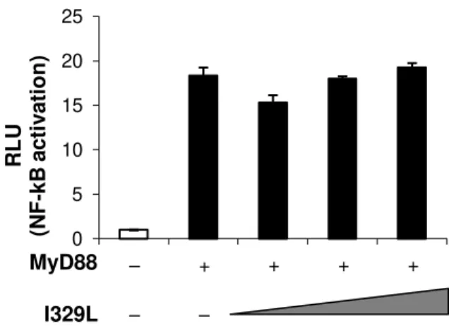
2.4.1.The inhibition of IFN-β induction by I329L is MyD88
independent and not through cytoplasmic sensors 108
2.4.2.The ASFV ORF I329L colocalizes to the early
endosome 111
2.4.3.The ASFV ORF I329L interacts with TRIF 113
2.4.4.The ASFV ORF I329L inhibits TLR3 signalling
pathway by two distinct mechanisms 117
2.4.5.Proteolytic processing of I329L 121
2.5.Discussion 128
β
3.1.Summary 135
3.2.Introduction 136
3.3.Materials and methods 139
3.3.1.Bioinformatic analysis 139
3.3.2.Cell culture 140
3.3.3.Plasmids 140
3.3.4.Lentivirus production 141
3.3.5.Lentivirus transduction of COS-1 cells 141
3.3.6.Luciferase reporter gene assay 142
3.3.7.Enzyme-linked Immunoabsorbent Assay (ELISA) 143
3.3.8.Western blot 143
3.3.9.Immunofluorescence 144
3.3.10.Immunoprecipitation 145
3.3.11.Statistical analysis 146
3.4.Results 146
3.4.1.MGF360-18R is a non-conserved ORF of the ASFV 146 3.4.2.The MGF360-18R colocalizes with the mitochondria 147 3.4.3.The inhibition of IFN-β transcription by ASFV ORF
MGF360-18R is NF-кB dependent 149
3.4.4.The ASFV MGF360-18R ORF inhibits induction of
IFN-β secretion 151
3.4.5.The ASFV MGF360-18R ORF inhibits activation of
IFN-β transcription by targeting MAVS 152
3.4.6.The MGF360-18R (Benin97/1) ASFV gene, but not the Ba971V variant, inhibits both TLR and cytosolic IFN-β
induction pathways, acting at the level of IRF-3 158
3.5.Discussion 163
4.1.Summary 173
4.2.Introduction 173
4.3.Materials and methods 176
4.3.1.Cell culture 176
4.3.2.Plasmids 176
4.3.3.Luciferase reporter gene assay 177
4.3.4.Western blot 178
4.3.5.Immunoprecipitation 179
4.3.6.Statistical analysis 179
4.4.Results 180
4.4.1.The ASFV MGF360-18R protein inhibits response
to both type I and type II IFN 180
4.4.2.The MGF360-18Rprotein diminishes the total
amount of cellular STAT1 but has no effect on STAT2 182 4.4.3.MG132, a proteasome inhibitor, reverts the
inhibition of GAS reporter by MGF360-18R 184
4.4.4.The proteasome inhibitor MG132 blocks
degradation of STAT1 in cells expressing MGF360-18R 185
4.5.Discussion 188
4.6.References 194
Chapter 5: Final Considerations 197
Figure 1.1.Activation of host-defence mechanisms 14
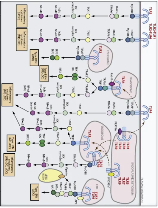
Figure 1.2.TLR trafficking and signalling 30
Figure 1.3.Cytosolic recognition pathways 34
Figure 1.4.Activation of classical Jak-STAT pathway by type I
and type II IFNs 42
Figure 2.1.ASFV I329L inhibition of Poly(I:C)-mediated IRF-3
and NF-кB activation is MyD88 independent 109
Figure 2.2.ASFV I329L inhibition of Poly(I:C)- mediated IRF-3
and NF-кB activation is independent of the RLR pathway 110
Figure 2.3.The ASFV ORF I329L inhibits Poly(I:C)-stimulated activation of IFN-β transcription in an NF-кB dependent
manner 111
Figure 2.4.The ASFV ORF I329L colocalizes to the early, but
not late, endosomes 112
Figure 2.5.The ASFV ORF I329L inhibits LPS-mediated
activation of NF-кB 114
Figure 2.6.The ASFV ORF I329L colocalizes to TRIF 115
Figure 2.7.The ASFV ORF I329L co-immunoprecipitates with
TRIF 116
Figure 2.8.Both domains of the ASFV ORF I329L inhibit Poly(I:C)-stimulated activation of IFN-β transcription in an NF
-кB dependent manner 118
Figure 2.9.The intracellular, but not the extracellular domain of the ASFV ORF I329L, inhibits ectopic TRIF stimulated activation of IFN-β transcription in an NF-кB dependent
manner 119
Figure 2.10.The ectodomain-transmembrane domain
fragment of I329L inhibits Poly(I:C)-mediated activation of the
is dependent on activation by either Poly(I:C) or ectopic
expression of TRIF and involves proteolytic cleavage 123
Figure 2.13.The ASFV mutant I329L gene has no effect on
IFN-β induction and NF-кB activation in cells stimulated by
ectopic expression of TRIF 125
Figure 2.14.The ASFV mutant I329L gene inhibits
Poly(I:C)-stimulated induction of IFN-β or NF-кB activation 126
Figure 2.15.The ASFV mutant I329L has no impact on secretion of IFN-β in cells induced by ectopic expression of
TRIF 127
Figure 3.1.MGF360-18R is a globular integral membrane
protein 147
Figure 3.2.Both variants of the ASFV MGF360-18R protein
colocalize with the mitochondria 148
Figure 3.3.Both variants of the ASFV MGF360-18R gene inhibit Poly(I:C)-mediated activation of IFN-β transcription in
an NF-кB dependent manner 150
Figure 3.4.MGF360-18R inhibits IFN-β secretion 151
Figure 3.5.Both variants of the ASFV MGF360-18R gene
inhibit activation of IFN-β transcription by MAVS 154
Figure 3.6.Overexpression of MAVS reverts MGF360-18R
mediated inhibition of the IFN-β transcription 155
Figure 3.7.Both variants of the MGF360-18R colocalize with
MAVS 156
Figure 3.8.The ASFV ORF MGF360-18R (Ba71V)
co-immunoprecipitates with MAVS 157
Figure 3.9.Expression of Benin97/1 variant of MGF360-18R,
but not Ba71V variant, inhibits the activation of IRF-3 159
Figure 3.10.The ASFV ORF MGF360-18R (Ba71V variant) does not inhibt IFN-β induction by ectopic expression of
TBK1 or IKKԑ 160
of phosphorylated and total IRF3, on activated cells
Figure 4.1.The two MGF360-18R variants inhibit signalling by
both type I and type II IFN receptors 181
Figure 4.2.Cells expressing MGF360-18R (Ba71V) protein
show a reduction in the levels of STAT1, but not STAT2 183
Figure 4.3.Addition of MG132 reverts MGF360-18R-mediated
inhibition of the impact of both type I and type II IFN 185
Figure 4.4.The proteasome inhibitor MG132 blocks degradation of STAT1 in cells expressing ASFV
MGF360-18R (Ba71V) 186
Figure 4.5. The proteasome inhibitor MG132 blocks degradation of STAT1 in cells expressing ASFV
MGF360-18R (Benin97/1) 187
Table 1.1.Viral PAMP detection by TLRs and other pRRs 8
Table 1.2.Phenotypic changes in IRF null mice 19
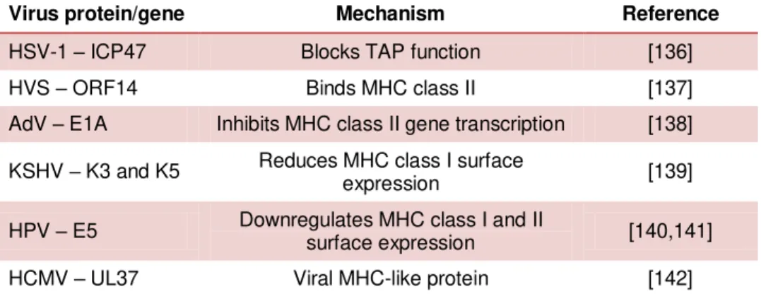
Table 1.3.Modulation of antigen presentation pathway 52
Table 1.4.Modulation of cytokines and cytokine responses 52
Table 1.5.Modulation of complement responses 53
Table 1.6.Modulation of apoptosis 53
Table 1.7.Inhibition of interferon production 60
Chapter 1
1. Introduction
The immune system of multicellular organisms evolved as a direct consequence of the selective pressure imposed by infectious microorganisms. The most ancient defence mechanisms, also known as the innate immune system, evolved to initiate an immediate and robust response against the invading microbes, and depend on relatively few germ-line encoded receptors. Emergence of the vertebrates was accompanied by the additional evolution of the adaptive immune system, which introduced a fundamental evolutionary advancement: more precise mechanisms of immune recognition and long-term immunological memory, based on an essentially infinite repertoire of receptors generated by gene rearrangements (reviewed by Hirano et al., 2011). [1]
1.1. Immune response to viruses
Viruses are particularly well adapted pathogens, capable of
parasitizing all cellular life forms, exploiting the host’s cellular
machinery for their survival and replication. Over millions of years of evolution, the organisms that are hosts to viruses have evolved anti-viral defences, but viruses have also responded through the evolution of multiple strategies to modulate, or inhibit host defences, so that the virus is able to complete the infectious process and then infect new hosts. The interactions between virus-infected cells and the host
defence mechanisms determine the harmful pathological
consequences that can occur during viral infection. These consequences reflect, not only the strategies that a given virus uses during infection, but also how the host resists infection. A highly virulent virus is not necessarily the most successful, as its very pathogenicity could lead to death of the host before the virus has spread. The concept that viruses benefit by mutating to less virulent forms, led to the assumption that virulence can be the result of incomplete adaptation of the virus to the host. A virus–host association that has existed for a long period is likely to have evolved a relationship in which the host suffers little or no harm. However, when a virus extends its host range into a new species, for example, recently emerged haemorrhagic viruses, typically will be much more virulent in the new host than in the old [3,4].
diagnostic reagents and techniques, and antiviral drugs. Virus host evasion mechanisms have also an enormous potential as a source of strategies for immunomodulation [3].
The host defence mechanism against viral infection consists of a complex relationship between components of the innate and the adaptive immune system. A key point in trying to understand this balance is the biology of the virus and its life style; as an obvious example, highly acute viruses, such as the recently virulent avian influenza, present a totally different challenge to the immune system than persistent viruses such as HIV, and this will not only dictate the most appropriate immune effector response necessary for effective protection, but also provide the rational framework for the development of novel therapeutic approaches.
The innate immune response represents a rapid first line of defence, and it is sometimes sufficient by itself to clear a viral infection. In these circumstances, innate immune mediators play a very important role in keeping the virus load low. Equally important, components of the innate response shape the adaptive immune response and direct the subsequent effector phase.
virus infection, and may result in associated immunopathology. Appropriate regulation of all these processes is necessary in order to have a successful immune response against an infection. Dissection of the critical cellular pathways that control these processes will eventually unveil opportunities for manipulating the host immune response to control viral infection and control pathogenesis (reviewed by Christensen & Thomsen, 2009) [5].
Both innate and adaptive immune responses and their role in protecting against viral infections will be briefly described below.
1.1.1. Innate immunity
Upon microbial infection, host survival critically depends on the establishment of a rapid and appropriate innate response. Characteristically, innate immune responses start within minutes or hours of infection, while activation of an effective antibody and activated cytotoxic lymphocyte response to the infectious agent takes several days. Initial control of viral spread is thus the responsibility of innate immunity. In addition, innate immunity, largely through the spectrum of secreted chemokines and cytokines, regulates the direction of the adaptive immune response. However, given that continued activation of the innate response may cause damage to the host, it must be itself tightly regulated and transient [6,7].
the epithelial cells lining the respiratory, gastrointestinal, and genitourinary tracts bear receptors that recognize conserved molecular structures expressed by a large variety of microbes, and so may also provide a component of innate immunity [6,8].
Cellular and humoral elements (complement system) of the innate immunity work together to constrain the virus spread and eliminate virus-infected cells, building an effective defence system against pathogenic microorganisms. The secretion of high levels of cytokines directs not only the activation and differentiation of the adaptive immune response but also the subsequent recruitment of antigen-primed effector T cells to the sites of viral replication (reviewed by Christensen & Thomsen, 2009) [5].
When exposed to a pathogen, epithelial cells and tissue resident macrophages are potent producers of cytokines. These are polypeptides that act as immunomodulating agents. They coordinate important aspects of the immune response, including inflammation, cellular recruitment, activation, proliferation and differentiation, being critical to the development and functioning of both the innate and adaptive immune responses. According to their function, cytokines can be divided into proinflammatory cytokines, anti-inflammatory cytokines and chemokines (chemoattraction mediators). Finally, cytokine expression is not restricted to cells of the immune system, e.g. epithelial cells can secrete cytokines such as IFN [6,9].
IFN-α and IFN-β, followed by TNF-α, IL-6, IL-12, IL-18 and type II interferon, IFN-. In addition to inducing a local antiviral response, cytokines also have a more general effect by inducing acute-phase proteins that are important and necessary for tissue damage repair and to clear infection (reviewed by Christensen & Thomsen, 2009) [5].
TNF-α, a proinflammatory cytokine, is able to regulate the expression
of adhesion molecules on the endothelium of nearby capillaries. This action induces changes which attract and facilitate the extravasation of leukocytes to the site of infection. When binding to receptors on infected cells, TNF-α is also able to induce an antiviral response that
eventually leads to apoptosis (reviewed by Rahman & McFadden, 2006) [10].
Type I IFNs are key contributors for both the innate and adaptive immune responses to viral infection. When an infected cell produces and releases type I IFNs, they will bind to the type I IFN receptors (IFNAR) of neighbouring cells. The consequent transcription of over 300 antiviral genes results in the inhibition of several steps of the viral life cycle. Furthermore, IFN-α/β are able to amplify the IFN original
signal, inducing an augmented antiviral state that result in secretion of high levels of cytokines and chemokines. Cells of the innate immune system are recruited to virus-infected tissues, where they are activated and in turn facilitate the induction of the adaptive immune response (reviewed by Le Bon & Tough, 2008) [11]
bound to the extracellular matrix or to cell surfaces through proteoglycans. Chemokines need to interact with specific cell surface receptors in order to exert their biological function. The two major families of chemokine receptors are the CXC chemokine receptors and the CC chemokine receptors (CCR), which bind CXC and CC chemokines, respectively (reviewed by Christensen & Thomsen, 2009) [5].
The intracellular signal transduction pathways responsible for expression of the multiple cytokines and chemokines released during viral infection, are activated as a consequence of pathogen recognition receptor (PRR) signalling in cells such as epithelial cells, macrophages and DCs. Germline-encoded PRRs are able to detect and distinguish between self and invariant microbial molecular structures (Pathogen Associated Molecular Patterns – PAMPs) shared by all pathogens of a given class. These molecular signatures are usually indispensable for the pathogen life cycle and are different from the molecular structures found in the host. In turn, the PRRs are similarly invariant. In response to PAMP recognition, PRRs execute the first line of host defensive responses and later participate in the control and direction of the second line of host defence, the adaptive immunity. The selective specificity of PRRs avoids activation of the immune system by self molecules. However, viruses usually replicate using host strategies and consequently generate molecular structures that resemble the molecular patterns found in the host. This poses a particular problem for innate recognition of viral infections (reviewed by Diebold, 2010) [12].
oligomerization domain (NOD)-like receptors (NLRs), C-type lectin receptors (CLRs) and DNA receptors (cytosolic DNA sensors). This variety of PRRs ensures the existence of multiple sensor systems that can detect and respond to almost any infection of the host. During viral infections, nucleic acid- and glycoprotein-PAMPs interact with particular classes of PRRs, which include certain Toll-like receptors, retinoic acid inducible gene-I (RIG-I), melanoma differentiation-associated gene 5 (MDA5), and the cytosolic DNA receptors. Interplay between TLRs and RLRs in different cell types during viral infection plays an important role in antiviral responses, as well in controlling adaptive immunity. While the cytoplasmic PRRs are responsible for limiting virus spread locally and for generating an inflammatory environment, the nucleic acid-sensing TLRs are crucial for orchestrating the adaptive anti-viral immune response that eventually leads to the elimination of the virus and virus-infected cells. To circumvent the fact that viral nucleic acids are structurally similar to eukaryotic nucleic acids, recognition occurs in a specialised endosomal compartment and not at the cell surface, in contrast to the TLR sensing bacterial, fungal and protozoan ligands (reviewed by Christensen & Thomsen, 2009, Diebold, 2010 and Kaway & Akira, 2011) [5,12,13].
PAMPs TLR usage PRRs involved in recognition
DNA TLR9 AIM2, DAI, IFI16
RNA TLR3, TLR7, TLR8 RIG-I, MDA5, NALP3
Structural protein TLR2, TLR4
Sensing of the invading viral pathogen through the appropriate PRR(s) triggers multiple and distinct signalling pathways, activating transcription factors such as interferon regulatory factor (IRF) 3 and 7 as well as Nuclear Factor-kappaB (NF-кB). Activation of these
pathways leads to the secretion of proinflammatory cytokines and chemokines that are involved in both innate and adaptive immunity. Some of the most critical mediators in the innate host response to viral infection are the type I IFNs (e.g. IFN-α and IFN-β). Type I IFNs induce
the expression of hundreds of IFN-stimulated genes that may have direct antiviral activity and/or modulate innate and adaptive immunity by activating immature DCs, enhancing NK-cell function and promoting survival and effector functions of T and B cells (reviewed by Christensen & Thomsen, 2009) [5].
1.1.2. Adaptive immunity
For T cells, two main functionally distinct sublineages exist, one
expressing an αβ T cell receptor (TCR) and the other expressing a δTCR. αβ TCRs specifically recognize antigens bound to MHC molecules, and participate in the activation of T cells in response to the presentation of antigen. The δ T cells, however, are thought to participate at the levels of both innate and adaptive immunity, and some, at least, recognize antigens directly, without the requirement for antigen presentation (reviewed by Born et al., 2011 and Chen, 2011) [15,16]
Immature αβ T cells leave the thymus as either CD4+ or CD8+ T cells. During viral infection, CD4+ T cells can differentiate into a variety of effector subsets, while CD8+ T cells differentiate into cytotoxic T lymphocyte (CTL), killing virus-infected cells, and also capable of releasing a range of effector cytokines. A subset of CD4+ T cells, known as regulatory T cells (Treg), regulates immune responses by suppressing them. T cells can only recognize peptides that have been degraded and bound to MHC class I or II. The MHC class I molecules (displaying endogenous peptides) are expressed on most somatic cells and interact with CD8+ T cells, whilst MHC class II (displaying exogenous, phagocytosed peptides) have a more limited expression, being restricted to professional APCs (such as DCs or B cells), and interact with CD4+ T cells (reviewed by Bonilla & Oettgen, 2010) [14].
express different profiles of cell-surface molecules that determine their effector cell capacity. The effector T cells had been thought to be terminally differentiated lineages, but it now appears that there is considerable plasticity allowing for conversion to other phenotypes (reviewed by Zhou et al., 2009) [17].
When immature CD8+ T cells interact with MHC class I – peptide complexes presented by professional APCs, they differentiate into CTLs that actively destroy any infected cells presenting the recognized foreign peptides. Activated CTLs up regulate perforin expression, which is stored in cytotoxic granules and released upon the recognition of an infected cell. Perforin is a pore-forming protein that leads to osmotic lysis of the target cells and subsequently enables granzymes to enter the target cells and initiate apoptotic cell death. In addition, the high levels of cytokines secreted by CD8+ T cells, induce an antiviral state in neighbouring cells, and apoptosis of the infected cell (reviewed by Smith-Garvin et al., 2009) [18]. Unfortunately, in cases of large-scale killing of virus-infected cells, the CTL activity may result in some degree of damage to the host organism.
pathways, naive B cells undergo clonal proliferation and terminal differentiation into short-lived antibody producing plasma cells or long lived memory B cells. Antibodies of all Ig classes can be produced in response to viral infections, and can significantly influence the outcome of the infection (reviewed by Bonilla & Oettgen, 2010) [14].
The function of antibodies in response to viral infections can be diverse. The major mechanism is antibody-mediated viral neutralization, occurring when antibodies bind the virus molecule that interacts with its cell-surface receptor, preventing virus attachment. Antibodies can also aggregate many infectious particles, resulting in their phagocytosis, therefore reducing the number of viruses that can effectively infect cells. Additionally, antibodies can act in concert with the complement system, IFN and other cytokines in order to clear viruses from persistently infected cells.
Following virus elimination, the pool of specific T and B cells substantially contracts, leaving a small population of antigen-primed memory cells, from which two major subsets of memory T cells are evidenced. A first line of specific defence is provided by effector memory T cells, in case of reinfection with the same or an antigenically related pathogen. Additionally, an expanded population of so-called central memory cells persists in the secondary lymphoid organs; these cells serve as a pool from which secondary waves of effector T cells may rapidly be derived, should the pathogen challenge overwhelm the forward defences (reviewed by Bonilla & Oettgen, 2010) [14].
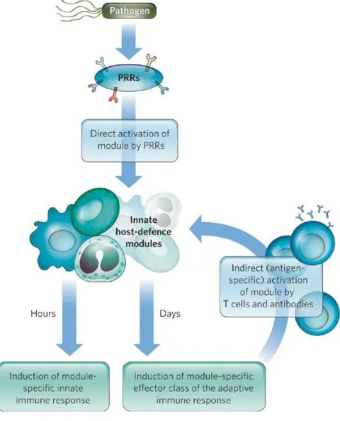
Figure 1.1 – Activation of host-defence mechanisms.
1.2. The Interferon System
In 1957, Isaacs and Lindenmann described the occurrence of a factor secreted by infected cells, able to inhibit viral replication in cells infected with homologous or heterologous viruses. They baptized this factor interferon (IFN) and conclusively demonstrated that IFN was a cellular product, acting to protect cells from viral infections [19,20,21].
The IFNs are now also recognized as central regulatory mediators of the immune response. The functions of IFNs are represented by three major biological activities: antiviral activity, antitumor activity and immunoregulatory activity. Fifty years have passed since the discovery of the interferon system, and much has been learnt about induction of IFN, IFN receptor signalling and IFN-dependent antiviral immunity. IFNs produced by infected cells are released and stimulate an antiviral state in neighbour cells, inducing the expression of proteins that interfere with viral processes, whereby viral replication is blocked or impaired. IFNs also have a major role in activation of the adaptive antiviral immune response. Immature plasmacytoid DCs (pDCs) are natural IFN-producing cells, and one of the key cells in the IFN-α
response to immune stimuli. pDCs differentiate into mature antigen-presenting DCs, which have a crucial role in T and B cell activation (reviewed by Fitzgerald-Bocarsly & Feng, 2007) [22].
Type I IFNs (IFN-α/β/ω/ɛ/κ in humans) possess strong antiviral activity,
and are able to induce a potent antiviral state in a wide variety of cells. However only IFN-α and IFN-β are induced directly in response to
virus infection. In humans, there are 30 genes coding for type I IFN, including 13 IFN-α genes, one IFN-β gene, one IFN-ω gene, one IFN-ɛ
gene, one IFN-κ gene and 13 additional pseudogenes of the IFN-α and
-ω families. The functional activities of this complex gene family are yet to be explored. These molecules signal through a ubiquitously expressed receptor composed of two chains: IFN-αR1 and IFN-αR2
(reviewed by Chelbi-Alix & Wietzerbin, 2007 and Hardy et al., 2004) [23,24].
There is only one type II IFN, known as IFN-, and which is secreted mostly by activated Th1 cells and NK cells and stimulates cell-mediated immune responses that are critical for the development of host protection against pathogenic intracellular microorganisms, such as the activation of macrophages for microbicidal activity. It also plays a central role in the development of antitumor immune responses, and it can amplify the induction of antiviral activity by IFN-α or -β, although
antiviral activity is not the primary biological function of IFN-. Type I and type II IFNs often work together to activate a variety of innate and adaptive immune responses that result in the induction of effective antitumor immunity and the elimination of viral infections. It signals via a ubiquitously expressed receptor composed of the IFN-R1 and
IFN-R2 subunits (reviewed by Young & Bream, 2007) [25].
and signal through a receptor complex composed of the unique
IFN-λR1 chain. Like the type I IFNs, their tissue distribution, specificity and regulatory mechanisms are not well understood (reviewed by Donnelly & Kotenko, 2010) [26].
Different human IFN-α’s and IFN-β, IFN-, and IFN-λ’s can establish
an anti-viral state in vitro; this is the essential signature of IFNs. However, the existence of three types of IFN, using three different receptors, raises the possibility of different roles in host defence against viruses and other pathogens.
1.2.1. Induction of IFN expression
Upon viral infection, cells are able to recognize the invading microorganism and, through multiple distinct routes that culminate in the induction of IFN, rapidly initiate antiviral mechanisms. The importance of any individual route of IFN induction depends upon the specific virus, the nature of the cell being infected or the stage of infection. Cells express PRRs that recognize viral PAMPs, differentiating them from self. Whenever a cell senses a virus infection, signal transduction pathways are activated, inducing the expression of type I IFN, a major component of the innate immune system. Type II IFN, in turn, is produced by activated lymphocytes and further amplifies the IFN response to infection (reviewed by Zhang et al., 2008) [27].
levels. The pathways leading to expression of either type of IFN are quite distinct, and so will be described separately.
1.2.1.1. Induction of Type I IFN
Virus infection of a cell induces the development of an antiviral state within the infected cell and due to the concomitant secretion of IFN, establishes an anti-viral state in nearby cells. The first potent IFN inducer to be identified was double-stranded RNA (dsRNA), a molecular pattern associated with viral infection, because it is produced by most viruses at some point of their replication [28]. It was postulated that dsRNA can mimic IFN induction by viruses. This finding facilitated investigations of the mechanism of IFN induction without the complexity of an associated viral infection [23]. Currently, the best studied model is the production of IFN-β in fibroblast cells in response
to either RNA viruses such as Sendai virus (SeV) or the synthetic chemical that mimics dsRNA, the polyinosine-polycytidylic acid (Poly (I:C)).
Type I IFN expression can be induced by several different mechanisms. However, the downstream kinases and transcription factors are common to all. Virus- or dsRNA-induced expression of type
I IFN is controlled by sequences present in the 5’ flanking region of the
IFN-α/β genes.
Two families of transcriptional factors play a major role in the transcriptional activation of type I IFN genes: the family of NF-кB and the family of interferon regulatory factors (IRF).
In unstimulated cells, NF-кB proteins (p65 and p50) exist as homo- or heterodimeric proteins which are retained in the cytoplasm by
stimuli derived from the antiviral response activate the IкB kinase
(IKK). This kinase is responsible for the phosphorylation of serine residues within the N-terminal destruction box of IкB proteins (e.g. S32 and S36 of IкBα). Phosphorylated IкB is subsequently ubiquitinated
and degraded by the proteasome, thus unmasking the nuclear localization signal of the NF-кB proteins, which translocate into the nucleus and bind to type I IFN promoter [29].
The family of IRF transcription factors mediate virus-, bacteria- and IFN-induced signalling pathways and as such play a critical role in antiviral defence, immune response, cell growth regulation and apoptosis. To date, IRF-1, IRF-3, IRF-5, IRF-7 and IRF-9 have been described as major regulators of type I IFN transcription, in concert with the transcription factor NF-кB [30].
The availability of genetically modified mice, which have distinct IRF deleted, has revealed the function of the members of the IRF family. Table 1.2 summarises what is known about IRF involved in the transcription of type I IFN genes:
IRF Defects
IRF-1 Apoptosis, iNOS, IL-12
IRF-3 Down modulation of type I IFN induction Increased susceptibility to infection
IRF-5 Induction of inflammatory cytokines (IL-6, TNF-α and IL-12) IRF-7 Block in the type I IFN induction
IRF-9 Type I and II IFN signalling, induction of IRF7, IFN-α and ISG
IRF-1 was identified by its ability to bind to the positive regulatory domain 1 (PRDI) in the virus-responsive element (VRE) of the IFN-β
gene, where it was assumed to function as an activator of transcription [31]. However, although IRF-1 is present in the IFN-α and IFN-β
enhanceosomes, binding to the respective promoter regions, it does not have a critical role in the virus stimulation of type I IFN genes. Instead, IRF-1 was shown to be involved in the antiviral defence mediated by IFN- and to play a critical role in the inducible expression of MHC class I and apoptosis [32,33]. More recently, it was demonstrated that IRF-1 is not required for IFN expression, but it is needed for expression of interferon-stimulated genes (ISGs) [34]. Thus, IRF-1 may uniquely control IFN-independent signalling events that lead to ISG expression and antiviral immunity.
IRF-3 and IRF-7 were identified by their ability to activate the promoters of IFN-α and -β genes. The identification of these two IRFs
and their role in the transcriptional activation of Type IFN genes had a major impact on the understanding of the molecular mechanism of the pathogen-induced innate antiviral response [35,36]. Although pathogen recognition may be mediated by distinct cellular receptors and signalling pathways, they all lead to the activation of IRF-3 or IRF-7 which are critical for the transcriptional activation of Type I IFN genes [37,38]. The IFN-β enhanceosome not only contains IRF-3 but also IRF-7 [39]. In addition, several authors have gathered evidence suggesting that relative levels of IRF-3 and IRF-7 in cells determine the levels of expression of individual IFN-α subtypes (reviewed by Paun & Pitha, 2007) [40].
TLR-3 and RIG-I/MDA-5 signalling pathways lead to the phosphorylation of IRF-3 at the carboxyl-terminal region (serines 385 and 386) and at the serine/threonine cluster (between region 396 and 405), by the IKK-related kinases, TANK-binding kinase (TBK)-1 and
IKKɛ. Serine 386, at C-terminal region, is critical for activation, as it is predicted to lead to a conformational change that allows IRF-3 to homo- or heterodimerize with IRF-7 [38,41,42]. Following translocation to the nucleus, IRF-3 associates with the co-activator CREB binding protein (CBP)/p300 and stimulates transcription of IFN-β[43], as well as some ISGs, such as CCL5/RANTES and ISG54 [44].
Several observations underline the importance of IRF-3 in the induction of the antiviral response. First, being ubiquitously expressed, IRF-3 is capable of stimulating the antiviral response and synthesis of IFN-β in all varieties of infected cells. Second, several viruses target
IRF-3, thus preventing the induction of Type I IFN. IRF-3 is required for type I IFN induction triggered by TLR3/TLR4, cytosolic RNA sensing or cytosolic DNA sensing pathways in many cell types, including cDCs, but it is not required for type I IFN induction in pDCs [40].
IFN-α production by an IKK-α (and not TBK1) dependent pathway
[46,47,48].
Constitutive expression of IRF-7 is restricted to some lymphoid cells, particularly pDCs that express high amounts of IFN-α in response to
TLR7/8 and TLR9 activation [40]. When induced to differentiate, monocytes express IRF-7, which was shown to be a key regulator of the differentiation of monocytes to macrophages [49]. Expression of IRF-7 can be induced in most cells types, not only by Type I IFN but also by TNF-α [50]. Finally, IRF-7 has a short half-life, a characteristic that may play a role in the regulation of the transient expression of IFN-α genes [51].
Constitutive expression of IRF-5 is restricted to few cell types, such as monocytes and DCs that express high levels of IFN-α upon viral
infection. IRF-5 is mainly expressed in the cytoplasm of non-infected cells and, upon viral infection, is phosphorylated and activated by distinct kinases. The activated IRF-5 forms either homodimers with itself or heterodimers with IRF-3 and then translocates into the nucleus. Both RNA and DNA viruses can activate IRF-5 nevertheless this activation is virus-specific [52]. Like IRF-7, MyD88-mediated activation of IRF-5 involves the formation of a complex composed by MyD88, IRAK4, IRAK1 and TRAF6. Most likely, this complex preferentially assembles with IRF-7 [46,53].
lymphocyte-chemotactic activity, it was suggested that IRF-5 may have an important role in lymphocyte trafficking [56,57]. Recently, a new role was described, in which IRF-5 expression in macrophages is responsible for initiating a potent Th1-Th17 response [58].
The transcription of the IFN-β gene requires an enhancer element
located upstream of the core promoter that is recognized by three distinct sets of transcription factors (NF-кB, IRFs and ATF-2/cJun) and by the high-mobility group (HMG) chromatin-associated protein HMGI(Y) [59]. This enhancer element is composed by four positive regulatory regions (PRDI-IV): PRDI and PRDIII sites are for binding of IRF-7 and IRF-3, the PRDII site is for binding of NF-кB, and PRDIV
site is for binding of ATF-2/cJun heterodimers. Virus infection leads to coordinated activation of all three types of transcription factors, which assemble on the IFN enhancer region to form a large, multi-subunit complex known as the IFN-β enhanceosome [32]. In a first phase, after being delivered to a single IFN allele, NF-кB plays a crucial role in the
recruitment of the remaining factors to the enhanceosome, leading to IFN-β transcriptional activation which, in turn, activates transcription of
IRF-7. At a second phase, the increasing levels of IRF-7 trigger enhanceosome assembly on multiple IFN-β alleles, thus amplifying the
production of IFN-β [60]. In order to have an optimal induction of the IFN-β promoter, cooperation between all transcription factors is
required. Since virus infection is the only known signal that can activate all of the IFN-β transcriptional activators simultaneously,
The promoter region of IFN-α genes contains only binding sites for
IRFs, lacking binding sites for NF-кB. Although the identity of the IRF
members that stimulate IFN-α transcription is uncertain, there is some
evidence that IRF-7 is required for induction. In pDCs, which constitutively express IRF-7 and induce the expression of massive amounts of type I IFN, the induction of IFN-α is not dependent on
primary induction of IFN-β and its feedback loop [62].
The activation of the different mechanisms leading to IFN expression requires, as a first step, recognition of the viral infection by the host cell. The discovery of pattern associated molecular patterns (PAMPs) and their recognition by cellular pattern recognition receptors (PRRs) has revolutionized our understanding of innate immunity, and explains how and why a virtually unlimited number of pathogens can be recognized by a small number of innate immune receptors, triggering anti-microbial responses.
There are at least two major complementary receptor systems (see Fig. 1.2 and 1.3) that detect most viral products: one class of receptors detects viral nucleic acids in endosomes of specialized cell types, whilst the second class of receptors are expressed ubiquitously and localized in the cytosol, where they are able to detect viral nucleic acids produced upon infection [63]. In addition to viral nucleic acids, several viral proteins have been shown to induce IFN, although this is not a general feature of viruses. For example, the fusion (F) protein of respiratory syncitial virus (RSV) and the glycoprotein G of vesicular stomatitis virus (VSV) can activate the synthesis of IFN type I through a TLR4-dependent pathway [64].
The distinct classes of antiviral PRRs and the strategies employed by the host for the successful detection of a viral infection will be briefly described.
1.2.1.1.1. The Toll like receptor pathway
Toll-like receptors (TLRs) are a family of PRRs that play central roles in innate immune defence against infection by binding to microbial molecules.
requires involvement of the TM domains and directs juxtaposition and activation of the intracellular domains. The intracellular signalling domains contain an intracellular Toll/IL-1R (TIR) motif, important in protein-protein interactions. This motif is also present in the signalling adaptors that are recruited to the ligand-activated TLR TIR domains, forming the first step in the signalling cascade leading to the expression of multiple genes involved in innate and adaptive immunity, including type I IFN (reviewed by Kang & Lee, 2011) [65].
TLRs are primarily expressed in sentinel APCs of the immune system such as macrophages and DCs, but can also be present in epithelial cells. The cellular expression of the different TLRs is heterogeneous: For example, TLR3 is expressed by mDCs and NK cells, whereas TLR7 and TLR9 are expressed by macrophages and both mDCs and pDCs, the latter being known to produce high levels of type I IFN in response to viral infection (reviewed by Moresco et al., 2011) [66].
CpG DNA motifs) prevents the recognition of self nucleic acids and activation of signalling pathways in the absence of infection. In addition to its intracellular expression, TLR3 was also detected on the surface of a few cell types, including fibroblasts, but until now no studies were published on the comparison of the physiological significance of intracellular versus cell surface TLR3 (reviewed by Barton & Kagan, 2011) [67]. Trafficking of endosomal receptors to endolysosomal compartments by UNC93B1 and proteolytic regulation of some TLRs (TLR7 and TLR9) are other strategies used to further control receptor activation [68,69,70].
In addition to recognizing distinct ligands, individual TLRs trigger different signal transduction pathways. This specificity is achieved by the engagement of different adaptors to different receptors, through interaction between the corresponding TIR domains. The particular signalling adaptor used determines which signalling pathway will be activated: TIR-containing adaptor MyD88 induces a pro-inflammatory response dependent on the activation of NF-кB and mitogen-activated protein (MAP) kinase, whereas TIR domain-containing adaptor protein inducing IFN-β (TRIF) is responsible for activation of IRF-3, IRF-7 and NF-кB, culminating in the induction of type I IFN and inflammatory
cytokines (reviewed by Moresco et al., 2011) [66].
being phosphorylated in an IRAK1-dependent manner, IRF-7 translocates to the nucleus and binds the promoter of the IFN-α gene
[46]. Combining the observations that pDCs have an efficient mechanism for retaining CpG DNA in the endosomes, and also have a high constitutive expression of IRF-7, it is not surprising that these cells express huge amounts of IFN-α.
The signalling of TLR3 is induced through TRIF, and not MyD88. Activated TRIF associates with TRAF3 and TRAF6 and subsequently, with the noncanonical kinases TBK-1 and IKKɛ (Fig.1.2), phosphorylates IRF-3 and IRF-7, leading to its dimerization and nuclear translocation to bind the promoter of type I IFN. TRIF also mediates the activation of NF-кB and activating protein 1 (AP-1) through the complex of kinases IKK-α/β/. These two transcription factors translocate into the nucleus, together with IRF-3 and IRF-7, and bind to the PRDI-IV positive regulatory elements of the IFN-β
enhancer region [72]. TLR3-mediated signalling also leads to phosphorylation of specific tyrosines and the recruitment of phosphatiylinositol 3-kinase (PI3K), essential for full activation of IRF-3 [73,74].
Virus infected cells mainly depend on TLR3, TLR7/TLR8 and TLR9 to induce the expression of type I IFN, following detection of viral nucleic acids. However, TLR4 is also capable of inducing type I IFN by the recognition of non-nucleic acid ligands, such as lipopolysaccharide (LPS). Upon LPS-binding, TLR4 initiates signalling transduction pathways through both MyD88 and TRIF adaptors. Signalling through MyD88 requires TRAM [71] and culminates in the activation of NF-кB,
TLR4 induces both a late pro-inflammatory response (NF-кB
dependent) and type I IFN expression.
Recently, a TLR2-dependent antiviral signalling pathway leading to the production of type I IFN was reported in inflammatory monocytes. Like TLR4, TLR2 recognizes certain viral proteins, and when it does so, it is internalized into endosomal compartments. However, in contrast with TLR4, all TLR2 signalling is MyD88 dependent. Thus, inflammatory monocytes are able to use TLR2 to activate unique MyD88-dependent pathways culminating in the activation of IRF3, IRF7 and NF-кB [77].
1.2.1.1.2. The cytosolic recognition pathway
All viruses, even those replicating within the nucleus (herpes viruses), include a cytoplasmic phase in their replication strategy; for example, viral genome amplification and/or mRNA metabolism and viral protein expression. Within the cytosol, there are specific PRRs that recognize viral nucleic acids, such as RIG-I-like receptors (RLRs), nucleotide binding and oligomerization domain (NOD)-like receptors (NLRs), and DNA receptors (cytosolic DNA sensors). Like TLRs, cytoplasmic sensors activate signalling transduction pathways leading to the production of type I IFN and pro-inflammatory cytokines (reviewed by Wilkins & Gale, 2010) [79,80]
The RLR family is composed by three RNA helicases, RIG-I, MDA-5 and Laboratory of Genetics and Physiology (LGP)-2. RIG-I and MDA5 are ubiquitously expressed in most cell types and are able to recognize viral RNA in the cytoplasm, leading to induction of IFN. On the other hand, LGP2 acts as a negative regulator of IFN gene expression, most probably by masking viral dsRNA from recognition by RIG-I and MDA5 [81,82].
Although structurally similar, RIG-I and MDA5 are not redundant and are responsible for IFN induction by different sets of viruses. This virus specificity may be the result of the distinct recognition of particular RNA structures or nucleotide composition by each sensor. For example, RIG-I specifically binds to a free 5´-triphosphate RNA structure. This feature probably allows for discrimination between self and non-self RNA, since 5´-ends of most endogenous RNAs are either capped or post-translationally modified to remove the 5’-triphosphate [73,82,83,84].
The adaptor for RIG-I and MDA-5 was identified and, although named differently by different groups, the recommended name is now mitochondrial antiviral signalling protein (MAVS) [37,84,85]. The MAVS protein is found in the outer mitochondrial membrane, a location essential for its function [38,85,86]. The interaction between the CARD domains of RIG-I and MDA5 with the CARD domain of mitochondrial MAVS leads to the activation of two IкB kinase-related kinases, TBK-1 and IKKɛ, responsible for the phosphorylation of IRF-3 and IRF-7. In addition, MAVS also activates NF-кB by a TRAF6 dependent pathway.
Recognition of viral DNA in the cytoplasm is carried out by specific DNA sensors. The first cytoplasmic DNA receptor to be identified was the DNA dependent activator of IFN-regulatory factors (DAI). This protein binds B-form DNA (particularly poly(dA:dT)), triggering activation of NF-кB, IRF-3 and possibly IRF-7, thus being responsible for DNA-dependent type I IFN induction in some cell types. However, cells that do not express DAI are still able to respond to viral DNA in the cytoplasm, suggesting that other DNA receptors must exist [90,91]. Recently, two additional proteins involved in the detection of cytosolic DNA and subsequent IFN induction have been identified: RNA polymerase III and interferon-inducible protein 16 (IFI16) (see Fig. 1.3) [92]. RNA polymerase III acts indirectly by transcribing AT-rich DNA
into uncapped 5′ triphosphate–bearing RNA, which serves as an agonist for RIG-I [93,94]. IFI16, a member of the pyrin and HIN200 domain (PYHIN)–containing protein family, is a sensor for intracellular non–AT-rich dsDNA [73]. These studies have shown that detection of cytosolic DNA probably requires multiple and possibly redundant sensors that converge on the signalling molecule STING and the kinase TBK-1 and lead to activation of the transcription factor NF-кB
Figure 1.3 – Cytosolic recognition pathways. (from Goubau et al., 2010)
1.2.1.2. Induction of type II IFN
1.2.1.3. Induction of type III IFN
In 2003, a novel class of IFNs has been identified, and named type III IFNs or IFN-λ. Type III IFNs have functional similarities with type I
IFNs, but unlike type I IFNs, which exert antiviral activity on all cell types, type III IFNs target primarily epithelial cells, and consequently play an important role in innate antiviral defences at epithelial surfaces, which constitute a major portal of entry for viral infections [97].
In addition to having similar functions, type I and type III IFNs also have similar expression patterns. In fact, it was determined that type III IFN genes are expressed in response to most classes of viruses and to a variety of TLR agonists, the same stimuli responsible for expression of type I IFN genes. Computer analysis of promoter sequences of type III IFN genes predicted the existence of potential binding sites for several transcription factors, some already known to be involved in the regulation of type I IFN genes transcription, e.g., AP-1, NF-кB, and various IRFs [26]. Accordingly, it was recently demonstrated that both classes of IFNs are induced by transcriptional mechanisms involving IRFs and NF-кB. However, while IFN-β
induction requires the coordinated action of a multifactor enhanceosome, and IFN-α expression is activated by multiple IRF -binding cis-promoter elements, the type III IFNs are induced through independent actions of IRFs and NF-кB. Hence, it was proposed that
IFN-λ expression is more flexible than IFN-α/β expression, which could
allow expression of type III IFNs in response to a wider range of stimuli compared with type I IFNs (reviewed by Iversen & Paludan, 2010) [98].
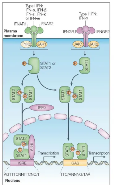
intracellular signalling pathway, regulating many of the same biological activities, including a range of antiviral immune responses [26]. The JAK-STAT pathway was the first signalling pathway shown to be activated by IFNs (Fig.1.4), and extensive studies over the years have firmly established its functional relevance in the interferon system.
Type I IFNs are secreted factors that are recognized by a cell surface transmembrane receptor – the type I IFN receptor. This protein is a heterodimer composed of two subunits, IFN-α receptor 1 (IFNAR1)
and IFNAR2, which cytoplasmic domains are associated with the inactive Janus tyrosine kinases, Tyk2 and Jak1, respectively. Prior to stimulation, IFNRA2 is also bound to STAT2 that is, in turn, weakly associated with STAT1. Upon IFN binding to the receptor and subsequent stimulation, the two subunits of the receptor associate and facilitate the activation of Tyk2 and Jak1. The phosphorylation of the tyrosine at position 466 (Tyr466) on IFNAR1 by Tyk2, creates a docking
site for the SH2 domain of STAT2, and its subsequent phosphorylation by Tyk2 at Tyr690, while Jak1 phosphorylates STAT1 on Tyr701
[64,99,100]. The activated STATs dissociate from the receptor forming a stable heterodimer and associate with IRF-9, forming the ISGF3 tertiary complex that translocates into the nucleus. In this complex, IRF-9 is the major DNA binding component and, in the nucleus, binds to IFN-stimulated response elements (ISRE) present in the promoter region of IFN-stimulated genes (ISGs), inducing their transcription. IRF-9 can also form a DNA binding complex with STAT1 homodimers and with STAT2 alone, and these complexes can bind to DNA with the same specificity as ISGF3 (reviewed by Paun & Pitha, 2007) [40].
protein (CBP)-mediated acetylation cascade, together with serine phosphorylation, also plays a critical role in type I IFN intracellular signalling. The cytoplasmic CBP protein is a mediator for the acetylation of cytokine receptors and their downstream signalling molecules, e.g. IFNRA2, IRF9, STAT1 and STAT2. Acetylation plays a major role in the complete formation and activation of the ISGF3 complex, thus mediating cytokine receptor signal transduction (reviewed by Tang et al., 2007) [101].
The type II IFN receptor is also a heterodimer composed of two subunits, the IFN- receptor 1 (IFNGR1), which associates with Jak1, and the IFNGR2, which constitutively associates with Jak2. Dimerization of the receptor, upon binding of IFN-, leads to association of Jak1 and Jak2 and subsequent activation of Jak2 which, in turn, phosphorylates Jak1. After being phosphorylated by activated Jak1 and Jak2, the C-terminus of IFNGR1 creates a pair of binding sites for STAT1, allowing for its phosphorylation at Tyr701. The
phosphorylated STAT1 homodimer dissociates from the receptor and translocates into the nucleus, where it binds to unique elements of IFN- stimulated genes, the gamma-activation sequence (GAS), and induces transcription. Of note is the fact that type I IFN stimulation is also able to form STAT1-homodimers, leading to the induction of ISGs containing GAS elements in their promoter region (reviewed by Goodbourn et al., 2000) [99].
IFN-λs exert their biological activities by signalling through a
heterodimeric receptor complex composed of IFN-λ receptor 1 (IFN
signal transduction cascade. Although IFN-λR1 is constitutively
expressed by a broad range of cell lines and tissues, there are many cell types that do not express IFN-λ receptors, and, as a consequence,
cannot respond to this cytokine (reviewed by Donnely et al., 2010) [26].
Type I and type II IFN also induce phosphorylation of STAT1 on Ser727,
and although this phosphorylation is not required for either nuclear translocation or DNA binding, it is essential for full transcriptional activity of STAT1. This reaction is catalysed by protein kinase C isoform PKC-δ, which is activated by the PI3K pathway, and directly
interacts with STAT1 [102,103].
An effective antiviral response requires a rapid and efficient induction of ISGs by the Jak-STAT pathway, in response to IFNs released by infected or activated cells. However, this response must be tightly regulated and terminated once the viral threat is over, in order to avoid damage to the host, and several negative regulators of the Jak-STAT signalling have been already described.
Phosphatases are important regulators of kinase based signalling cascades, inducing dephosphorylation of specific amino acid residues and, consequently negatively regulating activation of effector proteins. In particular, SH2-containing phosphatase (SHP)-1, SHP-2 and protein tyrosine phosphatase 1B (PTP1B) dephosphorylate tyrosine residues on the IFN receptors or on Janus kinases, preventing tyrosine phosphorylation of STATs and their subsequent activation [104,105].
protein turnover of the IFN receptor through ubiquitin-proteasome-mediated degradation, preventing STAT activation [104,106,107]. PIAS – proteins that inhibit activated STAT, are a family of proteins able to inhibit active STATs at the nuclear level. PIAS1 interacts with tyrosine phosphorylated STAT1, blocking its DNA binding ability, while PIASy acts as a transcriptional co-repressor of STAT1 [104,108]. PIAS proteins can also act as platforms to facilitate both removal and recruitment of other regulatory proteins, such as SUMO proteins [109].
STAT1 activity can be regulated by another cytoplasmic inhibitor, the small ubiquitin-like modifier (SUMO) proteins. SUMO posttranslational modification (SUMOylation) is a dynamic and reversible process that can both decrease STAT1 tyrosine phosphorylation and indirectly facilitate STAT1 dephosphorylation. Phosphorylation of STAT1 at Tyr701 and SUMOylation at the adjacent Lys703 are mutually exclusive, such that Tyr701-phosphorylated STAT1 remains unSUMOylated [110,111]. This results in the formation of semiphosphorylated STAT1 dimers that function as competitive polymerization inhibitors, increasing the solubility of fully active STAT1 molecules, thus inducing its dephosphorylation [110]. Together, these two mechanisms are able to diminish the amount of transcriptionally activate STAT1 in the cell nucleus, protecting cells against hyper responsiveness to IFN- [112].
STAT1β is a truncated form of STAT1, lacking the transactivation domain (TAD). This is a naturally occurring form of the protein, formed by differential splicing, that can replace STAT1 in homo- or
heterodimers formed following receptor activation. However, STAT1β