UNIVERSIDADE DE TRÁS-OS-MONTES E ALTO DOURO
Contribution to the study of emerging infectious
diseases in broiler breeders
Tese de doutoramento em Ciências Veterinárias
Sanidade Animal
Fernando Alberto Brandão Campos Lopes Moreira
Composição do Juri:
Prof. Dr. Vicente de Seixas e Sousa
Prof. Dr. Manuel Pizarro Díaz
Prof. Dr. Manuel Martins
Profª. Drª. Gertrude Thompson
Prof. Dr. Helder Cortes
Profª. Drª. Alexandra Esteves
Prof. Dr. João Simões
Prof. Dr. Nuno Alegria
Profª. Drª. Ana Cláudia Coelho
Supervision
Supervisor:Profª. Drª. Ana Cláudia Coelho
Department of Veterinary Sciences, School of Agrarian and Veterinary Sciences, University of Trás-os-Montes e Alto Douro (UTAD), Animal and Veterinary Science Center (CECAV) Quinta de Prados, 5001-801 Vila Real, Portugal
Co-supervisor:
Prof. Dr. Luís Cardoso
Department of Veterinary Sciences, School of Agrarian and Veterinary Sciences, University of Trás-os-Montes e Alto Douro (UTAD), Quinta de Prados, 5000-801 Vila Real – Portugal
This study was partially sponsored by the strategic research projects Pest-OR/AGR/UI0772/2011 and Pest-OE/AGR/UI0772/2014, financed by the Foundation for Science and Technology (FCT, Portugal).
“Os homens passam, as conquistas científicas permanecem ou
transformam-se. A história, que as arquiva, fará a sua crítica.
Sinto-me sombra a desvanecer-se nas gerações que se seguem. E
agora, ao despedir-me, ouso rematar: esforcei-me por bem
cumprir o meu dever.”
Egas Moniz, A última lição, 1944
Para a minha avó,
Maria Teresa Brandão
Acknowledgements
For all the support, I am very much thankful to:
My family: Fernando and Maria Paula, Frederica, Rolanda, Maria Teresa, António Alberto and the whole Moreira, Brandão, Campos and Lopes Family. To Fernando, Daniela, Emília, José Carlos, Rui, Susana, António, Francisco and the whole Peixoto and Tiago Family.
UTAD: Rector of UTAD Prof. Dr. António Fontaínhas Fernandes, Prof. Dra. Ana Cláudia Coelho, Prof. Dr. Luís Cardoso, Prof. Dra. Maria de Lurdes Pinto, Mrs. Fátima Fraga.
Lusiaves Group: Mr. Avelino Gaspar, Mrs. Susana Gaspar and the whole Gaspar Family, Eng. Pedro Ferreira, Eng. Filipe Ribeiro, Eng. Miguel Loureiro, Dr. Luís Ferreira, Eng. André Sousa, Eng. Henrique Pires, Eng. Alexandre Rolo, Eng. João Borges, Eng. Tiago Correia, Eng. Ângela Rodrigues, Dr. Ignácio Barragán, Mr. Vitor Oliveira, Eng. Sandra Bom, Eng. Nuno Ruivo, Mr. Paulo Amaral, Dr. Bruno Abreu, Eng. André Conde, Eng. António Tomas, Dr. Elisa Neves, Eng. Catarina Marques, Eng. Conceição Teodósio, Eng. Filipe Santos, Eng. Tiago Marques, Eng. Mónica Godinho, Eng. Cláudia Carreira, Eng. Luís Gaspar, Mr. Carlos Braz, Eng. Simão Pereira, Eng. Pedro Pinhão, Eng. Ruben Fonseca, Mr. Vitor Pinho, Eng. Luís Valente, Dr. André Leite, Eng. Ana Teles, Eng. Joana Pinheiro, Eng. Luís Antunes, Eng. Rafael Gonçalves, Eng. Pedro Palos, Eng. Mário Oliveira, Eng. Hugo Gomes, Eng. Andrea Figueiredo, Eng. Márcio Fernandes, Mr. Daniel Santos and Dr. Ricardo Batista.
Controlvet: Dr. Rui Sereno, Eng. Ana Patrícia, Eng. Dina Fernandes and Dr. Rita Silva.
Cobb Group: Dr. Juan Carlos Abad, Dr. Jaime Sarabia and Dr. Samuel Villares.
Ross Group: Eng. Juan António Játiva and Mr. António Marques.
PDRC: Dr. Blayne Mozisek.
Summary
This investigation, regarding important emerging infectious diseases, was carried out in one of the most important stages of the poultry industry, i.e. broiler breeders. This type of birds is susceptible to several agents that interfere with the immune system and predispose to infection. If the transmission of these pathogens to progeny is considered, their economic impact will be amplified in the broiler farms, compromising the overall production results. The construction of multi-age farms (farms with birds of different ages) poses a significant epidemiological risk. The fact is that farms have grown in size and density, and an ideal environment has been created for agents such as Mycoplasma synoviae, Aspergillus spp. and Reovirus to thrive.
The aim of the research described in this thesis has as its primary goal the acquisition of updated knowledge on these agents at broiler breeder level in Portugal. The creation of strategies for the control and prevention has also been considered. With this purpose, several studies were carried out and are described in the following chapters:
Chapter 1 – “Emerging infectious diseases in broiler breeders”. A general introduction comprising a review of the scientific literature concerning Mycoplasma synoviae, Aspergillus spp. and Reovirus, epidemiology, their economic importance, pathogenesis, lesions, clinical signs, diagnosis, control, treatment and prevention.
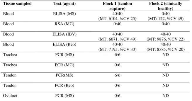
Chapter 2 – “Decreased production in broiler breeders due to tendon rupture by Mycoplasma synoviae”. In February 2013, an outbreak of lameness occurred in a broiler breeder flock (30,000 birds divided by four houses) of a multi-age farm with a total of two broiler breeder flocks (60,000 birds in total). The leg lesions began soon after transfer, between 22 and 24 weeks of age, and persisted for the rest of the flock’s life, until 64 weeks of age. Post mortem examination at the farm revealed lesions of arthritis and synovitis affecting the hock joint and the foot pads. Mycoplasma synoviae was detected in birds from the affected flock by serologic and molecular techniques. Treatments with fluoroquinolones in drinking water reduce the number of cases, but the morbidity continued and affected the standard production rates.
VIII
Chapter 3 – “Epidemiological survey on Mycoplasma synoviae infection in broiler breeder flocks by serology and polymerase chain reaction (PCR)”. Since the modernization and expansion of the poultry industry, infections with Mycoplasma spp. bacteria have been reported as a cause of considerable economic losses. The prevalence of M. synoviae infection in 974,000 Portuguese broiler breeders, belonging to 36 different flocks, was investigated from December 2008 to March 2012. The birds were analyzed by a commercial indirect ELISA (enzyme-linked immunosorbent assays), for serum antibodies, and a PCR (polymerase chain reaction), for tracheal tissue. Twenty-four flocks were positive (66.7%, 95% confidence interval [CI]: 43.5-76.9%). Infection with M. synoviae was confirmed by PCR in all of the 29 seropositive flocks. The Mycoplasma synoviae prevalence among chickens averaged 40.3% (483/1200), with values ranging from 0.0 to 83.3% per flock. The prevalence of M. synoviae farms with positive birds was determined in different poultry categories such as density, biosecurity, strains, offspring quality, premises’ age and other husbandry factors. Prevalence values were significantly higher among birds housed in new facilities, in birds with less than 3 years upon construction and also during the production period. The high prevalence of M. synoviae infection detected in the present study requires the adoption of appropriate control measures. Animal health authorities should be alert to the economic impact of M. synoviae, strategies for the planning and construction of new poultry premises and a raising awareness should be put into practice among the poultry industry.
Chapter 4 –“Presumptive Reovirus infection in broiler breeders”. Tenosynovitis is one of the pathological manifestations of avian Reovirus infections and are of economic importance, especially in broiler breeders, which are one of the most important stages of the poultry industry. In late May 2012, an outbreak of unilateral lameness occurred in a broiler breeder flock (15,000 birds divided by two houses) of a multi-age farm, with a total of four broiler breeder flocks (65,000 birds in total). Birds aged from 20 to 30 weeks presented joint lesions. Morbidity ranged between 5 and 10%. Routine post mortem examination of 30 birds revealed a range of visible joint lesions, typically unilateral in distribution. After post mortem examination, samples of tendons, heart and liver were collected for histopathology, bacteriological culture and PCR analysis. Microscopic examination of the tendons revealed changes consistent with a Reovirus infection and, to the authors’ knowledge, these are the first reported cases of viral arthritis in broiler breeders in Portugal.
Chapter 5 – “Assessment of Aspergillus spp. in a modern broiler breeder structure”. Members of the genus Aspergillus are ubiquitous opportunistic saprophytic fungi regarded as major respiratory pathogens in birds, which can cause significant economic losses in chickens, with mortality, decreased weight gain and reprobation at slaughterhouse. Initial contamination of broiler poultry farms often occurs through the use of contaminated litter or introduction of day-old birds that have received Aspergillus spp. conidia from hatchery facilities. This study evaluated different conditions at the broiler breeder level and tried to ascertain different risk factors connected to this contamination at the hatchery level. In the majority of modern broiler breeder reproduction farms in Portugal birds are kept on the ground and eggs are collected by means of an egg belt system. Two-hundred and ten samples were collected from 30 flocks (one sample per variable and per flock). In each flock, the following specimens were taken: 30 samples from surfaces (10 cm2) of eggs randomly collected from the floor (dirty eggs); 30 samples from eggs on the egg belt system (clean eggs); 30 samples from the egg belt system surface (10 cm2) and 30 samples from the egg belt system environment (plates); poultry feeds (n = 30); litter (n = 30); and birds (cloacal swabs, n = 30). Samples were tested using a swab soaked in distilled water and seeded on Rose Bengal (RB) medium for initial cultivation. The other sample from the egg belt system was performed putting the open RB plate on the running egg belt system for 5 min. Aspergillus spp. were detected in 46.2% of the samples (97/210), namely in 50.0% of clean eggs and 40% of dirty eggs. In the egg belt system 56.7% of the samples were positive. Regarding feed and litter samples, 43.3% and 33.3% accordingly were positive. Samples directly collected from birds were 23.3% positive. Specific broiler breeder conditions, feed stuffs and animals are potential sources of fungi contamination to the hatcheries and to the chick quality in broiler farms.
Chapter 6 – Final considerations. The main conclusions resulting from the experimental studies reported above are included and further investigation is pointed out for future research. The poultry industry is constantly evolving, especially at the broiler breeder level. The knowledge acquired from emerging infectious diseases will be a major contribution in advancing and achieving performances in the poultry business in Portugal.
Keywords: Aspergillus spp.; Broiler breeders; Infectious diseases; Mycoplasma synoviae; Reovirus.
Sumário
Esta investigação, efectuada sobre doenças infecciosas emergentes, foi realizada numa das mais importantes etapas da indústria avícola, o sector da reprodução. Este tipo de aves estão sujeitas a vários agentes que interferem com o sistema imunitário e as predispõem à infecção. Se a transmissão vertical aos descendentes é considerada, o impacto económico é ampliado para as explorações de frangos de engorda, comprometendo os resultados globais da produção. A construção de explorações de multi-idade (explorações com aves de diferentes idades) representa um risco epidemiológico importante. As explorações têm crescido em tamanho e densidade animal e, por esse motivo, foi criado um ambiente ideal para que agentes como Mycoplasma synoviae, os Aspergillus spp. e os Reovirus possam prosperar.
O objectivo desta investigação prende-se com a aquisição de conhecimentos actualizados destes agentes no sector de reprodução avícola em Portugal e a aquisição de estratégias para o seu controlo e prevenção. Com esse objectivo, foram realizados vários estudos descritos nos seguintes capítulos:
Capítulo 1 – “Doenças infecciosas emergentes em reprodutores de frango. Introdução geral”. Uma revisão da literatura a respeito de Mycoplasma synoviae, Aspergillus spp. e Reovirus, etiologia, importância económica, epidemiologia, patogenia, sinais clínicos, lesões, diagnostico, controlo, tratamento e prevenção.
Capítulo 2 – “Diminuição da produção em reprodutores de frango devido à ruptura de tendão por Mycoplasma synoviae”. Em Fevereiro de 2013, um surto de claudicação ocorreu num bando de reprodutores (30.000 aves divididas por quatro pavilhões) de uma exploração multi-idade, com um total de dois bandos (60.000 aves no total). As lesões nas patas começaram logo após a transferência entre 22 e as 24 semanas de idade, e persistiu durante o resto da vida do bando, 64 semanas de idade. O exame post mortem na exploração revelou lesões de artrite e sinovite que afectaram a articulação tibiotársica e as patas. Foi detectado Mycoplasma synoviae em aves do bando afectado através de exames sorológicos e moleculares. Foram efectuados tratamentos com fluoroquinolonas em água potável para
XII
reduzir o número de casos, mas a morbilidade continuou, o que afectou os indicadores de produção de referência.
Capítulo 3 – “Levantamento epidemiológico da infecção por Mycoplasma synoviae em reprodutores de frango através de sorologia e reacção em cadeia da polimerase (PCR)”. Desde a modernização e expansão da indústria avícola, as infecções bacterianas por Mycoplasma spp. têm sido relatadas como uma causa de perdas económicas consideráveis. A prevalência da infecção pelo Mycoplasma synoviae em 974 mil aves reprodutoras em Portugal, pertencentes a 36 bandos diferentes, foi investigada a partir de Dezembro de 2008 a Março de 2012. As aves foram analisadas por ELISA, para a detecção de anticorpos, e uma PCR no tecido traqueal. Vinte e quatro bandos foram positivos (66,7%, intervalo de confiança de 95% [IC]: 43,5-76,9%). A infecção com M. synoviae foi confirmada por PCR em todos os 29 bandos seropositivos. A prevalência M. synoviae nos reprodutores foi de 40,3% (483/1200), com valores variando de 0,0 a 83,3% por bando. A prevalência de M. synoviae em pavilhões com aves positivas foi determinada em diferentes categorias de aves como: densidade, biossegurança, estirpe, qualidade do pinto, idade dos pavilhões e outros factores de produção. Os valores de prevalência foram significativamente maiores nas aves alojadas em novas instalações, com menos de 3 anos e também foram significativamente superiores durante o período de produção. A elevada prevalência de infecção por M. synoviae detectado no presente estudo requer a adopção de medidas de controlo adequadas. As autoridades de saúde animal devem ser alertadas sobre o impacto económico de M. synoviae, novas estratégias devem ser planeadas para a construção de novas instalações de aves e todo o sector da avicultura deve ser sensibilizado para este problema.
Capítulo 4 – “Presumível infecção por Reovirus em reprodutores de frango”. A tenossinovite é uma das manifestações patológicas da infecção por Reovirus nas aves e tem uma elevada importância económica, especialmente se acontecer numa das etapas mais importantes da indústria avícola, ou seja, reprodutores. No final de Maio de 2012, um surto de claudicação unilateral ocorreu num bando de reprodutores (15.000 aves divididas em dois pavilhões) de uma exploração de multi-idade, com um total de quatro bandos (65 mil aves no total). As aves tinham idade compreendida entre as 20 e as 30 semanas e apresentavam lesões articulares. A morbilidade variou entre 5 e 10%. O exame post mortem de rotina em 30 aves revelou uma série de lesões articulares visíveis, geralmente unilaterais. Após o exame post
mortem, amostras de tendões, coração e fígado foram recolhidas para exame histopatológico, microbiologia e análise PCR. O exame microscópico dos tendões revelou alterações compatíveis com uma infecção por Reovirus e, pelo conhecimento dos autores, estes são os primeiros casos de artrite viral em reprodutores descritos em Portugal.
Capítulo 5 – “Avaliação de Aspergillus spp. numa moderna estrutura de reprodução avícola.” Membros do género Aspergillus são fungos saprófitas oportunistas omnipresentes, considerados como principais agentes patogénicos respiratórios em aves, que podem causar perdas económicas significativas, com a mortalidade, diminuição do ganho de peso e rejeitados ao abate. A contaminação inicial das explorações de frangos de engorda ocorre muitas vezes através do uso de camas ou da introdução de pintos do dia contaminados com Aspergillus spp. Este estudo avaliou diferentes condições a nível da unidade de reprodução e averiguou diferentes factores de risco associados à contaminação de ovos que pode disseminar e contaminar o centro de incubação. Na maioria das explorações de reprodutores de frango em Portugal as aves são mantidas no solo e os ovos são recolhidos através de um sistema automático de recolha (tapete automático de recolha de ovos). Duzentas e dez amostras foram recolhidas em 30 bandos (uma amostra por variável e por bando). Em cada bando, foi recolhido o seguinte material: 30 amostras de superfícies (10 cm2) de ovos recolhidos aleatoriamente do solo (ovos sujos); 30 amostras de ovos do sistema de recolha automático (ovos limpos); 30 amostras da superfície dos tapetes automáticos de recolha de ovos (10 cm2) e 30 amostras ambientais do sistema de recolha automático; ração de aves (n = 30); cama de aves (n = 30); e amostras das aves (zaragatoas cloacais, n = 30). As amostras foram testadas usando uma zaragatoa embebida em água destilada e semeado em Rose Bengal (RB) meio para a cultura inicial. A outra amostra do sistema de recolha automático foi realizada colocando a placa RB aberta no tapete automático em funcionamento durante 5 min. A presença de Aspergillus spp. foi detectada em 46,2% das amostras (97/210), 50,0% nos ovos limpos e 40% nos ovos sujos. No sistema automático de recolha, 56,7% das amostras foram positivas. Quanto às amostras de ração e de camas, 43,3% e 33,3% foram positivas. As amostras recolhidas directamente das aves, 23,3% foram positivas. Condições específicas de maneio, alimentação e animais, são potenciais fontes de contaminação de fungos para os centros de incubação e consequente alteração da qualidade do pinto do dia.
XIV
Capítulo 6 – Conclusões finais. Incluem-se as principais conclusões resultantes do trabalho experimental e são apontadas novas investigações para o futuro. A indústria avícola está em constante evolução, especialmente a nível de reprodutores de frango. O conhecimento adquirido a partir do estudo das doenças infecciosas emergentes apontadas traz um grande contributo para o avanço e cumprimento dos objectivos no negócio em Portugal.
Palavras-chave: Aspergillus spp.; Doenças infecciosas; Reprodutores de frangos; Mycoplasma synoviae; Reovirus.
Contents
Acknowledgements V
Summary VII
Sumário XI
Contents XV
List of figures XIX
List of tables XXI
Abbreviations XXIII
Publications resulting from the experimental work XXV
Free communications resulting from the experimental work XXVII
Chapter 1
Emerging infectious diseases in broiler breeders 1
1. Introduction 3
1.2. Mycoplasma synoviae in broiler breeders 4
1.2.1. Aetiology 5
1.2.2. Economic importance 6
1.2.3. Epidemiology and patohogenesis 7
1.2.4. Clinical signs and lesions 9
1.2.5. Diagnosis 9
1.2.6. Control, treatment and prevention 11
1.3. Reovirus in broiler breeders 12
1.3.1. Aetiology 12
1.3.2. Economic importance 13
1.3.3. Epidemiology and pathogenesis 13
1.3.4. Clinical signs and lesions 15
1.3.5. Diagnosis 16
1.3.6. Control, treatment and prevention 17
1.4. Aspergillosis in broiler breeders 18
1.4.1. Aetiology 18
XVI
1.4.3. Epidemiology and pathogenesis 19
1.4.4. Clinical signs and lesions 22
1.4.5. Diagnosis 22
1.4.6. Control, treatment and prevention 23
Chapter 2
Decreased production in broiler breeders due to tendon rupture by
Mycoplasma synoviae 25
2. Introduction 27
2.1. Case description 27
2.1.1. Post mortem examination 28
2.1.2. Serology 30 2.1.3. Bacteriology 30 2.1.4. Molecular biology 30 2.2. Results 32 2.3. Discussion 33 Chapter 3
Epidemiological survey on Mycoplasma synoviae infection in Portuguese
broiler breeder flocks 35
3. Introduction 37
3.1. Materials and methods 38
3.1.1. Flocks and birds 38
3.1.2. Serology 38
3.1.3. PCR 39
3.1.4. Variables 40
3.1.5. Data analysis 40
Chapter 4
Presumptive Reovirus infection in broiler breeders 47
4. Introduction 49 4.1. Case description 49 4.1.1. Histopathology 49 4.1.2. PCR 50 4.2. Results 51 4.3. Discussion 52 4.4. Conclusion 53 Chapter 5
Assessment of Aspergillus spp. in a modern broiler breeder structure 55
5. Introduction 57
5.1. Materials and methods 57
5.1.1. Flocks and birds 58
5.1.2. Samples and culture 58
5.1.3. Isolation 59 5.1.4. Data analysis 59 5.2. Results 60 5.3. Discussion 61 Chapter 6 Final considerations 65 References 71
List of figures
Figure 1. Broiler breeders (Chapter 1) 3
Figure 2. Multi-age broiler breeder farm (Chapter 1) 4
Figure 3. Egg with poor eggshell quality consistent with M. synoviae
contamination (Chapter 1) 7
Figure 4. Rupture of the gastrocnemius tendon consistent with Reovirus
infection (Chapter 1) 16
Figure 5. Fertile egg with Aspergillus spp. contamination (Chapter 1) 20
Figure 6. Pathological findings in the footpad of broiler breeders
(Chapter 2) 28
Figure 7. Incised swollen hocks joints with caseouses exudates and others
showing rupture of the gastrocnemius tendon (Chapter 2) 29
Figure 8. Microscopic examination of the tendons showing lymphocytic
List of tables
Table 1. Results of laboratory tests performed with samples from broiler
breeders with or without tendon rupture 33
Table 2. Prevalence of M. synoviae infection in Portuguese broiler breeder
flocks 41
Table 3. Factors affecting positivity to Aspergillus spp. in eggs 60
Table 4. Factors affecting positivity to Aspergillus spp. in feed, litter and
Abbreviations
AE avian encephalomyelitis
ARV avian reoviruses
bp basis pairs
bw body weight
CI confidence interval
DMSO dimethyl sulfoxide
DNases deoxyribonuclease
dNTP 2’-deoxynucleoside 5’-triphosphates
EDTA ethylenediaminetetraacetic acid
ELISA enzyme-linked immunosorbent assays
HI hemagglutination inhibition
HVT turkey herpesvirus
IBV infectious bronchitis virus
IFAT indirect fluorescent antibody test
ILT infectious laringotracheitis virus
Inac. inactivated
Inc. incorporation
LC light cycler
MG Mycoplasma gallisepticum
MS Mycoplasma synoviae
MSD Merck Sharp & Dohme
MT mean titer
ND not done
nm nanometer
NDV Newcastle disease virus
P p-value
PCR polymerase chain reaction
PDRC Poultry Department Research Center
PPLO pleuropneumonia-like organisms
RB rose Bengal
XXIV
RT-PCR reverse transcription polymerase chain reaction
SPA serum plate agglutination
STD standard
TBE buffer tris-borate-EDTA
U standard unit
UTAD University of Trás-os-Montes e Alto Douro
Publications resulting from the experimental work
Chapter 2
Moreira F.A., Cardoso L., Coelho A.C. (2014). Decreased production in broiler breeders due to tendon rupture by Mycoplasma synoviae. J Hellenic Vet Med Soc, 65(2), 109-114.
Chapter 3
Moreira F.A., Cardoso L., Coelho A.C. (2015). Epidemiological survey on Mycoplasma synoviae infection in Portuguese broiler breeder flocks. Vet Ital, 51(2), 93-98.
Chapter 4
Moreira F.A., Pinto M.L., Cardoso L., Coelho A.C. (2014). Presumptive Reovirus infection in broiler breeders. Am J Anim Vet Sci, 9(1), 53-55.
Chapter 5
Moreira F.A., Cardoso L., Coelho A.C. (2015) Assessment of Aspergillus spp. in a modern broiler breeder structure. Avian Biol Res, 8(1), 35-40.
Free communications resulting from the experimental work
Chapter 2
Moreira F.A., Játiva J.A., Cardoso L., Coelho A.C. (2013) Decreased production in broiler breeders due to tendon rupture by Mycoplasma synoviae. 50th Symposium Asociación Española de Ciencia Avícola – World’s Poultry Science Association, 2sd-4th October, Lleida, Spain. Poster.
Chapter 4
Moreira F.A., Pinto M.L., Cardoso L., Coelho A.C. (2013). Suspicion of Reovirus infection in broiler breeders in Portugal. XVIIth World’s Poultry Science Association Congress, 19th -23rd August, Nantes, France. Poster.
CHAPTER 1
1. INTRODUCTION
This investigation, regarding important emerging and re-emerging infectious diseases was carried out in one of the most important stages of the poultry industry, i.e. broiler breeders (Figure 1) (Butcher & Jacob, 2009).
Figure 1. Broiler breeders.
This type of birds stays long periods in the rearing and production sites. This means that they are susceptible to several agents that interfere with the defense system and predispose to infection. Very often, infections are apparently subclinical, but still induce damage in the infected hosts and may cause immunosuppression (Feberwee et al., 2008). If the transmission to progeny of these pathogens is considered, the economic impact will be amplified to the broiler farms, compromising the overall production results (Kleven, 2003; Cobb, 2011; Stipkovits et al., 2011). The construction of multi-age farms (farms with birds of different ages) (Figure 2), seen in the newest facilities, pose a significant epidemiological risk. Although very strict hygiene rules are being implemented, poultry farms built in the latest years are designed based on an economical perspective. Very rarely is disease prevention a primary consideration. The consequence is that farms have grown in size and
4
density, and an ideal environment was created for agents as Mycoplasma synoviae, Aspergillus spp. and Reovirus to thrive (Marois et al., 2005). It is necessary to determine new and more effective strategies to reduce losses due to these agents (Kleven, 2003). Economic losses reports increase every day, including leg pathology reports, day-old chick quality decrease and slaughter condemnations (Landman & Feberwee, 2001; Catania et al., 2010).
Figure 2. Multi-age broiler breeder farm.
1.2. MYCOPLASMA SYNOVIAE IN BROILER BREEDERS
Mycoplasma spp. are widespread in nature as parasites or commensals of eukaryotic hosts. They are very small prokaryotes devoid of cell walls, bounded by a plasma membrane only (Kleven, 2003; Vogl et al., 2008). These organisms, which were later identified as avian mycoplasmas were first isolated from chickens in 1935 by Nelson, 1936. Infections with Mycoplasma synoviae have been reported as endemic in the poultry industry of many countries worldwide in recent years, where they cause considerable economic losses to heavy breeders, broilers and layers (Butcher & Halabi, 2010; Seahorn & Hofacre, 2011; Moreira et al., 2015b). As a testament to the Mycoplasma’s resilience and adaptability, they continue to cause considerable economic losses to the poultry industry. The failure to eradicate M. synoviae from commercial poultry flocks has been largely due to the ability of these organisms to establish lifelong infections and to spread by horizontal and vertical
transmission among their hosts (Kleven, 1998; McAuliffe et al., 2006). The success of this fragile organism in infecting poultry flocks throughout the world indicates that evolving poultry management practices have facilitated the survival and transmission of this agent. Constructions of multiple age farms and the organism’s ability to cause lifelong infections and spread by horizontal and vertical transmission have also contributed to the prevalence of this “ever-present” pathogen (Butcher & Jacob, 2009).
1.2.1. Aetiology
Several mycoplasmas (genus Mycoplasma) are pathogens of mammals, birds, reptiles, fish and arthropods, causing a wide variety of diseases and having a predilection for the respiratory and the genital tracts as well as to joints (Vogl et al., 2008). Mycoplasma synoviae, of the genus Mycoplasma, is a species of the class Mollicutes and was designated as serotype S by Dierks et al. (1967). In general, are characterized by their small genome size and are thought to have undergone reductive evolution, losing many genes possessed by more complex bacteria in the process. They lack many genes, including those for cell wall synthesis and for the production of all 20 amino acids, as well as genes encoding enzymes of the citric acid cycle and the majority of all other biosynthetic genes. Presumably they can survive with a reduced genome as they have evolved in such a way as to acquire these products from their host in vivo devoid of cell walls (McAuliffe et al., 2006). This accounts for the “fried egg” type of colony morphology, resistance to antibiotics that affects cell wall synthesis, and complex nutritional requirements. Mycoplasmas tend to be quite host specific, some infect only a single species of animal, and others may have the ability to infect several different animal species (Ferguson-Noel, 2013). In many laboratories, identification of M. synoviae is based on typical colony and morphology, requirements for growth, biochemical characteristics and serological reactions. The complete genome sequence of a strain of M. synoviae is already available and genomes of other strains for comparison are likely to become assessable in a near future. The available information indicates a single serotype. There is considerable variation among field isolates. Sometimes they appear to be low virulent strains with mild clinical signs and other times field reports of M. synoviae high virulent strains (Ferguson-Noel & Noormohammadi, 2013).
6 1.2.2. Economic importance
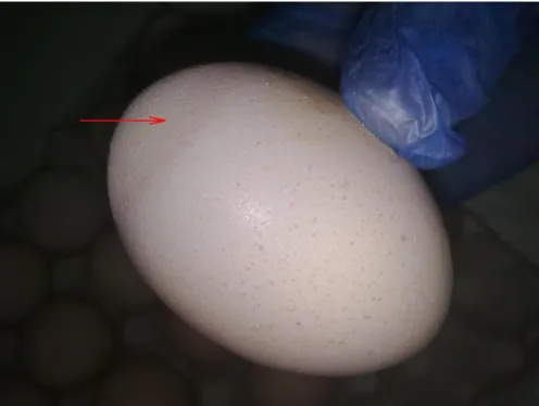
Infections with M. synoviae have been reported as endemic in the poultry industry of many countries worldwide, where they cause considerable economic losses to heavy breeders, broilers and layers (Noormohammadi, 2007; Kleven, 2008). Mycoplasma synoviae is responsible for infectious synovitis and causes economic losses because of decreased egg production, growth and hatchability rates, and downgrading of carcasses at slaughter due to airsacculitis and arthritis lesions (Marois et al., 2005; Peebles et al., 2011; Moreira et al., 2014a). Fertile eggs have a reduced hatchability due to late embryonic mortality (Butcher & Halabi, 2010; Moreira et al., 2014a). Mycoplasma synoviae infection is usually asymptomatic in broiler breeders but the possibility that it might play an important role in a complex respiratory disease in the offspring has motivated breeding companies to consider eradication (Fiorentin et al., 2003; Moreira et al., 2015b). In heavy breeders, the main problem associated with M. synoviae infection is a 5-15% decline in egg production (Moreira et al., 2014a). The effect on the quantity and quality of egg production will depend on the virulence of the M. synoviae strain, the degree of stress on the birds and the time of infection. If the infection occurs during the grow-out phase will typically result in minimal losses, while an infection during egg production, especially during the peak of production, will cause a dramatic decline in egg production. In most cases, the egg production will recover but remain below the standard curve (Stipkovits & Kempf, 1996). The clinical and economic relevance of M. synoviae seems to be increasing considering the number of worldwide publications and the emergence of strains affecting the eggshell quality (Figure 3) and egg production and the emergence in some countries of arthropatic and amyloidogenic strains (Landman, 2014).
Figure 3. Egg with poor eggshell quality consistent with M. synoviae infection.
1.2.3. Epidemiology and pathogenesis
Mycoplasma synoviae can be found in eggs laid by infected breeders. Vertical or transovarian transmission is not very efficient, as peak egg transmission from an infected breeder flock is low. If complicating factors are present such as immune suppression, for example, there may be a higher shed of the organism (Behbahan et al., 2005; Dhondt et al., 2007). Vertical transmission plays a major role in spreads of M. synoviae. When commercial breeder flocks became infected during egg production, the egg-transmission appears to be higher in the first 6 weeks after infection. After the chicks are hatched, the M. synoviae organisms are spread horizontally. Transmission occurs among birds by the aerosol route and by contamination of the feed and water within a house. The entire flock may be infected by 3 weeks of age (Kleven, 2003). Horizontal transmission occurs readily by direct contact. In general, M. synoviae appears to spread more rapidly than M. gallisepticum. M. synoviae can be present in the respiratory tract of infected chickens for 4 weeks and the spread between houses occurs (Ferguson-Noel & Noormohammadi, 2013).
Natural infection can be observed in the first week but acute infection is usually seen when chickens are adult. This fact suggests that the incubation period can be relatively short
8
but is generally considered 11-21 days. Chronic infection may or may not follow the acute phase at any age and may persist for the entire life of the flock (Kleven, 2003). Birds placed in contaminated environment are strongly suggested to became infected and remain carriers. Mycoplasma synoviae are also well known for their interactions with other infectious agents and environmental factors in producing clinical disease. Control of the clinical manifestations is simplified when concurrent infections are minimized and optimum environmental conditions are provided. It is well known that respiratory infections are significantly affected by environmental factors, and that disease severity is increased during the winter months. Temperature, ventilation, humidity, atmospheric ammonia, and dust all have important interactions with infectious agents in producing respiratory disease (Landman, 2014). However, there have been relatively few studies on the influence of environmental factors on the severity of mycoplasma infections (Moreira et al., 2015b). Atmospheric dust significantly increased the severity of air sac lesions and chickens maintained at environmental temperatures of 7 to 10 ºC were more susceptible to airsacculitis caused by M. synoviae than chickens maintained at 24 to 29 ºC (Kleven, 2003). Mycoplasmas also infect other domestic and wild avian species, so it is important to ensure they are not in contact with commercial chickens. Some data provide strong evidence that indirect transmission of Mycoplasma via contaminated feeders occurs (Feberwee et al., 2005b). Although M. synoviae can be transmitted via fomites, the birds infected in this way may quickly overcome mild disease and on recovery may be protected against more severe infections potentially acquired by direct bird-to-bird contact (Behbahan et al., 2005; Dhondt et al., 2007). Such indirect transmission is rather unexpected for wall-less bacteria, which are supposed to be sensitive to osmotic shock, heating or chemical treatments. However, M. synoviae may persist on feathers up to 2 or 3 days at room temperature and its high dissemination capacity has been demonstrated (Marois et al., 2005). Mycoplasmas are more likely to spread among farms by the mechanical route, which would include spread via contaminated equipment, shoes and other fomites (Kleven, 2003).
Mycoplasma synoviae infection most frequently occurs as a subclinical upper respiratory infection. It may cause air sac lesions when combined with other respiratory agents such as Newcastle Disease Virus (NDV), Infectious Bronchitis Virus (IBV), or both (Landman, 2014). At other times, M. synoviae becomes systemic and results in infectious synovitis, an acute to chronic infectious disease of chickens and turkeys, involving primarily the synovial membranes of joints and tendon sheaths producing an exudative synovitis,
tenovaginitis, or bursitis (Ferguson-Noel & Noormohammadi, 2013; Moreira et al., 2014a). Infectious sinusitis grossly distended infraorbital sinuses. There is fibrin, heterophils, epithelial cell hyperplasia, hypertrophy of mucous glands. Later there is lymphocytic infiltrates in the lamina propria or nodular formation. Tracheitis and airsacculitis can occur (Kleven, 2003). Pathogenicity of M. synoviae generally involves attachment and colonization of the respiratory tract and other additional factor can produce systemic invasion and clinical signs.
1.2.4. Clinical signs and lesions
Common disease signs like pale comb, lameness and retarded growth are the first observable signs. Disease progression debilitates the bird that became ruffled and swellings usually occur around joints, specially the hock and foot pads joints (Ferguson-Noel & Noormohammadi, 2013). Airsaculites may occur in chickens infected via respiratory tract at any age (Butcher & Jacob, 2009). In recent years, the occurrence of arthropathic and amyloidogenic strains of M. synoviae, as well as strains that induce eggshell apex abnormalities and egg production losses, has increased (Feberwee et al., 2008). Progeny of M. synoviae infected breeders may have increased condemnation, poor conversion rates and poor weight gain. Morbidity varies from 2 to 75% but is usually lower rounding 5 to 15% and mortality range between 1 and 10% (Kleven, 2003). As M. synoviae infection progresses caseous exudates involves tendon sheats and joints that became thinned over time and may evolve into the muscle and air sacs. In the respiratory form, airsacculitis may be seen.
1.2.5. Diagnosis
Diagnosis is based on epidemiological data, clinical signs, analysis of macroscopic lesions, specific serology, isolation and molecular characterization of M. synoviae. Monitoring must be part of control programs performed in breeder flocks and is mostly feasible by routine serology and PCR (Feberwee et al., 2005a; Luciano et al., 2011; Moreira et al., 2015b). Serologic procedures are useful for flock monitoring in M. synoviae control programs and to aid in diagnosis when infection is suspected. A positive serologic test, together with history and signs typical of the diseases, allows a presumptive diagnosis pending isolation and identification of the organisms. The tube agglutination test was a
10
common procedure, especially during the M. gallisepticum control program for turkeys in the 1960s and 1970s but is now rarely used. Serum plate agglutination (SPA) antigen, for the detection of antibodies to M. synoviae, is commercially available. Because the SPA test is quick, relatively inexpensive, and sensitive, it has been widely used as an initial screening test for flock monitoring and serodiagnosis. However, nonspecific reactors occur in some flocks infected with M. synoviae due to cross reactive antigens, or those recently vaccinated with oil-emulsion vaccines and/or vaccines of tissue-culture origin against various agents. The SPA test is highly efficient in detecting IgM antibody, which is the first class of immunoglobulins produced in response to infection (Kleven, 2003). The haemagglutination inhibition (HI) test has been commonly used to confirm reactors detected by SPA or, more recently, enzyme-linked immunosorbent assays (ELISA). However, the HI test is time consuming, the reagents are not commercially available and the test may lack sensitivity. ELISA assays were developed to increase testing efficiency and improve sensitivity and specificity of results relative to the SPA and HI tests. Commercial ELISA test kits are now commonly used for flock monitoring and serodiagnosis. In general, ELISA tests are slightly less sensitive but more specific than SPA tests and less specific but more sensitive than HI tests (Kleven, 2003; Ferguson-Noel & Noormohammadi, 2013). Ewing et al. (1998) reported that the SPA test missed infected commercial layer and breeder flocks that were detected by ELISA. Further confirmation of serologic results may be made by isolation and identification of M. synoviae from the upper respiratory tract or by polymerase chain reaction (PCR). However, few laboratories are equipped to culture this organism, as specialized culture media and specific incubator conditions are required. Techniques for the detection and analysis of DNA through PCR arise as a very interesting alternative of diagnosis method, because they offer sensitivity, specificity, capability of accomplishment of exams on a large scale (Nascimento et al., 1991; Silveira et al., 1996) and, nowadays, economic viability. The sensitivity observed in PCR is important for detection of pathogenic agents in clinical samples taken from asymptomatic animals, or those undergoing antibiotics treatment. Furthermore, it is possible to detect a pathogenic agent before the host’s immunologic response, or in host with immunity depression, demonstrating advantages also over the serologic tests (Garcia et al., 1995; Kempf, 1998; Buim et al., 2009).
1.2.6. Control, treatment and prevention
Antibiotic treatment of breeders is not effective against elimination of M. synoviae, although egg transmission level is reduced (Kleven, 2003). Macrolides like tylosin and tilmicosin and fluoroquinolones like enrofloxacin and difloxacin are among the antibiotic families most widely used in poultry in many countries (Jordan & Horrocks, 1996; Gerchman et al., 2011) but M. synoviae is susceptible in vitro to others several antibiotics including chlortetracycline, lincomycine, oxytetracicline, spectinomycine, tetracycline and tiamulin (Ferguson-Noel & Noormohammadi, 2013). In the past, mycoplasma eradication programs have been based on antibiotic or heat treatment of fertile eggs but more recently the intensive poultry industries rely heavily upon the application of vaccines for disease control (Zhang et al., 2010; Ferguson-Noel et al., 2012). Vaccination programs are presently being used to control outbreaks of the more virulent strains of M. synoviae (Ferguson-Noel & Noormohammadi, 2013). In relation to the presence of mycoplasmas in breeder farms, their concentration in some regions and inexistence of adequate sanitary barriers that may enable the isolation of farms are predisposing factors for the disease dissemination. Other contributing factors are related to the resistance to antimicrobial treatments, and to the immunologic system escape mechanisms that these pathogens make use of. The high M. synoviae occurrence in layer and breeder birds probably is due to the fact that the vaccine against is still very little used. The failure to eradicate M. synoviae from commercial poultry flocks has been largely due to the ability of these organisms to establish lifelong infections and to spread by horizontal and vertical transmission among their hosts (Kleven, 1998; McAuliffe et al., 2006). The primary objective for any poultry farm M. synoviae control is to prevent the introduction of the organism into a clean flock by use of a comprehensive biosecurity program (Butcher & Jacob, 2009).
12 1.3.REOVIRUS IN BROILER BREEDERS
Reoviruses (a name derived from Respiratory Enteric Orphan or REO) are members of the genus Orthoreovirus in the Reoviridae family (Rosenberger, 2003). Found to be ubiquitous among poultry flocks, avian reoviruses (ARV) have been isolated frequently from the gastrointestinal and respiratory tracts of chickens affected by several disease conditions, including viral arthritis/tenosynovitis, stunting syndrome, respiratory disease, enteric disease, immunosuppression, malabsorption syndrome or even unapparent infections (Jones, 2013a).
Originally, the REO abbreviation was used to identify virus groups that were not associated with any known disease (Jones, 2013a). Viral arthritis is an economically important disease of chickens that can be caused by different serotypes and pathotypes of ARV (Rosenberger & Olson, 1997). Tenosynovitis, defined by the changes in the tendons and their sheaths, can be considered different from the condition caused by M. synoviae and in some reoviruses that have arthrotropic characteristics include ruptured gastrocnemius tendons, pericarditis, myocarditis, hydropericardium, uneven growth and mortality (Olson & Kerr, 1966; Olson & Solomon, 1968).
Viral arthritis/tenosynovitis in poultry is one of the pathological manifestations of avian reovirus infection (Rosenberger, 2003). The Reovirus can act alone as a pathogenic agents or in combination with one or more other aetiological agents, such as M. synoviae or Staphylococcus spp., and this situation can lead to varied clinical pictures of arthritis/tenosynovitis (Rosenberger, 2003; Roussan et al., 2012). They can be isolated from birds without any signs of disease, but they are also associated with a variety of problems including viral arthritis/tenosynovitis, enteric disease and malabsorption syndrome (Jones, 2013c).
1.3.1. Aetiology
Reoviruses have a worldwide distribution in chickens but are more related to meat-type birds (Van der Heide, 1977). They are commonly found in the digestive and respiratory tracts of clinically normal chickens and turkeys. It is estimated that most of the reoviruses isolated from chickens are non-pathogenic. Several studies that were performed over the last years have revealed that ARV has unique properties, different to those displayed by mammals (Jones, 2013a).
ARV, which replicate in the cytoplasm, are non-enveloped with an icosahedral symmetry and a double-shelled capsid and are one of the few non-enveloped viruses that cause cell to fuse (Xu & Coombsa, 2009). This specific genome segments responsible for protein coding have been identified for the S1133 strain of ARV and differentiates them phylogenetically from most other animal reoviruses (Day, 2009). Another interest characteristic of the ARV is that is known to induce apoptosis in infected cells (Benavente & Martínez-Costas, 2007).
1.3.2. Economic importance
Avian Reovirus infections are of economic importance to the poultry industry (Savage & Jones, 2003). In meat-type chickens, economic losses are frequently associated with Reovirus infections. Increased mortality, viral arthritis/tenosynovitis and a general lack of performance are among the observed problems (Jones, 2013b). Breeder flocks that develop viral arthritis just prior to the onset of or during egg production may, in addition to lameness, be affected by increased mortality, decreased egg production, suboptimal hatchability/fertility and vertical transmission of the virus to progeny.
Infectious viral arthritis is currently the best defined and most readily diagnosed reovirosis (Ide & Dewitt, 1979; Rosenberger, 2003).
1.3.3. Epidemiology and pathogenesis
Reoviruses can be classified using serologic procedures or grouped according to their virulence. There are five serotypes of reoviruses from 77 isolates from intestines, respiratory tract, and synovial isolates (Kawamura & Tsubahara, 1966; Day, 2009). They are antigenically similar viruses and demonstrated clear strain differences based on virulence and virus persistence. There are considerable cross neutralization between heterologous serotypes (Islam et al., 1988). The ARV genome consists of 10 segments of double-stranded RNA: three large (L1, L2, L3), three medium (M1, M2, M3) and four small (S1, S2, S3, S4) (Jones, 2013b).
In general, ARV are associated with arthritis, but they have also been identified as the etiology of other diseases. Some examples are malabsorption syndrome conditions, pericarditis, myocarditis, hydropericardium, enteritis, hepatitis, bursal and thymic atrophy,
14
osteoporosis, and acute and chronic respiratory syndromes (Rosenberger, 2003). Although reoviruses have been found in many avian species, chickens and turkeys are the only recognized natural or experimental hosts for reovirus-induced arthritis (Pertilem et al., 1996). Other bird species from which reoviruses can be isolated are ducks, pigeons, geese and psittacine species (Watier, 2010).
Initially, the ARV replicates in the villi of the small intestine and in the bursa, and then spreads to other tissues. Generally, osmotic diarrhea appears due to villi blunting (Rosenberg, 2003b). When a bird is infected by reoviruses, these increase susceptibility to other infectious agents (Watier, 2010). This immunosuppression is due to lymphoid depletion and compromise of the immune system. Some authors report age-related resistance to reovirus-induced arthritis (Olson & Kerr, 1966; Jones & Georgian, 1984). Again, this age-associated susceptibility may be related to the inability of young birds to develop an effective immune response (Jones, 2013b). The virus can be spread laterally (horizontal transmission). In addition, vertical and egg-transmission are also possible, but at a lower rate (Robertson & Wilcox, 1986). ARV may be excreted from the intestinal or respiratory tracts for at least 10 days post-inoculation. This fact suggests fecal contamination as a primary source of contact (Jones, 2013b). Viral persistence can last for long periods, special in the caecal tonsils and hock joints (Savage & Jones, 2003). Birds that are infected at a young age are potential sources of infection (Rosenberger, 2003). Whether or not the disease occurs following infection with ARV, the incubation period ranges from 1 and 11 days and is highly dependent upon the virus pathotype, age of the host and route of exposure (footpad inoculation, intramuscular, intravenous, etc.) (Meng et al., 2012). Very often, infections are unapparent and demonstrable only by serology or virus isolation (Jones, 2013b).
Frequently, the virus is located in the flexor and extensor tendons of the pelvic limb and it is usually seen in young birds (1-2 months). Mortality is usually low, but morbidity can be as high as 100%. As already mentioned, the virus can persist for long periods in the tendons (Islam et al., 1988).
Avian reoviruses possess a group-specific antigen and a serotype-specific antigen. Humoral immunity (neutralizing antibodies) can be detected 7-10 days following infection. The presence of antibodies and its importance in establishing protection is not-well defined yet. Birds may become persistently infected in the presence of high levels of circulating antibodies. It is apparent, however, that maternal antibodies can afford a degree of protection to day-old chickens against naturally occurring and experimental challenges. From several
studies, the suppression of T-cell-mediated immunity by cyclosporin A resulted in increased mortality in reovirus-infected birds, but the relative severity of tendon lesions was not changed. Antibody protection is related to serotype homogeneity, virulence, host age and antibody titer (Rosenberger, 2003; Grande et al., 2002; Jones, 2013b). For cell mediated immunity, the CD8+ T-cells may play a role in pathogenesis and/or Reovirus clearance in the small intestine. Some authors have shown that challenging viruses are controlled in the absence of actively produced antibodies in B-cell immunosuppressed chicks (Day, 2009). This suggests that cellular immunity may be sufficient for protection of broilers (Jones, 2013b).
1.3.4. Clinical signs and lesions
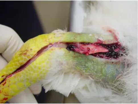
In an acute infection, lameness is generally present and some chickens are atrophied (Crespo & Shivaprasad, 2011). In chronic infection, lameness is even more pronounced and the percentage of infected chickens is small. Lameness in this type of lesion is due to enlargement in the area of the gastrocnemius or digital flexor tendons. In general, the rupture of the gastrocnemius tendon can be observed (Figure 4).
The swelling of the digital flexor and metatarsal extensor tendons is the more pronounced macroscopic lesion. Swelling of the foot pad and hock joint is less frequent and is marked be the edema of the tarsal and metatarsal tendon. Some petechial hemorrhages are frequent in the synovial membranes (Rosenberger, 2003; Jones, 2013b). In chronic infection, inflammation of tendon areas progresses, tendon sheaths become hard and in some cases they fuse. In the case of an early infection, recovery is quick but very often the tendon rupture occurs at transfer in the broiler breeders or with other kind of steady manipulation (Crespo & Shivaprasad, 2011).
In terms of microscopic lesions, the basic picture is edema, coagulation necrosis, accumulation of heterophilic material and perivascular infiltration. There is also hypertrophy and hyperplasia of synovial cells, infiltration of lymphocytes and macrophages, and a proliferation of reticular cells (Kerr & Olson, 1964; Hill et al., 1989). Lesions are strongly time-dependent and changes have been found in the type and number of positively staining cells. The synovial membranes develop villous processes, during the chronic phase, and lymphoid nodules are present. When the process becomes more chronic, the inflammatory picture changes, the amount of fibrous connective tissue increases, and a pronounced
16
infiltration or proliferation of reticular cells, lymphocytes, macrophages and plasma cells can also be seen. Irregular granulation tissues replace some tendons, and large villi appear on the synovial membrane (Jones, 2013b).
Figure 4. Rupture of the gastrocnemius tendon consistent with Reovirus infection.
1.3.5. Diagnosis
A presumptive diagnosis of viral arthritis may be made on the basis of signs and lesions. Primary involvement of the metatarsal extensor and digital flexor tendons, and heterophil infiltration in the heart, assist in differentiating the infection from bacterial and mycoplasmal synovitis (Jones, 2013b; Moreira et al., 2014b). Different diagnostic methods are available: fluorescent antibody techniques, virus isolation, typical physicochemical characteristics and the presence of a group-specific antigen demonstrable with the agar gel precipitin test. The immunoperoxidase procedures can be used, but they are not the first choice (Rosenberger, 2003).
Serology for reoviruses is routinely used and is based on group-specific antibodies that can be detected readily with the agar gel precipitin test or by IFAT indirect fluorescent antibody test (IFAT). In more recent years, the ELISA for detecting antibodies to avian reoviruses along with PCR has become more common (Bruhn et al., 2005).
1.3.6. Control, treatment and prevention
The ubiquitous nature of the avian reoviruses and their inherent stability, coupled with modern, high-density confinement rearing practices, suggests that elimination of virus exposure may be difficult (Jones, 2013c). Its resistance to inactivation may be frequently carried by mechanical means. Commercially available disinfectants should be validated for efficacy before use because of the avian Reovirus group relative stability (Rosenberger, 2003).
As far as it is known, chickens are most susceptible to pathogenic reoviruses at 1 day of age and then develop an age-associated resistance beginning as early as 2 weeks (Kerr & Olson, 1964). Vaccines and vaccination programs have evolved and can provide protection at 1 day of age. Active immunization can be achieved by vaccination with viable attenuated reoviruses, which is usually applied by the subcutaneous route (Giambrone & Clay, 1986). Reovirus vaccination of breeding stock can be carried out with live or inactivated vaccines. Inactivated vaccines are more effective if they are preceded by vaccination with a live vaccine. If a live vaccine is used, it should be administered prior to the onset of egg production, to prevent transovarian transmission of the vaccine virus (Jones, 2000; Cobb, 2011). The advantages of this type of immunization program include immediate protection of day-old progeny provided by maternal antibodies (Jones, 2013b). Vaccination of breeders is an efficacious method of controlling viral arthritis and other pathogenic reoviruses, but it should be recognized that protection is assured against homologous serotypes only (Rosenberger, 2003).
18 1.4.ASPERGILLOSIS IN BROILER BREEDERS
Aspergillosis in birds is usually confined to the lower pulmonary tract with florid lesions in air sacs and lungs. Manifestations of the disease depend on which organs or systems are involved. In adult birds, such as broiler breeders, the signs are unclear and most of the times the animals are subclinically infected (Beernaart et al., 2010).
Aspergillosis is an infection disease caused by members of the genus Aspergillus (Olias et al., 2010a). Aspergillus spp. are ubiquitous opportunistic saprophytic fungi considered as major respiratory pathogens in birds, which can cause considerable economic losses in chickens and turkeys, with mortality, decreased weight gain and slaughter condemnations (Kunkle, 2003; Fulleringer et al., 2006). This mycosis was described many years ago, but pathogenesis is still currently being discovered (Beernaert et al., 2010). Aspergillus spp. develops and sporulates easily in common poultry sources like feed, litter and other possible organic debris (Fulleringer et al., 2006; Arné et al., 2011). It is assumed that a compromised immune system and the inhalation of a considerable amount of spores are predisposing factors (Lugauskus et al., 2004; Martin et al., 2007; Beernaert et al., 2010). Poor ventilation systems and dusty conditions increase the risk of infection (Fulleringer et al., 2006; Arné et al., 2011). Fungal pneumonia is considered a major threat to the survival of young poultry (Olias et al., 2010b). Acute cases are more often seen in young birds following spores inhalation that commonly causes high morbidity and mortality. The chronic cases affect older birds and are more difficult to diagnose (Arné et al., 2011). The treatment of infected poultry is very difficult and may even be not effective in severe respiratory cases. Hygiene and prevention are the most important tools to protect poultry (Kunkle, 2003).
1.4.1. Aetiology
Aspergillosis is an infectious, non-contagious fungal disease caused by species in the ubiquitous opportunistic saprophytic genus Aspergillus, which comprises approximately 180 species (Dykstra et al., 2013). The predominant species is A. fumigatus, probably due to the small spore size when compared to Aspergillus flavus, Aspergillus niger, Aspergillus glaucus or Aspergillus nidulans. Other rarely isolated species are Aspergillus terreus and Eurotium amstelodami (Joseph et al., 2000; Tell et al., 2005; Beernaert et al., 2010).
1.4.2. Economic importance
The financial costs of aspergillosis are most readily apparent in broiler and turkey production (Baumel et al., 2000; Kunkle and Rimler, 1996). The incidence of chronic disease in adult birds like broiler breeders is unclear, but there are important economical losses when adult birds succumb to this disease (Akan et al., 2002; Dykstra et al., 2013). When the broiler breeder’s environment is contaminated with spores, although birds may appear healthy, birds may suffer losses of productivity including compromised hatchability and drop in egg production (Latgé, 1999). Other important factor is the contamination of the egg surface that will compromise the hatchery general environment. Chick quality is compromised when the egg surface is contaminated with fungi (Williams et al., 2000; Cortes et al., 2005; Moreira et al., 2015a).
1.4.3. Epidemiology and pathogenesis
Aspergillosis more common causative agent is A. fumigatus but all Aspergillus species are ubiquitous due to their broad nutritional capability (Kunkle, 2003). They usually occur in soil, grains, and organic matter. The spores’ concentration in the birds’ environment is an important predisposing factor along with others like ventilation, humidity, temperature, poor sanitation and feed quality (Redig, 1993; Tell, 2005). The initial contamination of poultry environments may occur through the use of contaminated litter or the introduction of day-old chickens that have retained spores from hatchery facilities (Arné et al., 2011). Hatcheries’ contaminated environment poses a major role in aspergillosis outbreaks in broilers. Frequently, the introduction of Aspergillus spp. in the hatchery occurs through contaminated egg shells (Figure 5) from contaminated breeder facilities (O’Meara & Chute, 1959; Williams et al., 2000; Planel et al., 2001; Moreira et al., 2015a).
20
Figure 5. Fertile egg with Aspergillus spp. contamination (red squares).
The conditions found in poultry structures, special humidity and temperature, promote the rapid growth of hyphae and efficient assexual multiplication, resulting in large amounts of conidia which can be easily inhaled by birds (Beernaert et al., 2010). The bird’s immune status can also be a predisposing factor (Beernaert et al., 2010). The pathological pattern of aspergillosis results more from the dose of inhaled conidia and host susceptibility than species virulence. Genus Aspergillus is among the most common mycotoxigenic genera, and it is well described that mycotoxins interfere with resistence to various infections. Toxins produced by pathogenic Aspergillus spp. may be involved in the pathogenesis in poultry. Gliotoxin is among several toxins produced by A. fumigatus and it is likely that this toxin is a virulence factor that contributes to the pathologic changes in aspergillosis. A discussion of the organism virulence factors has to be considered because this agent is a saprobe, opportunistic and noncontagious pathogen (Dykstra et al., 2013).
Birds’ susceptibility to Aspergillus spp. can be attributed to anatomical, physiological or respiratory immune system characteristics when compared to mammals (Tell, 2005). Immunocompetent mammals are naturally resistant to pulmonary aspergillosis unless they are exposed to overwhelming doses of conidia. In spite of this, aspergillosis in humans has increased a lot during recent years, in part due to complications of therapeutic