F
ACULDADE DE
E
NGENHARIA DA
U
NIVERSIDADE DO
P
ORTO
Dynamic Analysis of Upper Limbs
Movements after Breast Cancer Surgery
Ana Rita Carvalho Moreira
M
ASTER
T
HESIS
Mestrado Integrado em Bioengenharia
Supervisor: Hélder Filipe Pinto de Oliveira (PhD)
Co-Supervisor: Jaime dos Santos Cardoso (PhD)
Resumo
A qualidade de vida dos pacientes com cancro de mama tem vindo a tornar-se um fator de importante con-sideração aquando da escolha do tipo de tratamento a ser utilizado. Contudo, técnicas de tratamento comuns, como o caso da radioterapia ou da remoção cirúrgica dos nódulos linfáticos da axila, resultam em vários danos no funcionamento dos membros superior das mulheres. Estas deficiências incluem uma limitada mo-bilidade do braço e o seu inchaço, o que normalmente precede o aparecimento linfedema crónico. Estas morbidades afetam as várias atividades diárias dos pacientes e, consequentemente, contribuem para uma menor qualidade de vida.
Assim, é de extrema importância avaliar as restrições funcionais derivadas do tratamento do cancro, de modo a avaliar a qualidade dos procedimentos e a evitar complicações posteriores. Sendo assim, este trabalho tem como objetivo desenvolver um método eficaz para a avaliação do funcionamento dos membros superior do corpo, aplicável a pacientes com cancro da mama. Para este fim, é investigado o uso de informação de profundidade e de esqueleto, adquiridos com a Microsoft Kinect, para extrair atributos que caracteri-zam o movimento dos membros superiores. São utilizados algoritmos de classificação supervisionados para construir um modelo de classificação, obtendo-se resultados muito promissores, com elevada precisão de classificação. Deste modo, o método desenvolvido parece ser uma solução adequada ao objetivo proposto.
Após tratamento do cancro de mama, é fundamental para as mulheres manter uma atividade física con-tínua de modo a recuperar a mobilidade dos membros superiores. Dessa forma, normalmente é recomendado um conjunto de exercícios para efectuar em casa, mas as pacientes nem sempre os fazem como deveriam. Isto reforça a importância de um modelo de cuidado para reabilitação dos pacientes de cancro da mama, de modo a promover e apoiar a atividade física. Desta forma, neste trabalho foi também investigado um modelo de reabilitação para pacientes com cancro de mama. Recorrendo à Kinect, desenvolveu-se uma aplicação neste sentido, que instrui o paciente na execução dos exercícios e realiza uma avaliação da sua performance. Os resultados preliminares são bastante satisfatórios, mas ainda é necessário um trabalho mais aprofundado nesta área.
Abstract
The quality of life of breast cancer patients has increasingly become an important factor of consideration in choosing the type of treatment used. However, common treatment techniques, as the case of radiation therapy or the surgical removal of the axillary lymphatic nodes, result in several impairments in women’s upper-body function. These impairments include restricted shoulder mobility and arm swelling, which usually precedes chronic lymphedema. As consequence, several daily life activities of the women will be affected and, consequently, contribute to a decreased QOL.
Therefore, is of extreme importance to assess the functional restrictions caused by cancer treatment, in order to evaluate the quality of procedures and to avoid further complications. In this manner, the present work aims to develop an effective method for the evaluation of the upper-body function, suitable for breast cancer patients. For this purpose, it is investigated the use of both depth and skeleton data, provided by the Microsoft Kinect, to extract features that characterize the upper-limbs motion. Supervised classification algorithms are used to construct a predictive model of classification and very promising results are obtained, with high classification accuracy. Therefore, the developed method appears to be a proper solution for the proposed goal.
After breast cancer treatment, it is essential for the women to maintain a continuous physical activity in order to recover the upper-limb mobility. In that way, a home-base exercise program is normally recom-mended, but the patients not always perform the exercises as they should. This highlights the importance of a surveillance rehabilitation model for breast cancer patients to promote and support physical activity and exercise behaviors. Further in this research, it was investigated a rehabilitation model for breast cancer pa-tients. Taking advantage of the Kinect device, an application was developed in this direction, that instructs the patient on how to execute the exercises and makes an evaluation of their performance. Preliminary results are quite satisfactory, but further work is still needed.
Acknowledgments
This work would not be possible without the contribution, help and support of several people to whom I owe a sincere gratitude.
First of all, I am eternally grateful to Hélder, for his continuous support, motivation, friendship and for being always available. Without him nothing of this would be possible! Also, I have to thank to Professor Jaime for his helpful and pertinent advising, whenever needed. Furthermore, I am very thankful to Dr. André Magalhães from the Hospital São João, for the medical support. My gratitude goes also to INESC TEC and the VCMI (Visual Computing and Machine Intelligence) group, for the work environment provided, which allowed the development of this thesis.
To Inês, Sofia and Filipa, my daily mates, for working by me side, for all the lunches together and, more important, for all the confidences, chats and laughs. To M&B for all the things it taught me. For make me understand that the bacon is better than the egg. To 09, for those who entered with me and the did this journey by me side. To C’amelias for all the unforgettable moments we had together. All the tears and laughs and tears by laughs. All the dinners, beers and cheers. All the Porto and fados. From here I did friends for life. Some deserve a special word: to Xiló, for the unconditional friendship, always being there to hear me; to Joana, my timeless roommate; to Rita and Dani, or the embreast-it would not be complete. To João, despite everything, for all the inrideo ut pervenio lux. To all the others who accompanied me in these five years and made part of the best times of my life, a sincere thank you.
To Pedro, for being by my side every single day, for all the patience with my bad mood and my craziness moments, for making me stand up when I was almost falling. For all the love and encouragement, for everything that is not possible to thank.
To my mother, my role model, for all the efforts that I know she did so I could accomplish this, for always taking care of me, for the unconditional love and eternal support. To my father, for never stop believing on my capabilities and always trust that I could it. To my brother, for all the support and advisement and for never doubt of my success. To Lu, my second mother. To Koda and Woody, or the family would not be complete.
Rita Moreira
"We must try not to sink beneath our anguish, Harry, but battle on."
Albus Dumbledore
To my mother,
Contents
1 Introduction 1 1.1 Motivation . . . 1 1.2 Objectives . . . 2 1.3 Contributions . . . 2 1.4 Document Structure . . . 3 2 Breast Cancer 5 2.1 Breast Anatomy and Physiology . . . 52.1.1 Breast Lymphatic System . . . 6
2.2 Breast Carcinoma . . . 6
2.3 Breast Cancer Treatments . . . 7
2.3.1 Lymph Node Dissection . . . 7
2.4 Conclusion . . . 8
3 Literature Review 11 3.1 Functional Evaluation: Methods of Assessment . . . 11
3.1.1 Upper-Limb Volume Measurements . . . 11
3.1.2 Upper-Limb Motion Evaluation: Non-vision systems . . . 15
3.1.3 Upper-Limb Motion Evaluation: Vision-based systems . . . 18
3.2 Rehabilitation Model for Breast Cancer Patients . . . 22
3.2.1 Home-Based exercise intervention on breast cancer patients . . . 23
3.2.2 Virtual Reality in Upper-Body Function Rehabilitation . . . 24
3.2.3 Summary . . . 25
3.3 Conclusion . . . 26
4 Upper-Body Function Evaluation 29 4.1 Database . . . 29
4.1.1 Application for medical data acquisition . . . 29
4.1.2 Dataset . . . 31
4.2 Methodology . . . 33
4.2.1 Depth-map noise reduction . . . 33
4.2.2 Kinect Rotation Correction . . . 35
4.2.3 Patient Segmentation . . . 36
4.2.4 Arm Segmentation . . . 37
4.2.5 Feature Extraction . . . 39
4.2.6 Classification Models . . . 42
4.3 Results . . . 45
4.3.1 Depth map noise reduction . . . 45
4.3.2 Patient Segmentation . . . 45
4.3.3 Arm Segmentation . . . 47
4.3.4 Upper-Body Functional Evaluation . . . 48
4.4 Conclusion . . . 53
xii CONTENTS 5 Rehabilitation 55 5.1 Rehabilitation Model . . . 56 5.1.1 Avatar . . . 56 5.1.2 Windows Application . . . 56 5.2 Conclusion . . . 58 6 Conclusion 61 6.1 Future Work . . . 62 References 63 A Upper-body Function Evaluation 71 B Acquisition Protocol 77 B.1 Kinect System . . . 77 B.1.1 Hardware requirements . . . 77 B.1.2 Limits . . . 77 B.1.3 Skeleton Joints . . . 78 B.1.4 Position . . . 78 B.1.5 Acquisition Parameters . . . 78 B.1.6 Room environment . . . 78 B.2 Patient . . . 78 B.2.1 Arm Movement . . . 79 B.3 Data . . . 79 B.3.1 Files Organization . . . 79
List of Figures
2.1 Mammary gland anatomy. . . 5
2.2 Brest Lymphatic System. . . 6
2.3 Axillary Lymph Node Dissection. . . 8
3.1 Polhemus FastSCAN™ and Insignia™ laser scanner. . . 14
3.2 VICON and Codamotion capture systems. . . 18
3.3 Representation of a 2D stick-figure model with ribbons by Leung and Yang, and the demon-stration of Wren’s work. . . 19
3.4 Human tracking results using the approach proposed by Baumberg and Hogg. . . 20
3.5 Representation of an automated system proposed for motion capture. . . 21
3.6 System proposed to capture human motion: representation of the skeleton fitted to visual hulls (rendered as point sets) of a moving person. . . 21
3.7 Example of exercises that are normally advised to breast cancer patients in order to recover the the full range of movement. . . 23
3.8 T-WREX exoskeletons apparatus developed. . . 25
3.9 The Rehabilitation Gaming System proposed by Cameirão. . . 26
4.1 Skeleton Joints tracked by the Kinect device and Skeleton space axes. . . 30
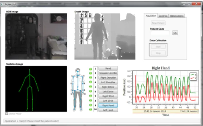
4.2 Graphical User Interface of the application developed for the data acquisition. . . 31
4.3 Skeleton joints saved on the database. . . 31
4.4 Data acquired using the Kinect device (Patient # 42). Color and corresponding depth-map image. . . 32
4.5 Morphological dilation. . . 34
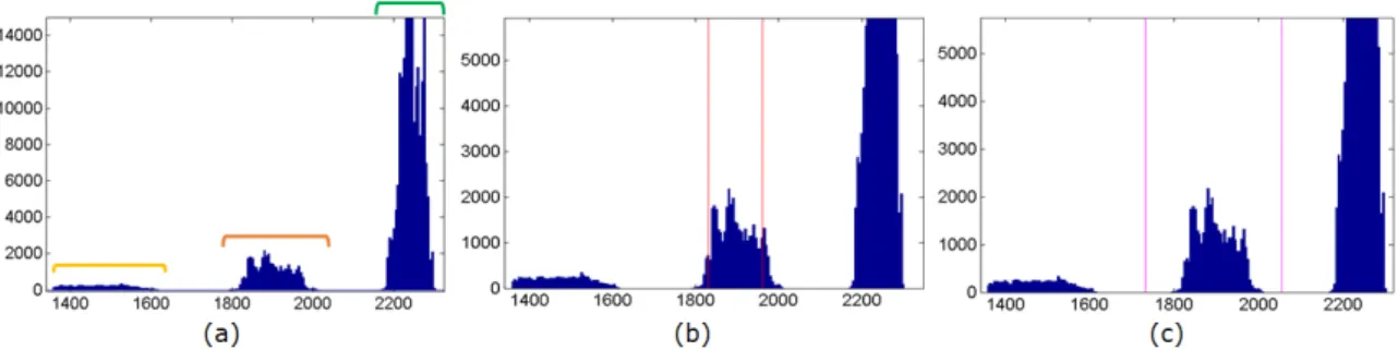
4.6 Histogram representation of depth values belonging to a neighboring region of a blob. . . 34
4.7 Bilateral Filter: the shape of the Gaussian kernel is dynamic based on difference of pixel intensity. . . 35
4.8 Image acquisition sketch when the Kinect is not parallel to the wall. . . 35
4.9 Method used to compensate the Kinect rotation. . . 36
4.10 Depth image histogram of Patient#42. . . 36
4.11 Patient segmentation process. . . 37
4.12 Areas of transition of the upper-limb and anatomical landmarks . . . 37
4.13 Delimitation points of the upper-arm area. . . 38
4.14 Result of the upper-arm segmentation. . . 39
4.15 Representation of the angle θ measured to evaluate the shoulder ROM . . . 40
4.16 Illustration of the volume measurement based on voxels . . . 40
4.17 Representation of the height (H) and width (W) measurements. . . 41
4.18 Flowchart of the process to obtain the hand instantaneous acceleration. . . 41
4.19 Representation of the angle computed to detected the elbow flexion. . . 42
4.20 Separating hyperplane and margins for an SVM trained with two classes samples. . . 44
4.21 Result of the bilateral filter with σr= 5, σd= 15 and with a 15 × 15 window. . . 45
4.22 Ground truth and body segmentation examples. . . 47
4.23 Arm contour detection examples. . . 48
xiv LIST OF FIGURES
5.1 Proposed rehabilitation model for breast cancer patients. . . 55
5.2 KMotion Capturer Software. . . 56
5.3 Biped skeleton with the skin mesh created in 3ds Max. . . 57
5.4 Rehabilitation App developed in this research. . . 58
5.5 Features evaluated in the rehabilitation model. . . 59
A.2 Delimitation points of the upper-arm area. . . 71
A.3 Delimitation points of the upper-arm area. . . 72
A.4 Bilateral filter with a 9x9 window. . . 73
A.5 Bilateral filter with a 15x15 window. . . 74
List of Tables
3.1 Comparison of the most significant methods for limb edema assessment. . . 12
3.2 Comparison of 3D methods proposed for arm edema assessment. . . 14
3.3 Self-report scales used for upper-body function assessment of breast cancer patients. . . 16
4.1 A comparison table for Natural User Interface libraries. . . 30
4.2 List of the skeleton joints saved on the database. . . 31
4.3 Medical information of the patients presented in the Database: type and year of surgery (Mast or BCS), surgery to axilla (SLN or ALND), use of radiotherapy (RT), lymphedema (Lymph.), physiotherapy (Phisio.) and the UEFI score. . . 32
4.4 Similarity Indexes used to evaluate the body segmentation. . . 46
4.5 Dice coefficient and Jaccard Index results of body segmentation. . . 46
4.6 Body contour detection error (in pixels) evaluated by the Hausdorff and average distance. . . 46
4.7 Dice coefficient and Jaccard Index results of arm segmentation. . . 47
4.8 Arm contour detection error (in pixels) evaluated by the Hausdorff and average distance. . . 47
4.9 Distribution of the 48 patients over the two classes. . . 48
4.10 Features used in the classification models. . . 49
4.11 Classification results for the different classifiers tested, using the lymphedema diagnosis as GT. 50 4.12 Confusion matrix for the LDA model. . . 50
4.13 Confusion matrix for the Naive Bayes model. . . 50
4.14 Confusion matrix for the linear SVM model. . . 51
4.15 Confusion matrix for the polynomial SVM model. . . 51
4.16 Confusion matrix for the RBF SVM model. . . 51
4.17 Classification results for the different classifiers tested, using the UEFI score as GT. . . 51
4.18 Confusion matrix for the LDA model. . . 51
4.19 Confusion matrix for the Naive Bayes model. . . 51
4.20 Confusion matrix for the linear SVM model. . . 52
4.21 Confusion matrix for the polynomial SVM model. . . 52
4.22 Confusion matrix for the RBF SVM model. . . 52
4.23 Classification results for the different classifiers tested, with the inclusion of the UEFI score as a feature. . . 52
4.24 Confusion matrix for the LDA model. . . 53
4.25 Confusion matrix for the Naive Bayes model. . . 53
4.26 Confusion matrix for the linear SVM model. . . 53
4.27 Confusion matrix for the polynomial SVM model. . . 53
4.28 Confusion matrix for the RBF SVM model. . . 53
Acronyms xvii
Acronyms
2D Two-Dimensional
3D Three-Dimensional
ALND Axillary Lymph Node Dissection
BCCT Breast Cancer Conservative Treatment
BCCT.core Breast Cancer Conservative Treatment.Cosmetic result
BCS Breast Conserving Surgery
BIS Bioelectrical Impedance Spectroscopy
CAD Computer Aided Design
CAML Computer Aided Measurement Laser
CCs Cue Circles
CT Computed tomography
DASH Disabilities of the Arm, Shoulder and Hand
DCIS Ductal Carcinoma in situ
DEXA Dual Energy X-ray Absorptiometry
DNA Deoxyribonucleic acid
EORTC QLQ BR23 European Organization for Research and Treatment of Cancer Quality of Life Questionnaire-Breast Cancer Module
FACT-B Functional Assessment of Cancer Therapy-Breast
FN False Negative
FP False Positive
GT Ground Truth
GUI Graphical User Interface
ICP Iterative Closest Points
IDC Invasive Ductal Carcinoma
ILC Invasive Lobular Carcinoma
IR Infra-Red
KAPS Kwan’s arm problem scale
LDA Linear Discriminant Analysis
MRI Magnetic Resonance Imaging
NUI Natural User Interface
QOL Quality-of-Life
RBF Radial Basis Function
ROM Range of Motion
RT Radiotherapy
PS Proximity Space
PSFS Patient-Specific Functional Scale
SDK Software Development Kit
SLN Sentinel Lymph Node
SLND Sentinel Lymph Node Dissection
SVM Support Vector Machine
TP True Positive
UBF Upper-Body Function
UEFI Upper Extremity Functional Index
Chapter 1
Introduction
Breast cancer is the most common cancer both in developed and developing regions, representing 23% of all cancers [35]. Nevertheless, due to effective earlier diagnosis methods and effective adjuvant therapies, the mortality rate is 27% or less in the more developed regions [35]. These high probabilities of long term disease survival mean that the women need to live daily with the consequences of the treatment. Consequently, the interest in the psychological adaptation and Quality of Life (QOL) of women after treatment has been growing. Increased psychiatric morbidity is associated with patients who experience unpleasant side effects of treatment for breast cancer. Women diagnosed with this disease have an higher risk of developing severe anxiety, depression and potential mood disorders, mainly due to worries regarding fear of death and altered body image, sexuality and attractiveness [115].
Approximately, 25% of women diagnosed with breast cancer present cancer cells in the axillary lymph node system [42]. Thus, besides the tumor removal, treatments generally includes the removal of axillary lymph nodes, as well as radiotherapy to the axilla [40]. However, this type of procedures are normally responsible for several upper-limb problems, including restricted shoulder mobility, lymphedema and/or arm/shoulder pain. Furthermore, at the 5 year follow-up the prevalence of arm/shoulder pain is 30-40%, of lymphedema 10–15%, and of restricted arm/shoulder mobility is 15–30% [80]. These upper-body mor-bidities are highly correlated with decreased QOL in breast cancer patients, since it disrupts valuable daily life activities [66, 102]. Therefore, studies support a timely screening as part of follow-up care and early management of the cancer-related physical impairments, with an appropriated rehabilitation program, can improve the the upper-limbs function and patients’ QOL [104].
1.1
Motivation
Breast cancer survivors normally experience long-term sequelae that include psychological distress, related with suboptimal cosmetic results, and physical impairments, which will contribute to a poor QOL. There is a diversity of strategies used is breast cancer treatment, both in surgery and in radiation therapy, which will result in different outcomes [83]. Therefore, it would be useful to have an objective and standard assessment of the final outcome of treatment, in order to identify which procedures have the less morbidity associated and to standardize these treatments. In this direction, a large research has been done and several methods were proposed for the evaluation of breast cancer treatment [15, 16, 32, 51, 82]. However, almost all the studies only focused on the cosmetic appearance, and the functional status has received much less attention. It is claimed by some authors [66, 102] that the upper-body morbidity, such as arm edema and restricted
2 Introduction
shoulder mobility, are correlated more strongly with QOL indicators than cosmetic status, due to their ability to disrupt valued life activities. So, it would be appropriate to include an Upper-Body Function (UBF) evaluation, besides the cosmetic assessment, in the evaluation of the breast cancer treatment.
The assessment of functional limitations in breast cancer patients can be performed by the identification of arm edema and by the evaluation of arm/shoulder mobility. However, this evaluation is not always done, and when it happens, the methods used have problems regarding the lack of objectivity or inaccuracy. The common procedures include subjective questionnaires [13], circumference measurements [103] or the use of a goniometer [38]. For that reason, there is a need of an simple, accurate, low-cost and reproducible method for the evaluation of UBF.
An early diagnosis of upper-limbs impairments is also important to have an identification of adequate therapies that can lead to greater success in managing functional morbidity, as well as prevention of pro-gression, with improved outcomes and QOL for breast cancer survivors. It has been well documented the benefits of physiotherapy and physical activity on the recovering of upper-extremity range-of-motion (ROM), strength, and function on women’s QOL [19, 26, 76, 77, 100]. Therefore, it would be also useful a compre-hensive model of care to identify exercise prescription and to guide the rehabilitation of breast cancer-related physical impairments.
1.2
Objectives
The main purpose of the present study is the development of a new methodology to assess objectively the upper-limbs functional status on breast cancer patients, using a low cost equipment, the Microsoft Kinect (Microsoft Corp., Redmond, WA, USA). On the other hand, it is also intended the use of the Kinect for the creation of a home-based rehabilitation system for physical impairments related to breast cancer treatment.
The Kinect sensor provides RGB and depth data, and allows a simplified skeleton tracking. To evaluate the upper-extremity function it will be assessed the limb volume and the temporal motion of the upper-arm and shoulder, using the depth camera and the tracking capabilities of the Kinect device. With this system it will be possible to extract movements’ features, including the upper-arm volume and the shoulder ROM, in order to evaluate the motion of the upper-limb over time. Thus, it will be possible to have a complete functional evaluation of the upper-body status of breast cancer patients, and identify reduced UBF caused by the treatment.
Also, it will be investigated a possible upper-body exercise system for home environment, using the Kinect device, that can be helpful in the rehabilitation of the arm/shoulder mobility and reduce the risk of lymphedema.
All the research depends on the availability of training and testing examples used in the development of the models. Therefore, this project included the collection of a data set with the help of an expert in breast cancer.
1.3
Contributions
The proposed work had four main contributions:
• Using the Kinect for Windows SDK it was developed a windows application for the collection of medical data. With this application is possible to acquire and record RGB and depth frames, as well as information about the skeleton’s joints positions over time.
1.4 Document Structure 3
• It was created a database comprising color, depth and skeleton data of breast cancer patients performing adduction/abduction movements. This database is a unique tool and can lead to new developments in the area.
• A new methodology is proposed, suitable for the evaluation of the upper-body function and lym-phedema detection in breast cancer patients.
• Finally, taking advantage of the Kinect capabilities, it was developed an application that can be used as home-based rehabilitation system to recover the upper-body function.
1.4
Document Structure
Besides the introduction, this document is composed by five more chapters. In chapter 2, a global intro-duction to the breast cancer problem is presented. In chapter 3, the literature review is provided on all the topics related to upper-body functional evaluation assessment. It includes methods used to measure the limb volume, subjective scales to evaluate the effects of injury in upper-body function and vision-based systems used to track human movements. Also, some insights are provided regarding virtual reality systems in home-based rehabilitation models. Chapter 4 describes the proposed method for upper-body function evaluation and the main results obtained. The application developed for rehabilitation is presented on Chapter 5. Finally, Chapter 6 serves as a conclusion to the presented research.
Chapter 2
Breast Cancer
2.1
Breast Anatomy and Physiology
The breast is a highly efficient organ mainly used to produce milk. It is a mass of glandular, fatty, and fibrous tissues positioned over the pectoralis major muscles of the chest wall [29]. The shape of the breast is similar to a tear-drop and has an extension toward the axilla, known as the tail of Spencer. Each adult female mammary gland usually consists of 15–20 glandular lobes. On the other hand, each lobe is composed by more than 40 smaller lobules, also known as the terminal ductal lobular units. The lobules terminate in many tiny bulbs which are the milk-secreting cells. The lobes, lobules and bulbs are all linked by the ducts [97] (see Figure 2.1).
Figure 2.1: Breast Anatomy: the mammary gland consists of lobes, which are made up of lobules. Ducts from the lobules converge to form lactiferous ducts. (a) Nonlactating breast, only with the duct system (b) Lactating breast, with alveoli at the ends of the ducts, which produce milk (From [97]).
6 Breast Cancer
2.1.1
Breast Lymphatic System
The lymphatic system is part of the body’s defense system against pathogens. In addition, it helps to maintain fluid balance in tissues and to absorb fat from the digestive tract. It is a network of tissue and organs that primarily consists of lymph vessels and lymph nodes [97]. Lymph nodes are small, oval-shaped structures distributed along the course of the lymphatic vessels. They filter the lymph, removing bacteria and other materials. The lymphatic vessels carry the lymph, which contains tissue fluid and waste products, as well as immune system cells.
In the upper-limbs, all the lymph vessels drain into the lymph nodes in the axilla. In addition, axillary nodes receive fluid from the upper back and shoulder, the lower neck, the chest, and the upper anterolateral abdominal wall. Regarding the breast, approximately 75% of the drainage of lymph fluid of the mammary gland is performed via lymphatic vessels into axillary nodes (see Figure 2.2) [29].
Figure 2.2: Brest Lymphatic System. Adapted from [120]
2.2
Breast Carcinoma
Cancer begins when cells in a part of the body start to grow out of control. These cells have a tightly regulated cell cycle that controls their growth, maturity, division and death. Cell division and growth is controlled by Deoxyribonucleic acid (DNA) and genes that lie within the cell’s nucleus, so any changes to DNA affects the cell. A cancer cell appears when a normal cell undergoes damage to the DNA that it is not repaired and the cell does not die, as it should. Instead, the cell undertakes division and the damage is propagated by the out-of-control growth of abnormal cells. This leads to formation of a tumor that may be benign (not dangerous to health) or malignant (has the potential to be dangerous) [57].
Breast cancer is a malignant tumor arising from the cells of the breast. Usually breast cancer either develops in the cells of the lobules or the lactiferous ducts. Less commonly, breast cancer can begin in the stromal tissues, which include the fatty and fibrous connective tissues of the breast [29].
There are different types of breast cancer, often divided into non-invasive and invasive. Non-invasive breast cancer, also known as carcinoma in situ, is when the cancer remain within the place of origin. The cancer do not grow into or invade normal tissues within or beyond the breast. One type of non-invasive cancer called ductal carcinoma in situ (DCIS) is considered a pre-cancerous lesion. This means that, although the abnormal cells have not spread out, they can eventually develop into invasive breast cancer. In invasive breast cancer, the abnormal cells spread outside the membrane that lines a duct or lobule, invading the surrounding
2.3 Breast Cancer Treatments 7
tissues. The cells can travel through the bloodstream or the lymphatic system to other parts of the body such as the bones, liver or lungs, creating metastasis. Invasive ductal carcinoma (IDC) and invasive lobular carcinoma (ILC) are the most common types of invasive breast cancer [69].
2.3
Breast Cancer Treatments
Besides disease control, breast cancer treatments aims to reduce the risk of distant metastases and/or local recurrence, obtain better aesthetic outcomes, relief of symptoms and restoring the QOL prior to diagno-sis [91]. The type of treatment chosen by the clinicians depends on several factors, such as women’s health and age, position and size of the cancer and how far it has spread. The treatments options normally include chemotherapy, radiotherapy and surgery.
Depending on factors such as the position and size of the cancer, the treatment chosen may be the surgical removal of the tumor by a mastectomy or a more conservative approach by Breast Cancer Conservative Treatment (BCCT). Non-surgical treatments, as chemotherapy and radiotherapy, may be used before surgery to help shrink the tumor or after surgery.
Mastectomy is the surgical removal of the entire breast. There are five different types: simple or total mastectomy, partial mastectomy, subcutaneous (nipple-sparing) mastectomy, modified radical mastectomy and radical mastectomy [56]. Total mastectomy involves removal of the breast, nipple, areola, and sentinel lymph nodes. The partial mastectomy removes the part of the breast that has cancer and some normal tissue around it. During subcutaneous mastectomy, all of the breast tissue is removed, but the nipple is left alone. A modified radical mastectomy consists in the removal of the entire breast, nipple, areola, and axillary lymph nodes but often leaves the chest wall intact. When the tumor is large and has spread to the muscles of the chest wall, a radical mastectomy may be necessary [57]. Although mastectomy may significantly reduce the recurrence risk of the cancer, it is a radical surgical intervention, so the psychological costs are very high. Patients who experienced a mastectomy fell less attractive, less sexually desirable and ashamed of their body. Other side effects may include weight gain, breast sensitivity, muscle stiffness and joint pain [4].
BCCT was created as an attempt to preserve the breast without compromising the survival of the patient. BCCT is defined as a combination of a breast conserving surgery (BCS) for resection of the primary tumour, followed by moderate-dose radiation therapy to eradicate any microscopic residual disease [91]. In breast conserving surgery it is only removed the breast tumor and some of the normal tissue that surrounds it, while preserving the natural shape and appearance of the breast.
Radiation therapy is normally used after the surgical removal of the tumor in order to eradicate any residual cancer cells. However, side effects of this treatment, caused by interference on lymphatic drainage, include swelling and heaviness of the arm, that will affect its mobility [34].
2.3.1
Lymph Node Dissection
The primary route of lymphatic drainage of breast is through the axillary lymph node group. The lymphatic system facilitate cancer spread, since cancer cells can enter lymphatic vessels and begin to grow in lymph nodes. In that case, there is a higher chance that the cancer metastasized to other places in the body.
About 40% of women diagnosed with breast cancer have cancer cells in their axillary lymph nodes [43]. Therefore, in addition to the removal of breast cancer through surgery, sometimes is needed the removal of one or more axillary lymph nodes to discover if the cancer has spread beyond the breast. Lymph node biopsy
8 Breast Cancer
and dissection is also important to determine the stage of the breast cancer and decide the type of treatment needed.
Axillary lymph node dissection (ALND) has been part of breast cancer surgery since the description of the radical mastectomy [40]. This technique involves the removal of, at least, six of the lymph nodes of axilla (see Figure 2.3).
Figure 2.3: Axillary Lymph Node Dissection (ALND). Adapted from [81].
Sentinel lymph node dissection (SLND) was designed to accurately stage tumor-draining axillary nodes with less morbidity than ALND [65]. The first lymph node to receive lymphatic drainage from the breast is called sentinel lymph node (SLN). Therefore, a tumor-free SLN can indicate the absence of cancer metastasis in the rest of the lymphatic system [88]. Otherwise, ALND remains the standard procedure of care for patients with metastases in SLN [40].
About 25% of women with breast cancer undergoing SLND presented cancer cells in the axillary lymph nodes and, therefore, needed a complete dissection of the remaining nodes [42]. These women subjected to ALND will probably be affected by severe morbidities in upper-extremity function since the removal of lymph nodes will affect the drainage of the limbs. The interruption of the axillary lymphatic system will result in the accumulation of fluid in subcutaneous tissue in the arm, with decreased distensibility of tissue around the joints and increased weight of the limb [34]. In this manner, significant impairments in UBF are associated with ALND, such as restricted arm and/or shoulder motion and arm edema [34, 88].
2.4
Conclusion
Breast cancer treatment approaches have been improving, moving towards a more conservative treatment. Following this line of thought, surgical procedures as the radical mastectomy and ALND were replaced by a conservative treatment and the use of a SLND to reduce the number of unnecessary lymph node dissections. However, these less extensive procedures still result in considerable morbidity in several patients, such as restricted upper-body function, caused either by the lymph node removal [34, 109] or by the use of additional radiation therapy of the axilla [34], since both procedures will interfere with the axillary lymphatic system.
Upper-body impairments include reduced motion of the arm/shoulder, strength and flexibility, arm/shoulder pain and/or arm edema. Restricted UBF is typically associated with alterations in the use and function of the upper-body and adverse physical, psychosocial, and social implications that profoundly influence all aspects of daily life and, therefore, QOL [48, 62].
An extensive research has been done regarding the effect of BCCT in the physical appearance of the breast, and several methods were proposed to objectively evaluate the aesthetic results of the treatment [83], as a mean to improve patients’ QOL. However, upper-extremity functional impairments are, in most cases, considered for the treatment evaluation. Some authors [66, 102] defend that upper-body morbidity caused
2.4 Conclusion 9
by breast cancer treatments, as indexed by arm edema, are correlated more strongly with QOL indicators than cosmetic status, due to their ability to disrupt valued life activities. Thereby, although concerns related to the impact of BCCT on appearance may be important, the upper-body functional limitations also deserve attention. Unfortunately, limited research and conflicting results characterize the work undertaken to assess the UBF problems related to breast cancer treatment [13, 45].
In other words, there is a lack of practical and cost-effective methodologies to assess changes in upper-body function caused by BCCT, that have a relevant influence in women’s QOL. Therefore, the objective evaluation of UBF restrictions can be an helpful tool in the determination and quantification of the sequel related to breast cancer treatment. This functional evaluation is essential to identify which procedures have the less morbidity associated and to standardize these treatments. Moreover, with an early identification and diagnosis of impairments, therapeutic interventions can lead to greater success in managing upper-limb morbidity, with improved outcomes and QOL for breast cancer survivors [8].
Chapter 3
Literature Review
In the previous Chapter it was discussed the main causes and consequences of the breast cancer treatments regarding the UBF. It was stated the importance of a functional evaluation of the upper-body motion, in order to identify the procedures that present better results and, more important, to have a timely diagnosis in order to prevent further complications and, thereby, improve women’s QOL. In this way, this Chapter discusses several methods that are, or could be, used to address this purpose.
3.1
Functional Evaluation: Methods of Assessment
The assessment of UBF alterations caused by breast cancer treatment can be divided in two different ways: 1. Firstly, it can be evaluated the change of upper-limbs shape, namely, the identification of an higher
volume in the affected limb. These findings can indicate the presence of a arm swelling related to lymphedema.
2. Secondly, although objective methods of functional evaluation has focus on arm swelling detection, it is possible to identify other aspects of interest in functional evaluation [80]. Other limitations, as the restricted shoulder ROM and the reduced strength and flexibility, affect upper-extremity functional capacity and, therefore, daily-life activities. Thereby, it is also important to assess motion limitations. When deciding which method of assessment is the most adequate, several factors should be considered, such as the sensitivity and specificity of the measure, if it is able to identify early edema (before patient report advanced symptoms), and if the method is affordable, transportable, practical for clinical use, non invasive and time efficient [47].
3.1.1
Upper-Limb Volume Measurements
For many women diagnosed with breast cancer, lymphedema is one of the several morbidities associated to the upper-limbs and that adversely affect the function status and QOL [108]. Lymphedema is regarded as incurable, progressive, disfiguring and disabling disorder that is difficult to treat [46]. Therefore, the early detection of arm edema indicators allow an early intervention, with an appropriated therapy, in acute lymphedema that can be reversible, reducing the risk of chronic lymphedema development [3, 84].
Lymphedema was previously diagnosed clinically by medical history and physical examination [34,107]. However, this type of diagnosis is only effective for advanced sustained disease since the detection is more
12 Literature Review
difficult to ascertain in the early stages, particularly when edema is mild or intermittent [107]. Currently, methodologies for edema diagnosis often focus on limb volume assessment [9]. Ideally, the evaluation should be performed by the comparison between measurements of the limb before and after the treatment. However, there is no habit of performing these measures before the surgery, thereby bilateral limb comparisons are usually made [9].
3.1.1.1 Methods for Limb Edema Assessment
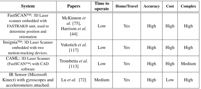
Lymphedema detection is normally assessed by the comparison of the limb volume with the unaffected limb. Several volume methodologies currently used for this goal include water displacement, circumference measurements, bioimpedance or imaging techniques (see Table 3.1).
Table 3.1: Comparison of the most significant methods for limb edema assessment. Adapted from [72].
System Time to
operate Home/Travel Accuracy Cost Complex
Water Displacement:The limb is immersed into a container and the
amount of the displaced water represents the volume of the limb [108].
High No High Low Medium
Circumferential Measurements:The volume can be estimated assuming cylindrical/conic volumes between several measures taken along the
limb [103].
High Yes Low Low Low
Perometer ® :The device scans the limb with IR light and assess limb
volume at small intervals [21].
Medium No High Medium High
CT:Determines of the overall cross-section area and quantify the
density of the tissues [27].
Low No High High High
DEXA:Uses a tissue-specific mode with attenuation of X-ray dependent on
the thickness, density, and chemical structure of the tissue examined [92].
Low No High High High
BIS:Small current passes through the body. Measures volumes by comparing impedance values of both arms [22].
Medium No Medium High Medium
The water displacement is based in a simple physical principle. The limb is immersed into a container and, therefore, the amount of the displaced water will represent the volume of the limb. When performed properly, water displacement is accurate [103], however, it is time-consuming, non portable and can be nonhygienic [108]. On the other hand, there are difficulties in the definition and implementation of the upper level for immersion [108].
The volume can also be obtained indirectly from multiple circumferential measurements of the limb by a tape measure, assuming cylindrical/conic shapes. Accuracy will depend on the spacing between the measurements. Sources of error in this method arise from the assumption of circular cross-section of the limb and from the way the operator uses it [103].
Arm circumferences can also be measured with the use of a Perometer ® [21]. The Perometer ® is an opto-electronic device that depends on the interruption of infra-red (IR) light beams by the limb. The arm
3.1 Functional Evaluation: Methods of Assessment 13
is positioned in a frame with a mobile source of IR light: emitting diodes on two adjacent sides and rows of corresponding sensors on the opposite two sides. The movement of the frame along the limb allows the automatic calculation of the volume from a large number of vertical and horizontal diameter measurements at 0.31 mm interval. This method allows reliable and highly reproducible measurements [108], however the size and the cost of the equipment limits its usability and portability.
In the review of Stanton et al. [103] addressing non-invasive methods for lymphedema detection, it was also assessed the use of imaging techniques for this purpose. Computed tomography (CT) has been used to evaluate limb swelling, since this technique allows the determination of the overall cross-section area and quantify the density of the tissues. In lymphedema, CT has shown that subcutaneous compartment increases in volume [27]. The radiation dose is a particular drawback for the repeated use of CT. Magnetic resonance imaging (MRI) was compared with CT for the investigation of limb swelling after breast cancer treatment [27]. Differences between the two methods were small and, although no use of radiation with MRI, the use of CT is cheaper and more readily available.
Dual energy x-ray absorptiometry (DEXA) is typically used to study soft tissue composition as well as bone mineral density. It uses a tissue-specific mode (fat, lean tissue, and bone) with attenuation of X-ray dependent on the thickness, density, and chemical structure of the tissue examined [92]. A criticism similar with CT is applied in this case because of the radiation dose that is used.
Bioelectrical impedance spectroscopy (BIS) as a lymphedema measure has been previously well de-scribed [22,103]. The procedure involves passing an small current through the body at a range of frequencies that can be used to provide information on the amount of total body water and extracellular water. The working principle assumes that extracellular and intracellular fluids act as a network of resistors with the cell membranes behaving as an imperfect capacitor [103]. Comparing the impedance values between the treated and untreated sides, it is possible to measure lymphedema in a accurate manner [22].
3.1.1.2 Three-dimensional approaches for Limb Edema Assessment
Some of the methods described above can provide an objective and accurate measure of the limb volume, however they are time-consuming, complicated or expensive. Therefore, the search for an accurate, repro-ducible, low-cost and easy-to-use system is still on going.
The fast evolution of 3D technology over the last decade allowed the development of several efficient and cost-effective applications in medicine and health care. Therefore, traditional methods to assess health status, are being replaced by the use of more sophisticated systems. Low-cost, non-invasive and ease of use 3D body-surface scanners are transforming the ability to accurately measure the body size, shape, and skin-surface area. These features make them appealing for widespread clinical applications [112].
In the recent years, some systems comprising 3D laser scanning for limb volume measurement in edema detection were proposed (see Table 3.2).
In 2007, McKinnon et al. [75] evaluated the use of digital scanning (Polhemus FastSCAN™ [59]) (Figure 3.1) for lymphedema measurement by comparison to the method of water displacement. The Polhemus FastSCAN system combines laser scanning with 3D spatial orientation. McKinnon concluded that laser scanning is a method that combines precision and reproducibility in tissue volume measurement and may have clinical utility for measuring lymphedema. Harrison et al. [44] also validated the clinically use of FastSCAN™ for the assessment of postoperative facial swelling.
More recently, the use of Insignia™ laser scanning system [87] (Figure 3.1) was assessed by Vukotich et al.[117] as a mean to obtain limb volume. In comparison with water displacement method, the use of the scanner proved to be suitable for assessing volume in any patient.
14 Literature Review
Table 3.2: Comparison of 3D methods proposed for arm edema assessment.
System Papers Time to
operate Home/Travel Accuracy Cost Complex FastSCAN™:3D Laser
scanner embedded with FASTRAK® unit, used to
determine position and orientation
McKinnon et al.[75], Harrison et al.
[44]
Low Yes High High High
Insignia™:3D Laser Scanner embedded with two motion-tracking devices.
Vukotich et al.
[117] Low Yes High High High
CAML:3D Laser Scanner (FastSCAN™) with CAD
software
Trombetta et al.
[113] Low Yes High High Medium
IR Sensor (Microsoft Kinect) with gyroscopes and
accelerometers attached.
Lu et al. [72] Medium Yes High Low High
In 2012, the use of a Computer Aided Measurement Laser (CAML) technique was proposed by Trombetta et al.[113] to quantify post-surgery lymphedema. They defend that the use of IR laser scanning and computer aided design (CAD) is a more sensitive and accurate method that provides a fast, precise and non invasive technique to quantify arm edema. The 3D scanner analyzes the limb and collects data on its size, shape and appearance. This data is processed in CAD software to create a model through which it is possible to determine circumferential and limb volume measurements. In their study, the FastSCAN™ was used, in conjugation with a CAD software, to acquire circumferential and volume data for phantoms and upper arm of enrolled patients. The data was compared with circumferential measurements and minimal errors were obtained.
Figure 3.1: (a) Polhemus FastSCAN™ [59] and (b) Insignia™ laser scanner [87].
Lu et al. [72], on the other hand, proposed a method for measuring limb volume and for detecting early swelling that relies in IR imaging sensors, such as Microsoft Kinect. The IR sensor is used to capture different views of the human arm while its moved around the subject. A constrained imposed in this method is the fact that the user needs to hold the sensor at 80 cm of the target limb, approximately, while moving the device around. After image acquisition, it is needed to perform a coarse and a fine registration in order to register pairs of consecutive depth images into the same 3D coordinate frame, and iteratively to register all pairs into a single reference coordinate frame. The coarse registration is performed manually (the user needs to click on a set of four corresponding points on two consecutive images), but the authors intend to automate this method with the implementation of gyroscopes and accelerometers attached to the sensor. The fine registration is
3.1 Functional Evaluation: Methods of Assessment 15
accomplished by the use of Iterative Closest Points (ICP) algorithm. The first results confirm the robustness of the system and the ability to detect small and localized differences in limb volume. But in a future work the authors still intend to compare the results with tradition methodologies for limb volume measurement, such as water displacement method.
Several other identical approaches were studied, including 3D scanners and 3D image acquisition, for body swelling detection and volume measurement with different goals than lymphedema evaluation. For example, Kau et al. [63], described the use of laser scanners for monitoring facial swelling following orthog-nathic surgery. In a different manner, Hayn et al. [49] assessed the use of a 3D camera-based measurement in order to detect and quantify leg edema. The goal of their study was the evaluation 3D imaging techniques used as an extension of home monitoring for heart failure patients.
3.1.1.3 Summary
Traditional methods for the assessment of arm edema include water displacement, circumference measure-ments, bioimpedance or imaging techniques. Although accurate, water displacement methods are time-consuming and the apparatus limits its portability. Methods based on multiple circumferential measurements, on the other hand, have limited accuracy and several sources of error. The Perometer has more accurate re-sults but is more expensive. Imaging techniques, such as CT or MRI, require equipment that is not commonly available in clinics due to cost and the need for specialized training. Also, BIS involve the use of several elec-trodes placed along the arm, which leads to a high lifetime operational cost.
Therefore, all these constrains limit the practical use on clinical settings of these procedures. In this way, research is been done to test the application of the arising 3D technologies on body volume estimation. Several authors [75, 113, 117] validated the used of 3D scanners for limb volume assessment, as the case of Polhemus FastSCAN™ and Insignia™ laser scanner. These 3D scanners allow an easy and efficient way for upper-limb modeling and volume assessment, however they are very expensive devices and are quite difficult to be handled by non-professionals. On the other hand, Lu et al. [72] explored the use of a low-cost system, the Microsoft Kinect, for arm modeling and volume measurement. However, the proposed method is quite complex since it depends on gyroscopes and accelerometers attached to the sensor, and its time-consuming since it is necessary to move the device around the limb. Moreover, it is operator-dependent, so its reproducibility is limited.
It remains to find the most suitable method for clinical use, low-cost and easy to operate, in order to assess limb volume and identify arm edema in breast cancer patients. But the investigation of these new 3D technologies, with great potential application in health care, may be the right path to follow.
3.1.2
Upper-Limb Motion Evaluation: Non-vision systems
The assessment of functional status after breast cancer treatment has been concentrated on lymphedema de-tection [80]. However, it is also important to go beyond the limb size and evaluate the UBF impairments, activity limitations and participation restriction that patients of breast cancer normally experience. In 2003, the study of Engel et al. [33] about long-term upper-body morbidity and patient QOL, found that restrictions on upper-limbs motion is the most important source of decreased QOL after breast cancer treatment. There-fore, more clinical attention should be given to other aspects of UBF morbidity, such as limited shoulder ROM.
In that way, as for edema detection, there is a need for an objective, reproducible and low-cost method for evaluation of women’s upper-body motion after breast cancer treatment. This method should be sensitive to
16 Literature Review
the unique issues of breast cancer patients, this is shoulder mobility, and responsive to change in the patients’ status [13].
3.1.2.1 Subjective Methods for UBF Evaluation
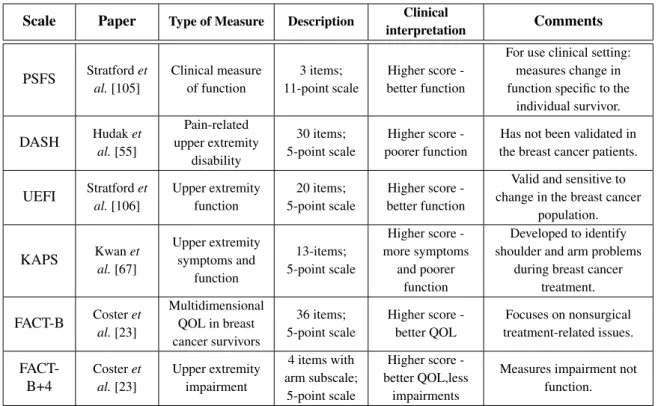
Normally, UBF evaluation rely on subjective measurements of patients experiences, symptomatology and function limitation [13]. In that way, several generic self-report questionnaires have been developed to cap-ture the effects of injury in upper-body function (see Table 3.3).
Table 3.3: Self-report scales used for upper-body function assessment of breast cancer patients. Adapted from [13]
Scale Paper Type of Measure Description Clinical
interpretation Comments PSFS Stratford et al.[105] Clinical measure of function 3 items; 11-point scale Higher score -better function
For use clinical setting: measures change in function specific to the
individual survivor. DASH Hudak et al.[55] Pain-related upper extremity disability 30 items; 5-point scale Higher score -poorer function
Has not been validated in the breast cancer patients.
UEFI Stratford et al.[106] Upper extremity function 20 items; 5-point scale Higher score -better function
Valid and sensitive to change in the breast cancer
population. KAPS Kwan et al.[67] Upper extremity symptoms and function 13-items; 5-point scale Higher score -more symptoms and poorer function Developed to identify shoulder and arm problems
during breast cancer treatment. FACT-B Coster et al.[23] Multidimensional QOL in breast cancer survivors 36 items; 5-point scale Higher score -better QOL Focuses on nonsurgical treatment-related issues. FACT-B+4 Coster et al.[23] Upper extremity impairment 4 items with arm subscale; 5-point scale Higher score -better QOL,less impairments
Measures impairment not function.
Specific scales for UBF assessment include the Patient-Specific Functional Scale (PSFS) [105], devel-oped as a clinical measure of functional status limitations related to the effect of a treatment/intervention. Patients are asked to identify up to 5 daily life activities which they are unable to perform or are having difficulty with as a consequence of the treatment. Indicated items are rated according to the current level of difficulty and are followed to provide a comparison of activities level performance over time.
Disabilities of the Arm, Shoulder and Hand (DASH) scale [55], in turn, was designed to measure physical function and symptoms in patients with any disorder affecting the upper-extremity. This self-report covers symptoms such as pain, weakness and numbness, and the degree of difficult performing work and recreational activities [13].
The Upper Extremity Functional Index (UEFI) [106] aims to evaluate patients’ upper-extremity func-tional status in a variety of activities. On the other hand, Kwan’s arm problem scale (KAPS) [67] was developed to identify shoulder and arm problem in breast cancer patients, including arm/shoulder motion, pain, stiffness, swelling and impairments performing daily activities.
Self-report questionnaires are also used to evaluate QOL indicators of breast cancer survivors. As an example, there is the Functional Assessment of Cancer Therapy-Breast (FACT-B) scale [23]. It is composed
3.1 Functional Evaluation: Methods of Assessment 17
with a Breast Cancer Subscale, which complements the general scale. The FACT-B+4 [23] is a subscale of the FACT-B designed to capture the impact of arm morbidity in a greater extent. Other examples include the European Organization for Research and Treatment of Cancer Quality of Life Questionnaire-Breast Cancer Module (EORTC QLQ BR23) [101] and the BREAST-Q [89].
3.1.2.2 Objective Methods for UBF Evaluation
For the objective assessment of upper-extremity functional limitations there are several methods that include tests of flexibility, strength and endurance. From these tests, the most common for the evaluation of UBF in breast cancer patients is the goniometry [38, 80, 109], used to assess active and passive shoulder ROM in all planes. Comparing measurements of the affected and unaffected limb is possible to detect restricted mobility or impaired shoulder function. The movements that are normally evaluated are: abduction, flexion, extension, internal rotation and external rotation [38].
Other approaches for upper-extremity functional measurement can include the evaluation of strength and endurance assessed by the use of isometric and isokinetic dynamometry and/or maximal performance of a set of tasks/exercises using the repetition maximum method [45, 47, 48]. So, strength and endurance can be measured by means of an incremental exercise protocol where each stage lasts one minute in duration and increments are made by increasing speed of movement and weight held [48]. The movements normally combine a traditional "upright row" and "shoulder press", with the range of movements specified for each patient and each arm [45, 48]. The use of a isokinetic dynamometer for strength and endurance assessment was also reported [45]. The patients are asked to perform sets of repetitions, with 15 seconds of rest between sets, using the dynamometer. The assessment of the grip strength can also be relevant since this is an impor-tant requisite for good arm function. A standard hand dynamometer can be used for this purpose, where the patient is normally asked to perform a maximum contraction three times for each side [45, 48].
There are several other methods to test speed and accuracy movement. Box and Block, for example, is used to measure gross unilateral manual dexterity. It is asked to the patient to move the maximum number of small blocks from one compartment of a box to another within 1 minute [25]. On other hand, Nine-hole Peg Test is used to assess dexterity and upper-extremity function. The requirement is to insert 9 dowels into a board and then removes them in the shortest time possible [25]. However, these methods, and other similar, were not tested in breast cancer context, so it is not known its sensitivity for functional limitations related to breast cancer treatment.
3.1.2.3 Summary
Normally, the assessment of upper-limb function in women diagnosed with breast cancer is performed through broad-based questionnaires that measures psychological, social, and physical functioning aspects of QOL in diverse patient populations, including breast cancer survivors. Although easy-to-use and useful to provide reference data, these subjective methods are generally not accurate and adequately sensitive to UBF issues of breast cancer patients. Moreover, a qualitative evaluation like this has problems related to impartiality and poor reproducibility. Nevertheless, these self-reports can be an useful tool as a validation method for the results obtained in this study.
There is also some studies that report the use of flexibility, strength and endurance tests for the evaluation of UBF in breast cancer patients. However, the use of these methods is not a clinical standard in breast cancer so, it is not well known its sensitivity for functional limitations related to the treatment. The one
18 Literature Review
that is most commonly used is the ROM assessment by a manual goniometer. But, it is operator-dependent, time-consuming, it presents several sources of error, and has low accuracy and reliability.
Thus, to overcome these limitations, other methods should be considered in order to have an efficient, time-adequate and objective method for assessment of patient’s UBF after breast cancer treatment.
3.1.3
Upper-Limb Motion Evaluation: Vision-based systems
As stated before, traditional clinical methods for upper-extremity function evaluation can include either sub-jective scales obtained by patient’s self-report, or more obsub-jective measures, such as range of motion and strength tests, which still present several sources of error and low reliability.
So, to have a better understanding on upper-limb function, a more objective and accurate analysis of motion is needed. Human movement tracking systems have demonstrated to be able to to generate real-time data that dynamically represents the human movement. This field has been in constant research in the last decades, due to its promising application in many areas, including medical diagnosis. In that way, this area of research should be considered, since it can provide objective information on the upper-limb movement patterns. Human movements are normally detected using systems with visual sensors (e.g. cameras) either assisted by visual markers placed upon the human body or marker-free [125].
3.1.3.1 Marker-based human motion tracking systems
In marker-based tracking systems, cameras are used to track human movements, with identifiers placed upon the body [125]. The work of Johansson in 1973 [60] is the milestone in human motion tracking. He used his Moving Light Display system to perceive human motion. The tracking during trajectories was possible due to small reflective markers attached in the joints of human subjects.
Nowadays, several marker-tracking systems are commercially available. For example, VICON [71] (see Figure 3.2) is a passive optical system, designed for motion capture in virtual and immersive environments. It uses several cameras emitting a beam of IR light and a set of reflective markers placed on the objected to be tracked. The markers reflect the IR radiation that is recognized by the system. In that way, is pos-sible to construct a 3D representation of the object. A similar technique is used in Qualisys system [90]. Codamotion [70] (see Figure 3.2), on the other hand, is an active visual tracking system. This technology uses miniature infra-red markers, to track the key positions on the subject. Signals from these markers are emitted to three linear arrays inside a CODA unit which provides an immediate and precise 3D measurement. Another example is Polaris [58], an optical systems that measure the 3D positions of either active or passive markers affixed to a object. Using this information, each Polaris System is able to determine the position and orientation of the object based on the information received from the markers.
3.1 Functional Evaluation: Methods of Assessment 19
However, all these technologies have several drawbacks associated. The use of markers can be unreliable, they can move and wobble, giving rise to noisy data. Also, all the systems require calibration and professional intervention. Moreover, these methods are expensive and very complex, and the space requirements often limit their usability in clinical settings [125]. To overcome these restrictions, a lot of research has been done in order to develop marker-free motion tracking systems.
3.1.3.2 2D Markerless human motion tracking systems
Markerless tracking systems only exploit optical sensors to measure movements of the human body. On 2D motion tracking, it is only considered the human movement in an image plane. This approach can be employed with or without the use of explicit shape models.
Model-based approaches use a priori an human body model to match with the acquired image data. This method uses the knowledge of the movement in 2D for feature correspondence and body structure recovery. The models used are usually stick figures (see Figure 3.3), the simplest representation of a human body, which consists of line segments linked by joints, wrapped around with ribbons or blobs [39].
An early attempt to segment and track body parts was made in Akita work [1]. It assumes that the order of human movement and the spatial relationships between the body parts can be approximated by the use of a key frame sequence of stick figure poses. The stick figure contain the legs, head, arms, and trunk elements, and the cone model is used to provide knowledge of the rough shape of the body parts.
Leung and Yang [68], on the other hand, applied a 2D ribbon model to gather motion information of a moving human object. The system implements two main processes. The first, extracts moving human outlines from an image sequence using a 2D ribbon model. The second, interprets the outline and determines if an extracted 2D ribbon belongs to a part of the body or to the background.
Wren et al. [122] proposed a region-based approach, where the human body is considered as a set of blobs that can be described using a spacial and color Gaussian distribution (see Figure 3.3).
In the work of Ju et al. [61] it is assumed that a person can be represented by a set of connected planar patches: the cardboard person mode. The parametrized image motion of these patches is constrained to enforce articulated motion. The recovered motion parameters provide a good description of the movement that can be used for recognition.
Figure 3.3: Representation of (a) 2D stick-figure model with ribbons by Leung and Yang [68], and the demonstration of the work by Wren et al. [122]: (b) Video input; (c) Segmentation; (d) 2D representation of the blob statistics.
In a different manner, other approaches were described without the use of shape models. In this case, the pose recovery step is ignored, and the human movement is characterized in terms of a simple low-level, 2D features from a region of interest. The motion models are then described in statistical terms normally derived from the low-level features. An example is the work of Baumberg and Hogg [5], where active shape models
20 Literature Review
are applied to track pedestrians (see Figure 3.4). The tracking is initiated in the foreground region, obtained by background subtraction. A Kalman filter is used for spatio-temporal control, similar to the work of Blake et al.[6].
Figure 3.4: Human tracking results using the approach proposed by Baumberg and Hogg [5].
Chang et al. [17] proposed a method for tracking cyclic human motion based on decomposing complex cyclic motion into components and maintaining coupling between components. The decomposition reduces the dimensionality of the problem and enables a graphical modeling of the articulated human body.
Finally, in the work of Wong and Wong [121], is presented a system were is used a wavelet estimator. The human body is located within a small search window, using color and motion as heuristics.
3.1.3.3 3D Markerless human motion tracking systems
2D approaches have several restrictions for the addressed purposed, due to their viewing angle. On the contrary, the use of 3D techniques for human motion identification has the advantage of the knowledge available a priori about the kinematic and shape properties of the human body.
The use of 3D shape models simplify the tracking process and allows the prediction of events such as (self) occlusion and (self) collision. Model-based methods contain stick figures, volumetric, and a mixture of models [125]. In this case, the stick figure is regarded as a collection of segments and joint angles with various degree of freedom at the articulation sites.
In the work of Huber [54], for 3D segment tracking and recognition of human pose and gestures, it is used a stick figure representation, where the joints are connected by line segments. The author studies the behavior of a Proximity Space (PS) method, developed for tracking objects, in the recognition of human poses and gestures as a person moves through a cluttered environment. The PS method uses LoG filtered images and relies on stereo measurements to spatially distinguish between objects in 3D.
Ronfard et al. [94] developed a method to find people in static video frames using learned models of both the appearance of body parts (head, limbs, hands), and of the geometry of their assemblies. The system is built on Forsyth & Fleck’s general "body plan" methodology and Felzenszwalb & Huttenlocher’s dynamic programming approach for efficiently assembling candidate parts into "pictorial structures".
Instead of stick figures, it can also be used volumetric models, such as elliptical cylinders. Ivana et al. [78] presented an automated system for motion capture that includes both the model acquisition and the motion tracking, using multiple synchronized video streams (see Figure 3.5). It is computed the 3D voxel reconstructions of the body shape at each frame which then are used as input to the model acquisition and tracking. The human body model consists of ellipsoids and cylinders. Model acquisition starts with localization of body part based on template fitting and growing, which uses prior knowledge of average body part shapes and dimensions. This initial model is then refined using a Bayesian network that imposes human body proportions onto the body part size estimation. The tracking procedure is an extended Kalman filter that estimates model parameters based on the measurements made on the labeled voxel data.
3.1 Functional Evaluation: Methods of Assessment 21
Figure 3.5: Representation of the automated system for motion capture proposed by Ivana et al. [78].
Rohr [93] introduced a model-based approach for the recognition of pedestrians. The human body is represented by a 3D-model of cylinders. The estimation of model parameters in consecutive images is done by applying a Kalman filter.
Gonçalves et al. [41] studied the position and motion of a human arm in 3D without any constraints on its behavior. The arm was modeled as two truncated right-circular cones connected with spherical joints. A recursive estimator for arm position is used to predict the appearance of the arm on the image, and the difference between the predicted and actual images is then used as an error measurement for the estimator.
In the work of Chung et al. [20], on the other hand, the human body is simplified as a stick model represented by connections of several circular cylinders. The authors proposed a new image feature called cue circles (CCs). Using CCs, 3D motion of the human body is measured from a sequence of boundary contour image pairs, which is obtained by an active binocular sensor system. Stereo matching for recovering the body model is carried out by finding pairs of CCs between the pair of contour images under consideration. Theobalt et al. [110] described a system to capture human motion, where is applied a 2D feature tracking algorithm and a silhouette-based 3D volumetric scene reconstruction (see Figure 3.6). The person is recorded by multiple synchronized cameras, and a multi-layer hierarchical kinematic skeleton is fitted to each frame in a two-stage process. The pose of a first model layer at every time step is determined from the tracked 3D locations of hands, head and feet. A more sophisticated second skeleton layer is fitted to the motion data by applying a volume registration technique.
Figure 3.6: System proposed by Theobalt et al. [110] to capture human motion: representation of the skeleton fitted to visual hulls (rendered as point sets) of a moving person.

![Table 3.1: Comparison of the most significant methods for limb edema assessment. Adapted from [72].](https://thumb-eu.123doks.com/thumbv2/123dok_br/15850424.1085508/32.892.108.771.426.868/table-comparison-significant-methods-limb-edema-assessment-adapted.webp)

![Table 4.1: A comparison table for Natural User Interface (NUI) libraries. Adapted from [99].](https://thumb-eu.123doks.com/thumbv2/123dok_br/15850424.1085508/50.892.107.762.550.955/table-comparison-table-natural-user-interface-libraries-adapted.webp)