Universidade de Lisboa
Faculdade de Farmácia
THE REFERENCE INFLIXIMAB MEDICINE AND BIOSIMILAR CT-P13 INDUCE
A SIMILAR IMMUNE RESPONSE IN INFLAMMATORY BOWEL DISEASE
PATIENS
Miguel Roque Santos
Dissertation supervised by Professor Doutor João Gonçalves
Master in Biopharmaceutical Sciences
Universidade de Lisboa
Faculdade de Farmácia
THE REFERENCE INFLIXIMAB MEDICINE AND BIOSIMILAR CT-P13 INDUCE
A SIMILAR IMMUNE RESPONSE IN INFLAMMATORY BOWEL DISEASE
PATIENS
Miguel Roque Santos
Dissertation supervised by Professor Doutor João Gonçalves
Master in Biopharmaceutical Sciences
À minha mãe Maria Roque Santos
Pelo eterno orgulho de nossa caminhada, pelo apoio, compreensão, ajuda, e, em especial, por todo carinho ao longo deste percurso.
Ao meu irmão Hugo Santos
Pelo carinho, compreensão e pela grande ajuda
Aos meus amigos e colegas de curso, Pela cumplicidade, ajuda e amizade.
Ao professor Doutor João Gonçalves
The use of therapeutic mAbs as the anti-TNFa agents has been known not to cure, but treat auto-immune diseases such as the inflammatory bowel diseases (IBD), improving the overall quality of life of patients. These therapeutic mAbs are characterized by their high specificity allowing to block therapeutic targets as pro-inflammatory cytokines such as TNFa. However, as mAbs are biological drugs, called proteins. Their fundamental disadvantage is the risk of inducing immunogenicity, which can affect the effectiveness and safety of the drug, leading to loss of therapeutic response in patients. This phenomenon is called LoR. For that reason, it is necessary to monitor the treatment of these drugs, also referred to as TDM. Developing therapeutic mAbs is a complex and expensive process due to the complexity of the protein molecular structure. It requires carefully production, storage and quality control, which reflects to their high cost. Since the patent of therapeutic mAbs has expired, many manufacturers have taken the opportunity to introduce biosimilar drugs to the market. This has allowed for national health systems to spend less resources, making it accessible to more patients.
CT-P13 was the first biosimilar drug of Infliximab to be approved by EMA in 2013, after the drug showed similar effectiveness and safety in rheumatoid arthritis and ankylosing spondylitis patients in two main studies called PLANETRA and PLANETRAS. Based on the results obtained in these studies the therapeutic indication of CT-P13 was extrapolated to the IBD in 2014 which leads to some mistrust in relation of the efficacy and safety of biosimilar in IBD patients.
Doubts of extrapolation seem to be a non-issue nowadays since the post-commercialize studies attest to the similarity of CT-P13 face to the original drug. However, doubts relating to switching, multiple-switching, reverse-switching and cross-switching still persist. To evaluate if these drugs are similar, a comparison is done of drug and other levels and ADAs. The ELISA essays have been widely used and stand out due to it’s simplicity.To measure a patients ADAs level bELISA, which is a type of ELISA can be used, even though bELISA has it’s limitations. The presence of drug and IgG4 subtype ADAs are described as being factors that difficult the ADAs detection which makes with the same detection depending on the sensibility of each commercial kit, although these differences may not be significant.
The aim of this study is to evaluate immunogenic response in patients treated with biosimilar CT-P13 through the detection of the drug and CT-P13 in order to verify how the presence of ADAs interferes with pharmacokinetics. Mapping epitopes of biosimilar and anti-Remicade ADAs with resource to an in house ELISA study, can verify which drugs are most immunogenic between IBD patients.
Another in house ELISA study was done in order to map epitopes of anti-biosimilar and anti-Remicade ADAs subtype IgG4. Ultimately, a neutralization study was done to detect the presence of neutralizing antibodies (NADAs).
In general, the data presented in this study suggests that CT-P13 and Remicade are similar in inducing an immune response in IBD patients, corroborating with others studies described. Therefore the switching between Remicade and biosimilar should not be motive of mistrust in terms of effectiveness and safety.
O uso de mAbs terapêuticos tais como os agentes anti-TNFa têm permitido, não curar mas sim tratar, doenças auto-imunes como as doenças inflamatórias intestinais (DII), melhorando assim a qualidade de vida dos doentes que as vivenciam. Estes mAbs terapêuticos caracterizam-se pela sua alta especificidade permitindo bloquear alvos terapêuticos como citocinas pró-inflamatórias tais como o TNFa. Porém os mAbs terapêuticos uma vez tratando-se de medicamentos biológicos, nomeadamente proteínas, têm como fundamental desvantagem poder induzir nos doentes imunogenicidade que pode afectar a eficácia e a segurança destes fazendo com que os doentes deixem de responder à terapêutica, fenómeno designado por LoR que torna necessário a monitorização da terapêutica destes fármacos designada por TDM. Desenvolver mAbs terapêuticos é um processo complexo e dispendioso devido à complexidade da estrutura molecular das proteínas que requer cuidados na produção, armazenamento e controlo de qualidade o que se reflecte no seu custo elevado. A queda das patentes dos mAbs terapêuticos deu oportunidade à introdução de medicamentos biosimilares no mercado que por não terem de ser submetidos a todos os estudos a que os originais tiveram de ser sujeitos, são mais económicos permitindo que os sistemas nacionais de saúde possam economizar verba e com que mais doentes possam usufruir da terapêutica.
O CT-P13 foi o primeiro biosimilar do Infliximab original a ser aprovado pela EMA em 2013 após o fármaco ter demonstrado eficácia e segurança similares nos doentes com atreite reumatóide e espondilite anquilosante em dois estudos principais denominados de PLANETRA e PLANETRAS. Com base nos resultados obtidos nestes estudos a indicação terapêutica do CT-P13 foi extrapolada para a DII em 2014, o que levantou alguma desconfiança em relação à eficácia e segurança do biosimilar nos doentes com DII. A questão da extrapolação parece estar ultrapassada nos dias que decorrem uma vez que estudos de pós comercialização atestam a similaridade do CT-P13 face ao produto de referência. Porém dúvidas persistem em relação à substituição (switching), múltiplo-switching, o switching -reverso e switching-cruzado.
A avaliação da similaridade destes fármacos tem sido efectuada por medição do perfil de fármaco e de anticorpos antifármaco (ADAs). Para medir estes parâmetros destacam-se os ensaios de ELISA que têm sido vastamente utilizados devido à simplicidade de execução desse tipo de ensaio. Na medição de ADAs pode ser salientado bELISA, contudo está documentado algumas limitações deste tipo de ensaio na detecção de ADAs. A presença de fármaco e de ADAs do subtipo IgG4 estão descritas como sendo factores que dificultam a detecção de ADAs o que faz com que essa mesma detecção dependa da sensibilidade de cada kit comercial embora as diferenças possam não ser significativas.
O objectivo deste estudo foi avaliar a resposta imunogénica dos doentes tratados com o biosimilar CT-13 pela detecção de fármaco e de CT-P13 a fim de verificar como a presença de ADAs interfere com a farmacocinética. Mapear os epitopos dos ADAs anti-biosimilar e anti-Remicade com recurso a um ensaio de ELISA in house, de modo a verificar qual dos fármacos é mais imunogénico entre os doentes com DII, sendo que à parte foi optimizado e realizado outro ELISA in house no sentido de mapear epitopos de anti biosimilar e anti Remicade ADAs do subtipo IgG4. Por último realizou-se um ensaio de neutralização para deteção da presença de anticorpos neutralizantes (NADAs).
No geral os dados apresentados neste trabalho sugerem que o CT-P13 e Remicade são similares na resposta imune que induzem nos doentes com DII o que corrobora com outros estudos descritos. Assim sendo assim o switching entre o Remicade e o biosimilar não deverá ser motivo de desconfiança em termos de segurança e efectividade.
INDEX
1. Introduction ...23
1.1 Biologic medicines ...24
1.2 Therapeutic mAbs ...24
1.3 Type of mAbs ...27
1.4 Pharmacokinetics of therapeutic mAbs ...28
1.5 Biosimilars ...29
1.6 How original mAbs and biosimilars have been compared ...30
1.7 Infliximab ...31
1.8 Remicade dosage and routine of administration. ...32
1.9 Biosimilar CT-P13 ...33
1.10 Therapeutic drug monitoring of IBD patients under IFX treatment ...34
1.11 The importance of the Therapeutic Drug Monitoring to evaluate efficacy and safety of CT-P13 and the original Infliximab. ...36
2 Aims ...39
2.1 Overall goals and specific aims ...40
3 Methods ...42
3.1 Patients and healthy controls ...43
3.2 Immunogenic response of IBD patients to the IFX biosimilar CT-P13 drug based on IFX and ADA TL-serum profile...43
3.3 Infliximab synthetic peptides ...44
3.4 Development of two in house ELISA assays to screen which the synthetic peptides bind to ordinary IgGs ADAs and IgG4 ADAs, contained in the positive serum samples from the IBD patients under the biosimilar treatment and the IBD patients under the original IFX treatment. ...47
3.5 Screening epitopes of the anti-Infliximab (anti-Remicade and anti-biosimilar) IgG antibodies ...48
3.7 Screening of Infliximab epitopes of the Infliximab (Remicade and anti-biosimilar) IgG4 antibodies ...50
3.8 Identification of neutralizing anti-infliximab profile ...52
4 Results ...54
4.1 Screening of anti-Remicade IgG epitopes ...55
4.2 Screening of anti-biosimilar IgG epitopes ...56
4.3 Screening of anti-Remicade IgG4 epitopes ...57
4.4 Screening of anti-biosimilar IgG4 antibodies ...57
4.5 Identification of neutralizing anti-Remicade antibodies profile ...59
5 Discussion ...64
6 Conclusion ...69
7 References ...71
8 Appendix...78
8.1 Scheme of LISA Duo Infliximab TRACKER® ELISA kits (Theradiag, France) ...79
TABLE INDEX
Table 1 – Representative scheme of the Infliximab infusion routine. ...33
Table 2 – Representation of different possible results obtained by drug and ADA serum-TL measurement. ...35
Table 3 - Patients and health controls. ...43
Table 4 – Infliximab commercialized synthetized peptides ...44
Table 5 – ELISA assay optimization ...47
Table 6 – ELISA Results of the drug and ADA serum-TL found in among the IBD patients. ...55
Table 7 – OD values of negative controls and the white. ...60
Table 8 – OD values of IFX measured on IBD patients with Immunogenicity ...60
Table 9 – OD values of IFX measured after the neutralizing assay. ...60
Table 10 – OD values of negative controls and the white. ...62
Table 11 – OD values of Biosimilar measured on IBD patients with Immunogenicity ...62
FIGURE INDEX
Figure 1 - Antibody structure. ...26
Figure 2 - IgGs Subclasses ...27
Figure 3 - Schematic representation and nomenclature of mAbs in clinical use in IBD. ....28
Figure 4 - Pharmacokinetic of multiple infusions of therapeutic mAbs ...29
Figure 5 - Localization of the infliximab synthetized peptides on chain H FAB IFX ...45
Figure 6 - Localization of the infliximab synthesized peptides on chain L FAB IFX. ...46
Figure 7 - Localization of the synthetized peptides on the FC IFX ...46
Figure 8 – Scheme of the IFX peptides coated on 96-wells plates and the indication how serum samples was added. ...48
Figure 9 - Detection of anti-infliximab IgG antibodies ...50
Figure 10 - Detection of anti-infliximab IgG4 antibodies ...52
Figure 11 - Optical Density values of seropositive patients which them serum contains the anti-Remicade antibodies against Remicade ...52
Figure 12 - Optical Density values of seropositive patients which them serum contains the anti-biosimilar antibodies against biosimilar drug. ...56
Figure 13 - Optical Density values of seropositive patients which them serum contains the anti-Remicade IgG4 antibodies against Remicade. ...56
Figure 14 - Optical Density values of seropositive patients which them serum contains the anti-biosimilar IgG4 antibodies against biosimilar. ...56
Figure 15 - Average of the mapping ELISA assay to detect IgG ADA subtype epitopes. 568 Figure 16 - Average of the mapping ELISA assay to detect the IgG4 ADA subtype epitopes. ...56
Figure 17 - Optical Density values of Remicade detected in ELISA assay after the neutralizing process with positive serum to anti-Remicade antibodies. ...61
Figure 18 - Optical Density values of the biosimilar detected on ELISA assay after the neutralizing process with positive serum to the anti-Remicade antibodies ...63
ABBREVIATIONS
µg Microgram µL Microliter Aa Amino acid Ab Antibody
ABAbs Anti-biosimilar antibodies
ABDAb Anti-biosimilar drug antibodies Abs Antibodies
ADA Adalimumab
ADAb Anti-drug antibodies
AIAbs Anti-infliximab antibodies
AIDAb Anti-infliximab drug antibodies
ARAbs Anti-Remicade antibodies
ARDAb Anti-Remicade drug antibodies
AS Ankylosing spondylitis
AUC Area under the curve
AUCss Area under the curve at steady state
bELISA bridging ELISA
BSA Bovine Serum Albumin
Cavss Average steady state plasma drug concentration CD Crohn disease
CDRs Complementarity-determining regions
CL Constant light chain
Clss Clearance at steady state
Cmax Maximum concentration
Cmaxss Maximum concentration at steady state
Cmin Minimum concentration
Cminss Minimum concentration at steady state
CRP C-reactive protein
Css Concentration at steady state
DN Double negative result
DNA Acid deoxyribonucleic
DP Double positive result
EIA Enzymatic immunoassay
ELISA Enzyme-Linked ImmunoabSorbent Assay Fab Antibody variable region
Fc Constant fragment
FcR Constant fragment receptor
Fv Variable fragment
HCL Hydrochloric acid
HRP Horseradish Peroxidase
IA Immunoassay
IBD Inflammatory bowel disease IFX Infliximab
IFX-B Infliximab biosimilar drug IFX-R Infliximab Remicade drug
IFX-TL Infliximab sérum trough levels Ig Immunoglobulin
IgG Immunoglobulin G IgG4 Immunoglobulin IgG4 IgM Immunoglobulin M
IV Intravenous
KDa Kilodalton
LoR Loss of response
mAb Monoclonal antibodie mAbs Monoclonal Antibodies Mg Milligram
NADA Neutralizing anti-drug antibody
NADAs Neutralizing anti-drug antibodies
NK Natural killer cell OD Optical Density ON Over night PA Psoriatic arthritis PS Psoriase P55 Receptor 55 of TNFα P75 Receptor 75 of TNFα PBS Phosphate Buffer Saline
PBST Phosphate Buffer Saline Tween
PK Pharmacokinetics
RIA Radioimmunoassay
RMP Reference medicinal products
ss Steady state
TDM Therapeutic Drug Monitoring
TL Trough levels
Tmax Time to reach the maximum concentration TMB 3,3’5,5’ tetramethylbenzidine
TNFα Tumor necrosis factor alpha UC Ulcerative Colitis
VH Variable heavy chain VL Variable light chain
Vss Volume of distribution at steady state
α alpha δ delta ϒ gama ε epsilon μ mu κ kappa λ lambda
1. Introduction
24 1.1 Biologic medicines
Biologic medicines also designated biopharmaceuticals are a class of medicines obtained by living organisms. These kinds of medicines are polymers of amino acids protein molecules that include protein molecules or peptides. How proteins are huge macromolecules are more difficult to characterize comparatively with the general of chemical drugs (Berghout, 2011; Peres, Padilha & Quental, 2012). Due to the complexity of the tri-dimensional protein structure small differences can occur during the production process because the cell lines that generate the protein of interest are prone to generate variations in protein products which can influence their quality, efficacy and safety. These variations can be present in every new batch that is produced, and so, a major control of quality, efficacy and safety is necessary which makes these medicines more difficult to develop, justifying their high prices (de Mora & Fauser, 2017; Mulcahy, Hlavka & Case, 2018). The first generation of biologics was launched in the late 1970s and early 1980s, and this class of drugs is today one of the high growing sectors of the pharmaceutical industry (Gabbani, Deiana, Annese; 2017). The great success of the biologic medicines is because these class of drugs are designed to block specific pathways of action of some disease, so they are more specific and selective compared with conventional drugs and this can be considerable an advantage. Some examples of these drugs are vaccines, blood components, allergenics, somatic cells, gene therapy, tissues and recombinant therapeutic proteins (Kennedy, Oliveira , Granja, Sacramento, 2018). About the recombinant therapeutic proteins, these treatments has the disadvantage to be prone to provoke intolerable immune response in some patients that recognize the biologic agent as a strange molecule to the organism, and so generating anti-drug antibodies (ADAs) that can affect the efficacy of the biologic drug and also development of side effects can occur. The mechanism whereby biologic protein drugs have the ability to induce an immune response mediated by ADAs is named immunogenicity and it can manifests with different intensities once it is a result of multifactorial consequences such as genetic background, severity of the disease that the biologic are design to treat, routine of administration, condition of the drug that comprehends aggregation and denaturation of the therapeutic protein which could have a significant impact on efficacy and safety of biological therapies as well (Dingermann, 2008).
1.2 Therapeutic mAbs
Some biologic medicines are monoclonal antibodies (mAbs) that are Abs produced by unique B-cell lymphocyte which produce only one antibody (Ab) type that have equivalent
25 biological and physicochemical proprieties and so that a same affinity and avidity and they bind to only one epitope on the antigen (Dingermann, 2008). MAbs where firstly obtained by the hybridom technic developed by Geores Köhler and Cesar Milstein in 1975. The technic consist in the production of a hybridom cell by the fusion between a lymphocyte B cell from a mouse pre induced to generate an immune response against the antigen of interested with a myeloma that is a kind of cancer cell. From these fusion results a hybrid immortalized cell line named hybridoma that produce mAbs. The greatness of the mAbs achievement was that contrary to the polyclonal Abs that are generated against different epitopes of the same antigen, mAbs once are generated only to the same epitope are much more specific. The exploration of mAbs specificity was a big step for the application of mAbs as therapeutic drugs. Nowadays mAbs are obtained with resource to biotechnology process such as recombinant DNA, protein engineering or phage display. After all mAbs obtainment opened new doors to the immunology science progress (Martin & Bugelski, 2012). Their application allowed the development or enhanced of assays such as radioimmunoassay, enzymatic immune assay (EIA) or flow-cytometry based on specific mAbs conjugated to fluorophores and so that, more specifics. This improvement became easier the identification of biomarkers, cell population and cell path ways. Therefore mAbs are antibodies which are glycosylated proteins with “Y-shape” composed by two heavy chains (CH) and two light chains (CL). Abs also has a constant fragment (FC) and a variable fragment (FV). The variable portion of the light chain (VL) can be kappa (K) or lambda (λ) type depending of some differences in polypeptide sequence. What codify if an antibody displays a K or a λ chain are two rearranged DNA sequences (VJ) while the variable regions of the CH are codified by three rearranged DNA sequences (VDJ). Farther each variable region contains three hypervariable sub-regions designed complementary determining regions (CDR1, CDR2 and CDR3) that form the paratope that means the antigen biding site which hold the epitope region on the antigen. An antibody Ab is composed by four chains held together by disulfide bounds. About these four chains, two of them are identical light chains with a mass about 22 KDa each and the other two are identical heavy chains with a mass about 53 KDa (Arosa, Cardoso, & Pacheco, 2012).
26
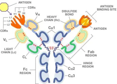
Figure 1 - Antibody structure.
Representation of an immunoglobulin structure. In dark blue is represented the constant regions of the heavy chains (CH1, CH2, CH3). The clear blue represents the variable region of the heavy chain (VH). The dark green is the constant region of the light chain (CL) and the clear green the variable region of the light chain
(VL). In orange is represented the paratopes with their complementarity-determining region (CDRs) and the
yellow represents the antigen.
Source: Novimmune -- Science: Antibodies. (2016). Novimmune.com. Retrieved 26 October 2016, from http://www.novimmune.com/science/antibodies.html
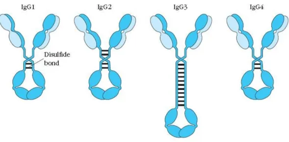
There are 5 classes of Abs (IgA, IgD, IgG, IgE and IgM) according to the type of Fc region. IgA has alpha (α) Fc region, IgD has delta (δ) Fc, IgG has gama (γ) Fc, IgE has epsilon (ε) Fc and finally IgM takes mu (µ) Fc. Further the Abs have kappa (k) or lambda (λ) light chains. IgGs are the Ig which perform more activity in the immune system. They have a mass of about 150 KDa and are the main Igs in the blood stream and lymph. The half-life of 23 days which is the major among all five Abs classes, form 15% of total serum proteins and exist in the blood as a monomer. Further there are different isotypes of IgGs. IgG1, IgG2a, IgG2b, IgG3 and IgG4. IgGs and have functions like opsonization and agglutination that are important to facilitate the phagocytosis process by macrophages because macrophages express Fc receptors (FcR) which links to the Fc portion of IgG, on the other hand the Fab region links to specific regions on the antigens in bacteria or other pathogens named epitopes. Another efficiency of IgGs are the activation of the complement system, neutralization of toxins and virus and the opsonization of damage cells promoting the connection with the NK cells that also express FcR and thus contributing to the elimination of damaged cells by cytotoxicity (Arosa et al, 2012).
27
Figure 2 - IgGs Subclasses
The different subtypes of IgG antibodies that vary depending of the number of disulfide bounds in the hinge region and also of their amino acid sequence. IgG1) Immunoglobulin 1. IgG2) Immunoglobulin 2. IgG3) Immunoglobulin 3. IgG4) Immunoglobulin 4.
Source: Owen, Punt, Stranford, Jones, & Kuby, 2013. Kuby immunology (p. 89). New York: W.H. Freeman
1.3 Type of mAbs
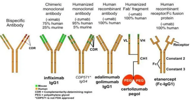
The mAbs can be classified as mouse, chimeric, humanized and human depending of the percentage of murine and human sequences and that determines the mAb name. Theoretically the mAb type can have impact on immunogenicity, once murine mAbs are expected to be more immunogenic then chimeric mAbs, and chimeric mAbs more immunogenic then humanized that finally is supposed to be more immunogenic then full human mAbs although the idea that full human mAbs are non-immunogenic is wrong because even full human mAbs have the ability to induce immunogenicity. By the other hand chimeric mAbs are well tolerated among some patients. Is important to have in mind that the answer how human the mAb is or how murine the mAb is, is just one of the different aspects that influence immunogenicity, however there are other aspects that influence immunogenicity as previously mentioned that also can have impact (Atzeni et al, 2013).
28
Figure 3 - Schematic representation and nomenclature of mAbs in clinical use in IBD.
The figure represents the therapeutic mAbs nomenclature, regarding to humanization level.
Source: Adapted from Lee, Chinen, & Kavanaugh, 2010. Immunomodulator therapy: monoclonal antibodies, fusion proteins, cytokines, and immunoglobulins. Journal of Allergy and Clinical Immunology, 125(2), S314-S323.
1.4 Pharmacokinetics of therapeutic mAbs
Therapeutic mAbs are proteins and needs to be administrated by intravenous or subcutaneous infusions. Once these drugs are used to treat chronic conditions in general, multiple infusions are needed to achieve the remission of symptoms and to maintain the diseases under control. Multiple infusion regimes implies that physicians have to establish a right dosage and a right interval of drug administration, for each patients and at the beginning of the treatment some changings in these two parameters could be necessary until the steady state (ss) be achieved. Due to the multiple infusions the pharmacokinetics of therapeutic mAbs fits with the bolus graphic (Brandse et al, 2017).
29
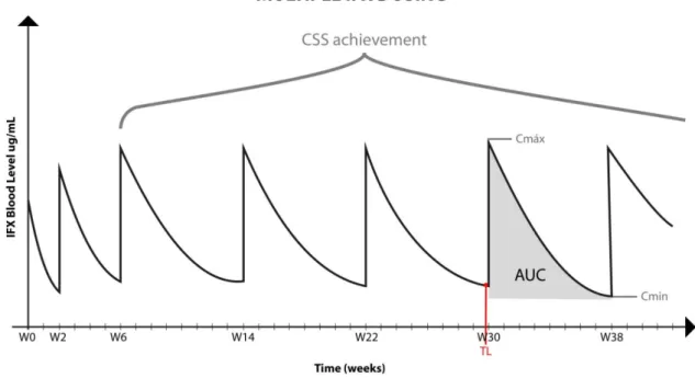
Figure 4 - Pharmacokinetic of multiple infusions of therapeutic mAbs
Represent the infliximab pharmacokinetic of multiple infusions bolus graph. Concentration of IFX at the steady state (Css), maximum serum concentration (Cmax), minimum serum concentration (Cmin), area under the curve (AUC), trough levels (TL).
Source: Adapted from Maini et al, 1999; Yoo et al, 2013.
To reach the steady state the physician should evaluate the area under the curve (AUC) of the continues infusions and when the multiple infusions start showing a similar AUC geometry and also showing same values of minimums concentration (Cmin) (Brandse et al, 2017).
1.5 Biosimilars
When the patents of conventional drugs expired, generic copies of their reference products can be commercialized, however regarding to the biologic medicines is not possible to reproduce precisely copies because the complexity of the protein macromolecules. This is the reason why biologic copies are not considerate generic copies but biosimilar ones. An approved biosimilar is by definition highly similar to the reference drug such that any molecular or structural dissimilarities or potential differences in the underlying mechanisms do not affect the quality, efficacy and safety. A Biosimilar drug depends of the manufacture processing that includes: line cells, purification process, growth, storage, and transport conditions, so there’re always differences of glycosylation,
30 phosphorylation, sulfation and other post-translational modifications that can ultimately affect the drug efficacy and so have an impact regarding to immunogenicity. Is important to keep on mind that these differences are present not only between the reference drug and his biosimilar but also between different batches of the same reference or biosimilar biologic and product (Jahnsen, 2016; Park, Lee, Yun & Yoo, 2015).
How the development of a biosimilar does not need so many studies to clarify the quality, efficacy and safety, the cost of production is cheaper than a new biologic drug and for this reason attracts the interesting of countries governments because the changeability of a RMPs to biosimilars allows to reduce costs for the countries healthcare systems and consequently increase the accessibility of patients to these drugs. Although some concerns among physicians regarding to the quality, efficacy and safety are included in the equation and not only the financial aspect. The issues that have been emerging are if biosimilars have the same quality safety and efficacy, once perfect copies of some biologic agent are too difficult to reproduce. Other concern is if the changeability of a RMP by their biosimilar has impact on immunogenicity, and if so, if can have impact on efficacy and safety (Ben-Horin et al, 2015).
1.6 How original mAbs and biosimilars have been compared
The studies aimed to compare pharmacokinetic parameters such as maxim drug concentration at the ss (Cmax,ss); minimums drug concentration at ss (Cmin,ss); AUC at the ss (AUC,ss); time to reach Cmax (Tmax); the swing (Cmax,ss - Cmin,ss)/ Cmin,ss); degree of fluctuation (Cmax,ss - Cmin,ss)/average steady state plasma drug concentration (Cav,ss); mean residence time (MRT); terminal elimination half-life (T(1/2)); total body clearance at steady state (CI,ss); volume of distribution at ss (V,ss). Biosimilars also
should show similar immunogenicity, comparatively with the RMP, by accessing the ADAs profile, that are generated to the therapeutic mAb influencing the pharmacokinetic (PK) by neutralizing and increasing the drug clearance. The blood drug levels and ADAs are detected with resource to immuno-assays (IAs) based on the linkage antigen-antibody (Park et al, 2013). Radioimmunoassay (RIA) and the enzymatic immunoassay (EIA) are two types of immune-assays widely described to detect serum drug levels and ADAs. RIA is a more sensitive assay then EIA, however it has the disadvantage to be a more complex technic which requires special laboratories prepared to the handling radioactivity and so, carrying more risks. Further, RIA assay is a more expensive technic then EIA. The enzymatic-linked immunosorbent assay (ELISA) is characterized to be simple to perform and cheaper comparatively to RIA. There are different types of ELISA such as direct
31 ELISA, indirect ELISA, sandwich ELISA competition ELISA and bridging ELISA (bELISA). The bELISA have been used for mAbs and ADAs detection, important parameters to monitoring patients, and to test the similarity between RMPs and biosimilars (Svenson, Geborek& Saxane, 2017). ADAs detection allows performing cross-react assays and neutralization assays. Cross react-react assays consists in verify if the ADAs generated by the patients that are under the original treatment recognize the biosimilar mAb, and to verify if the ADAs generated by patients treated with respective biosimilar recognize the original therapeutic mAb. When both situations happens is considerate that exists cross-reactivity and it is a high indicative of similarity. The ability of the ADAs to neutralize the drug and affecting the effectiveness of the treatment is important to cooperate to test similarity in terms of effectiveness (Ben-Horin et al, 2015).
Other way to evaluate ADAs is to study their response trough analyzing their biding sites. These technic consist in mapping the drug mAb to identify the regions where the epitopes are spotted. Mapping epitopes on the therapeutic mAb provide information about the regions of the drug that are more and the regions that are less immunogenic. Further, in the case that the epitopes are mostly placed at CDR regions, these data can be crossed with the results of the neutralization studies to verify if both studies are in agreement, once is expected that, if ADAs bind to CDRs region, also are neutralizing the drug activity that will not be able to link to their target. (Kosmač, Avčin & Toplak, 2011).
1.7 Infliximab
Infliximab (IFX) is a chimeric IgG1 mAb against soluble and transmembrane forms of tumor necrosis factor alpha (TNFα) containing about 25% murine Fv region and 75% human. IFX is able to bind TNFα an important pro-inflammatory cytokine. The biding of IFX to TNFα blocks the pro-inflammatory pathway decreasing inflammation. The class of mAbs which blocks TNFα is named as anti-TNFα agents. Anti-TNFα agents are indicated to treat chronic inflammatory diseases when patients stop responding to the conventional drugs (Lichtenstein, 2013). IFX was produced in murine hybridoma cell lines (Sp2/0-AG1) purchased from ATCC, Manassas, VA, USA) and commercialized with the brand name Remicade by Jassen Biotech and indicated as an anti-inflammatory medicine (Gabbani et al, 2017). Remicade is prescribed when the conventional treatments stop doing effect and is indicated to treat chronic inflammatory disease such as rheumatoid arthritis (RA) an immune condition which provokes inflammation of the joints, inflammatory bowel diseases (IBD) which includes crohn’s disease which cause inflammation of the
32 digestive tract associated with formation of fistulae, abnormal passageways between the gut and other organs. IFX are also indicated to treat ankylosing spondylitis (AS), a disease which consists in inflammation and pain in the joints of the spine and finally psoriatic arthritis (PA) and psoriasis (PS). PA is characterized by emerging red scaly patches on the skin and also the inflammation of the joints while PS disease doesn’t includes inflammation of joints as a symptom. Patients with IBD which the conventional drugs such as immunosuppressors or corticosteroids stops being effective’s starts doing biological treatments like IFX that allow patients achieve the remission state of respective chronic disease and then be prescribed as a maintenance dosage (EMA, 2018).
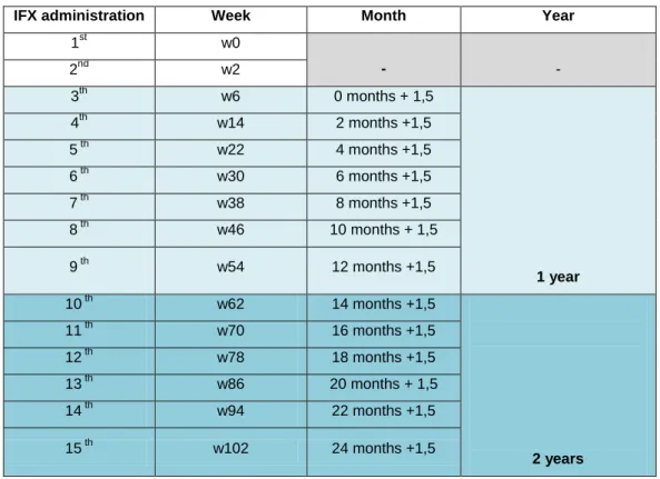
1.8 Remicade dosage and routine of administration.
Remicade was approved with a posology of intravenous (IV). The European Commission granted a marketing authorization valid throughout the European Union on 13 Aguste 1999. It is administrated by multiple intravenous infusions with the dosage of 3-5 mg/kg initially at week (w) 0, w2 and w6, with the propose to achieve a fast clinical improvement of symptoms and after the week 6 is given in intervals of 4-8 weeks with dosage of 3mg/kg for RA and 5mg/kg for the others indications based on several multicenter, randomized, double blind clinical trials for evaluation clinical efficacy and safety including IMPACT I, IMPACT II, ACCENT I, ACCENT II, ATTRACT, ASPIRE and START studies which established the optimal dosage and intervals of administration to get a better response and a minimum risk of adverse reactions or living treatment. (EMA, 2018); Gabbani et al, 2017). The dosage could be raised until 10mg/kg in patients who show a weak response. Remicade is administrated as an infusion lasting one-two hours and after the administration they are monitored for more one-two hours afterwards to prevent undesirable reactions especially situations of infusion-related reactions like anaphylactic shock that can endanger the patient's life and if so, it must be insured that the place where the drug administration is performed provides the necessary conditions to act in such a situation, so the administration of Remicade is limited to hospitals. How the patients under Remicade treatment needs to take multiple infusions to maintenance the disease in remission, the physician aim is to achieve the steady state by analyzing the IFX concentration contained in serum samples collected immediately before to the next Remicade infusion. So this serum samples are obtained when the IFX concentration are minimums and are designed as serum TL. When the serum TL shows the same value the physician can interpret that the patient achieved the steady state for IFX treatment. If some patient are keeping a maintenance dosage of IFX every 8 weeks and the actual
33 value of serum drug TL is higher than previous TL measurement, it can be interpreted by the clinic as an increased of the AUC and this indicate that the dosage of the present infusion should be lower or the dose interval of every 8 weeks reduced. By the other hand if the actual infliximab trough level (IFX-TL) value is minor then the previous measurement it suggests that the AUC decreased and due that the Remicade dosage should be incised or the infusions intervals expanded. The clinic can also consider that multiple Remicade infusions achieved Css when the AUC geometry is the same at the same interval of time that use to be as a standard interval of every 8 weeks. Farther the values of TL for every 8 weeks, if was that the interval previously determined by the clinic for some patient under Remicade treatment, should be the same (Park et al, 2015).
Table 1 – Representative scheme of the Infliximab infusion routine.
IFX administration Week Month Year
1st w0 - - 2nd w2 3th w6 0 months + 1,5 1 year 4th w14 2 months +1,5 5 th w22 4 months +1,5 6 th w30 6 months +1,5 7 th w38 8 months +1,5 8 th w46 10 months + 1,5 9 th w54 12 months +1,5 10 th w62 14 months +1,5 2 years 11 th w70 16 months +1,5 12 th w78 18 months +1,5 13 th w86 20 months + 1,5 14 th w94 22 months +1,5 15 th w102 24 months +1,5
The information contained on the table was adapted from Maini et al, 1999,Yoo et el, 2013.
1.9 Biosimilar CT-P13
CT-P13 is the IFX biosimilar approved in 2013 by EMA and is commercialized as Remsima in Europe and Inflectra in USA. The authorization to be commercialized as the IFX biosimilar to treat AS and RA conditions was given after CT-P13 shown comparable efficacy and safety in two pivotal studies. The PLANETAS with AS patients and the
34 PLANETRAS study with RA patients. Both studies published in 2013 shown comparable efficacy and safety among the AS and RA patients. These results obtained for AS and RA diseases, was considerable enough for EMA have extended the marketing authorization for IBD, PS and psoriatic arthritis PA. That extended step is called extrapolation. The extrapolation of data from some diseases to other diseases was criticized for different authors among the scientific community in 2014. The main concern was about if among patients with IBD the efficacy and the safety also are comparable for CT-P13 and original IFX (Casteele & Sandborn, 2015; Ha & Kornbluth, 2016; Hlavaty & Letkovsky, 2014). The results of CT-P13, in IBD, were obtained a bit later with post-marketing records which suggesting similar effectiveness and safety comparing with original IFX (Farkas et al, 2017). Nowadays doubts related to the efficacy and safety of CT-P13 is not a concern anymore. However the concerns now remain regarding to the changeability of the RMP, the Remicade by the CT-P13 in patients that had started the Remicade treatment already. The issues are related to if changeability can affect immunogenicity and pharmacodynamics parameters of the patients that have been tolerated well Remicade. Despite documented studies have shown comparable efficacy and safety of CT-P13 and original IFX among the different diseases that are both indicated to treat, concerns tend to stay present among physicians (Fiorino et al, 2018). Immunogenicity is the most common mentioned concern mainly in patients which the reference product is changed by a biosimilar. Is a fact that biosimilar medication is susceptible to be immunogenic in some patients. The intensity of the immunogenicity among patients is variable because it depends of the immunologic features of each patient that always have differences. More over different biosimilars agents can present different immunogenicity standards so the comparison of immunogenicity levels between biosimilars and original drugs became a necessity by the importance of monitoring patient by the clinic to optimize the dosage route and minimize the risk of lack response to the treatment (Jahnsen, 2016).
1.10 Therapeutic drug monitoring of IBD patients under IFX treatment
The application of therapeutic drug monitoring (TDM) of patients who are being treated with IFX or CT-P13, provides an important tool for physicians enable a more effective and safe use of both medicine by their patients. TDM is consists in evaluating IFX and ADA serum TL by measuring serum TL levels. TDM is important because a better use of the medicine is related to a better quality of life improvement. TDM also helps physicians to identify the patients who will benefit and who don’t will benefit from
35 treatment and as well help physician to make decisions related increase or decrease dosage, changing infusions intervals or change the class of therapeutic mAb. The measurement of serum TL levels of patients been treated with IFX provide for different results possible (Bendtzen, Ainsworth, Steenholdt, Thomsen, & Brynskov, 2009,
Table 2 – Representation of different possible results obtained by drug and ADA serum-TL measurement.
Drug presence ADA presence Interpretation
+ -
The infliximab is being effective, and no immunogenicity detected. This result indicates that patient should keep continuing with treatment.
- +
No drug levels detected and positive result for ADAs presence. These results indicate that the ADAs presence are neutralizing IFX and increasing clearance. In that situation the patient should stop with IFX treatment and mAb switching is recommended once worse clinical outcomes are documented to be related this result.
+ +
Double positive result (DP) means that the ADAs that are being
generated to the IFX are non-neutralizing ADAs and so don’t
affect the PK. The treatment can be maintained if the effectiveness or safety doesn’t be compromised. Is reported that ADAs detected in a double positive result can be transient and stop be detected in the future.
36
- -
Double negative result (DN) is more difficult to interpret because there’s more than one situation that can be happen. One the one hand ADAs may be present and affecting PK by increasing the drug clearance; however these ADAs are not being detected at the ELISA assay because are forming complexes with IFX that for the same reason are not being detected as well. Is well documented the interference of the drug levels in the detection of ADAs because the complexes formed and is for that reason that the ADA detection should be performed with serum-TL because are collected at the Cmin. If patient are displaying high concentrations of IFX serum-TL could be the reason for the DN result and so the IFX treatment should be stopped. By the other hand if the patient is not generating ADAs the null levels of IFX can occur by other reasons that are provoking a fast clearance of IFX. Other factors such as high baseline C-reactive protein (CRP) levels, low albumin blood levels, high body site, sex (males have shown higher clearance then women). When null IFX trough levels are not related with ADAs the clinic decision should be increase the dosage or decrease dosage interval.
Information based on Bendze et al, 2009; Khanna, Levesque, Sandborn & Feagan, 2014.
1.11 The importance of the Therapeutic Drug Monitoring to evaluate efficacy and safety of CT-P13 and the original Infliximab.
The therapeutic drug monitoring (TDM) is a useful tool to identify or prevent patients who will not respond to the treatment. The monitoring is achieved by studding TL serum concentrations of drug serum concentration and ADAs, obtained widely by bridging ELISA assay. Loss of response (LoR) is associated with non-efficacy and non-safety of the drug, and these patients are more likely to develop LoR against the new drugs that will be administrated and more likely to have to do surgery. When the loss of respond happens in the beginning of treatment and the LoR are not associated with the presence of ADAs. This kind of LoR is called primary loss of response LoR. The patients which the treatment is efficient by a sustainable period of time and after achieved the remission phase starts developing ADAs that are responsible for LoR, that particular situation is named secondary LoR. Secondary LoR is a phenomenon that is associated with the development of neutralizing ADAs (NADAs) TDM is also useful to compare the immunogenic response to the drugs and so reference medicinal products (RMPs) can be
37 compared in terms of similarity with their biosimilars. That’s the reason why TDM is related with save costs with no impact on efficacy when helps to prove that the changeability by RMP to a biosimilar is feasible (Atzeni et al, 2013). With TDM the changeability by RMP to the similar can be tested and is designed as switching but also the switching of the bsimilar by the RMP can be tested and is called reverse switching. The change of one biosimilar for other biosimilar is named cross switching and TDM can contribute to test the similarity of the cross switching once new biosimilars should be launched in the market in the next years. Further TDM is a way to optimize serum drug levels and dose intervals, especially in patients with LoR (Michell et al, 2016).
Secondary LoR are present in 40% of patients and also associated with low IFX-TL. However secondary LoR are more difficult to determine when patients show double negative result. The bELISA used to detect IFX levels is also designed as double antigen ELISA because to detect anti-infliximab antibodies (AIAbs) the IFX is used as antigen where the ATIs recognize and link, and as well IFX is used to detect ATIs (Ungar et al, 2015). The patients that present DN result with double antigen ELISA, 50% of these DN results are false negative DN, because in patients that the immunogenic response is a predominate mediated by monovalent IgG4 AIAbs, due the monovalent nature the IgG4 are only able to establish one link to the IFX fixed on the ELISA bottom well, but can’t be detected by the labeled IFX used to detect AIAbs. This limitation of double antigen ELISA technic can be overcome by the anti-lambda ELISA assay. Anti-lambda ELISA assay use as a secondary Ab an anti-lambda able to detect IgG4 ATIs that contains light lambda chains. How IFX have kappa light chain, the secondary anti-lambda antibody doesn’t detect IFX and so is specific to detect IgG4 AIAbs. For patients that present DN result with double antigen ELISA assay and negative result for IgG4 AIAbs. detection, the low or null IFX levels should be considerate a cause of a non-immunogenic clearance. Monitoring IgG4 ADA profile is a way to distinguish between DN false negatives from DN positives and so helping physicians to choose the right decisions for the right patient. Evaluating DN results is a way to understand better how patients respond to the drug. And a better understanding of this subject can also be used to compare the immune response between RMP and biosimilar to test their similarity (Shapiro et al, 2011; Afonso et al, 2016).
The extrapolation of the data obtained with CT-P13 with AS and RA patients, for IBD patients, was a concern in 2014 among the scientific community. The treatment with CT-P13 biosimilar, among IBD patients is nowadays known as feasible and safe (Blair & Deeks, 2016). Today the concern remains about if Remicade is more or less immunogenic than CT-P13 and so if the switch, cross switch and reverse switch are feasible among IBD patients without having impact in secondary LoR rates. The data
38 obtained here can also be compared with results obtained with different commercialized kits. That aspect is relevant once bridging ELISA assays have limitations and comparing the data obtained with different kits allows verifying if the different results are compatible. As well, rates of patients having secondary LoR due to the ADAs outcome can be compared not only between patients treated with the original and the biosimilar but with the rates obtained with other anti-TNF mAbs such as the humanized class of therapeutic mAbs because the controversy about who defends the implementation of switching of chimeric mAbs by humanized mAbs, arguing that humanized mAbs are likely less immunogenic then chimeric mAbs once they have less murine domains (Ilias, Gonzi, Kurti & Lakatos, 2018).
39
2 Aims
40 2.1 Overall goals and specific aims
The authorization of biosimilar CT-P13 to treat patients with IBD was extrapolated from two pivotal studies performed with patients with RA and AS conditions, the PLANETRA and PLANETAS studies. The lack of published data about CT-P13 regarding to IBD patients in 2014 generated some concerns regarding similarity equivalency in IBD patients’ immune response.
Nowadays the concern is not about the effectiveness and safety of CT-P13 in IBD but the switch, the reverse switch and inter-switch. Monitoring the therapeutic drug CT-P13 has contributed to clarify these doubts, and to provide a more efficient use of the medicine among IBD patients. Thus helping physicians have better control over the patient’s PK and emerging ADAs responsible for secondary LoR. However, several essays still provide different sensibilities in drug and ADA detection, through which false negative results for ADA can occur.
The lack of ADA detection associated to DN (IFX-/ADA-) result can be related to IFX-ADA complex formation, that the bELISA should not be able to detect, but also could be related with a limitation of bELISA to detect monovalent IgG4 ADA subtypes.
The results obtained with the therapeutic drug monitoring are important to be compared with the ones obtained with Remicade for a better understand about the similarity of both medicines.
The identification of ADAs epitopes can be relevant once it can provide information about which infliximab regions are more immunogenic, suggesting the presence of immunodominant epitopes which can be relevant to the development of anti-immunogenicity medicines. Further, mapping assay is a way to compare the immune response intensity of CT-P13 and Remicade, and so testing if both drugs offer comparable safety in terms of immunogenicity.
The immunogenicity can have more impact on the effectiveness when the ADAs generated are directed to CDR regions because that blocks the interaction of infliximab with TNFa. NADAs which have such ability are responsible for a highest LoR. Due that is important to compare the ability of biosimilar and Remicade to induce the formation of NADAs with propose of evaluating the efficacy and safety.
The aim of this study is to evaluate the similarity of the biosimilar CT-P13 with the original Infliximab, the Remicade drug, among IBD patients aiming to know if the
41 changeability of the Remicade for the biosimilar CT-P13 can be implemented successfully in patients with IBD and so providing a cheaper treatment.
To achieve these goals four tasks were established:
Task 1: Measure CT-P13 and ADAs TL-serum levels to verify how immunogenicity affects the CT-P13 pharmacokinetic of IBD patients.
Task2: Mapping the epitopes of ADAs directed to the biosimilar and epitopes of ADAs directed to Remicade to verify if the immune response of IBD patients treated with CT-P13, is similar to IBD patients under the original Remicade.
Task3: Mapping the epitopes of IgG4 ADAs subtypes, directed to the biosimilar and epitopes of IgG4 ADAs directed to Remicade to verify if the immune response of IBD patients treated with CT-P13, is similar to IBD patients under the original Remicade.
Task4: Comparison of the similarity between a biosimilar and original product regarding the intensity of the immunogenicity and perform a neutralizing essay to detect the presence of NADAs among IBD patients treated with CT-P13 and Remsima.
42
3 Methods
43 All assays and laboratory procedures here described in this chapter were optimized and systematically revised in order to minimize inter and intra-assay variability.
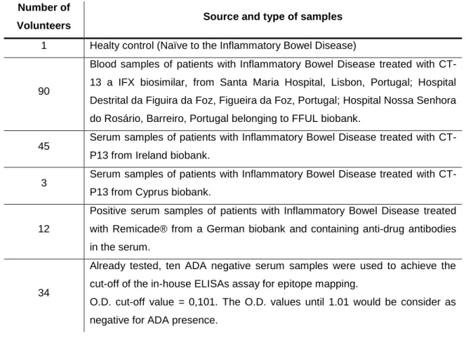
3.1 Patients and healthy controls
Patients and healthy controls are evidenced in the Table 3. Table 3 - Patients and health controls.
Number of
Volunteers Source and type of samples
1 Healty control (Naïve to the Inflammatory Bowel Disease)
90
Blood samples of patients with Inflammatory Bowel Disease treated with CT-13 a IFX biosimilar, from Santa Maria Hospital, Lisbon, Portugal; Hospital Destrital da Figuira da Foz, Figueira da Foz, Portugal; Hospital Nossa Senhora do Rosário, Barreiro, Portugal belonging to FFUL biobank.
45 Serum samples of patients with Inflammatory Bowel Disease treated with CT-P13 from Ireland biobank.
3 Serum samples of patients with Inflammatory Bowel Disease treated with CT-P13 from Cyprus biobank.
12
Positive serum samples of patients with Inflammatory Bowel Disease treated with Remicade® from a German biobank and containing anti-drug antibodies in the serum.
34
Already tested, ten ADA negative serum samples were used to achieve the cut-off of the in-house ELISAs assay for epitope mapping.
O.D. cut-off value = 0,101. The O.D. values until 1.01 would be consider as negative for ADA presence.
Blood samples from patients with Inflammatory Bowel Disease and healthy subjects were collected and serum was isolated and stored at -20º C. until analysis.
3.2 Immunogenic response of IBD patients to the IFX biosimilar CT-P13 drug based on IFX and ADA TL-serum profile.
The identification of positive serum samples, from the IBD patients under the biosimilar Remsima, containing the ADAs to the biosimilar CT-P13, was detected by a commercial kit (Theradiag LISA-TRACKER Duo Infliximab, France).The commercial kit
44 can measure two parameters, the Infliximab and the anti-Infliximab ADAs levels. IFX drug is detected based an indirect ELISA and ADAs by bringing ELISA model (Appendix 8.1). 3.3 Infliximab synthetic peptides
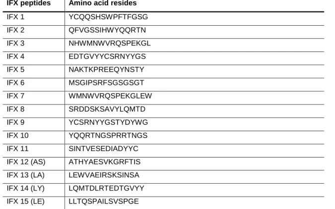
Based on literature ADAbs bind preferentially on the murine portions of the IFX drug spotted in the Fab fragments of IFX drug. To map the epitopes of ADAbs on IFX mlecule, 21 peptides of the IFX Fab fragment were commercially synthetized (ProteoGenix; Bachem). These 21 peptides were previous identified by phage display technology for being potential epitopes where the anti-Infliximab antibodies (AIAbs) attach. Here it was intended to verify which infliximab peptides are epitopes of the anti-Remicade and anti-Biosimilar (CT-P13) ADAs. The reactivity between the peptides, which mimics potential epitopes and the ADAs of positive serum samples suggest immunogenicity and so the intensity of the immunogenic response to the biosimilar and original IFX can be evaluated. The assay as well allows verifying to understand the regions of the IFX molecule that are more immunogenic for the anti-Remicade ADAs and for anti-biosimilar ADAs.
The IFX peptides amino acid sequences are evidenced in Table 4 and their localization on the IFX molecule are represented on Figure 5, Figure 6 and Figure 7. Table 4 – Infliximab commercialized synthetized peptides
IFX peptides Amino acid resides
IFX 1 YCQQSHSWPFTFGSG IFX 2 QFVGSSIHWYQQRTN IFX 3 NHWMNWVRQSPEKGL IFX 4 EDTGVYYCSRNYYGS IFX 5 NAKTKPREEQYNSTY IFX 6 MSGIPSRFSGSGSGT IFX 7 WMNWVRQSPEKGLEW IFX 8 SRDDSKSAVYLQMTD IFX 9 YCSRNYYGSTYDYWG IFX 10 YQQRTNGSPRRTNGS IFX 11 SINTVESEDIADYYC
IFX 12 (AS) ATHYAESVKGRFTIS
IFX 13 (LA) LEWVAEIRSKSINSA
IFX 14 (LY) LQMTDLRTEDTGVYY
45 IFX 16 (PV) PGERVSFSCRASQFV IFX 17 (SG) SPRLLIKYASESMSG IFX 18 (SN) SSSLGTQTYICNVHN IFX 19 (KV) KDTLMISRTPEVTCV IFX 20 (ST) SGSGSGTDFTLSINT IFX 21 (GF) GGSMKLSCVASGFIF
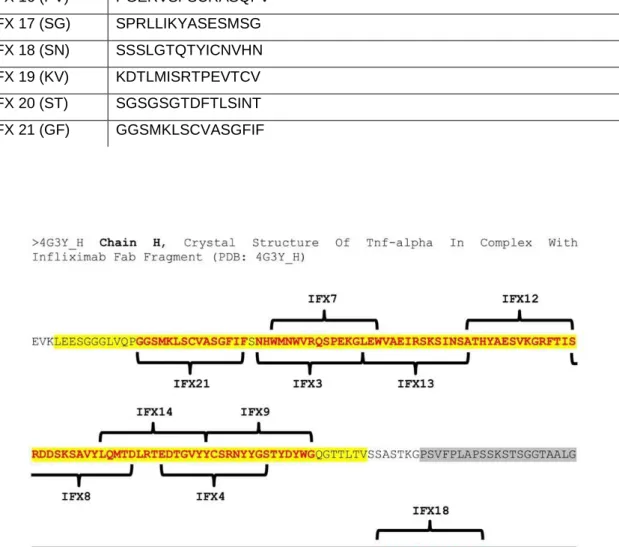
Figure 5 - Localization of the infliximab synthetized peptides on chain H FAB IFX
In yellow is represented the sequence of the variable heavy chain (VH) and in grey the constant heavy chain 1
(CH1). The red words represent the sequence of IFX which the synthetized peptides (IFX3, IFX4, IFX7, IFX8, IFX9, IFX12, IFX13, IFX14, IFX18, IFX21) are located.
Source: Chain H, Crystal Structure Of Tnf-alpha In Complex With Infliximab Fab – Protein - NCBI. (2016).
Ncbi.nlm.nih.gov. Retrieved 29 October 2016, from https://www.ncbi.nlm.nih.gov/protein/471270577.
46
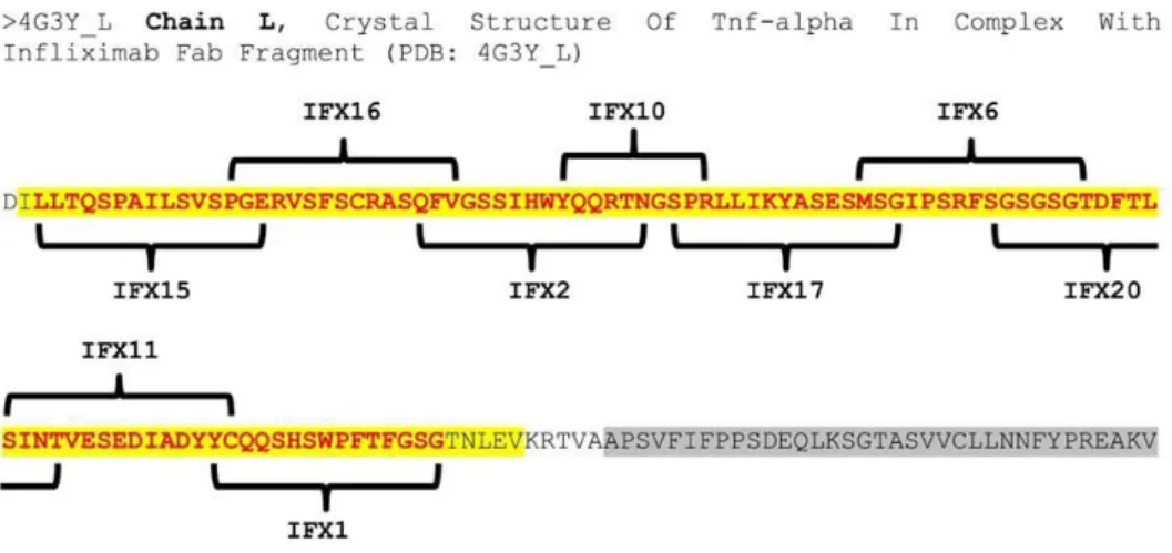
Figure 6 - Localization of the infliximab synthesized peptides on chain L FAB IFX.
In yellow is represented the sequence of the variable light chain (VL) and in grey the constant heavy chain 1
(CH1). The red words represent the sequence of IFX which the synthetized peptides (IFX1, IFX2, IFX6, IFX10, IFX11, IFX15, IFX16, IFX17, IFX20) are located.
Source: Chain L, Crystal Structure Of Tnf-alpha In Complex With Infliximab Fab - Protein - NCBI. (2016). Ncbi.nlm.nih.gov. Retrieved 29 October 2016, from https://www.ncbi.nlm.nih.gov/protein/471270576.
Figure 7 - Localization of the synthetized peptides on the FC IFX
In grey is represented the sequence of the constant heavy chain 2 (CH2) and in black the constant heavy chain 3 (CH3). The white words represent the sequence of IFX which the synthetized peptides (IFX19, IFX5) are located.
47
Source: IgG [Homo sapiens] - Protein - NCBI. (2016). Ncbi.nlm.nih.gov. Retrieved 29 October 2016, from https://www.ncbi.nlm.nih.gov/protein/185362.
Lyophilized peptides were diluted in PBS 1x to a final concentration of 1mg/mL and stored at -80Cº away from light according with the guideline of peptides solutions provided by the peptides supplier.
3.4 Development of two in house ELISA assays to screen which the synthetic peptides bind to ordinary IgGs ADAs and IgG4 ADAs, contained in the positive serum samples from the IBD patients under the biosimilar treatment and the IBD patients under the original IFX treatment.
An ELISA assay was developed to screen which synthetic peptides bind to the ADAs contained in the serum of patients with Inflammatory IBD, developing immunogenicity. The ELISA optimization process included testing different peptides concentrations, sample dilutions, coating, blocking and washing buffers, conjugated antibody as evidenced in Table 5:
Table 5 – ELISA assay optimization
ELISA step Optimization
Samples PBST 0.1%
PBS 1X
Blocking Buffer
PBS 1X containing 1% bovine serum albumin PBS 1X containing 3% bovine serum albumin PBS 1X containing 5% bovine serum albumin PBS 1X containing 1% Caseine PBS 1X containing 3% Caseine PBS 1X containing 5% Caseine PBS 1X containing 1% Pierce PBS 1X containing 3% Pierce PBS 1X containing 5% Pierce Washing Buffer PBS 1X PBS 1X containing 0.05% Tween 20 Anti-Human IgG Horseradish
Peroxidase (HRP) Antibody
Dilution: 1:5000 / 1:25000
PBS 1X containing 1% bovine serum albumin
Anti-Lambda IgG-HRP Antibody
Dilution: 1.5000 / 1.10000 / 1:20000 / 1:30000 PBS 1X containing 1% bovine serum albumin
48 Anti-human IgG4 biotinylated
Antibody
Conjugated with: Streptavidin
Anti-mouse IgG Antibody (Dilution: 1:1000 / 1:5000/ 1:10000)
Dilution: 1:5000 / 1:10000 / 1:20000
PBS 1X containing 1% bovine serum albumin
3.5 Screening epitopes of the anti-Infliximab (anti-Remicade and anti-biosimilar) IgG antibodies
An ELISA assay was developed to identify which infliximab peptides are recognized by the anti-Remicade antibodies (ARAbs) and anti-biosimilar IgGs antibodies (ABAbs) from the IBD patients that developed immunogenicity against the respective IFX drug.
Two 96-wells plates (Nunc Immobilizer Amino Plates; Thermo Scientific) for the anti-Remicade IgG antibodies detection and another two for the anti-biosimilar IgG antibodies were coated with 100µg/µL synthetized infliximab peptides (ProteoGenix; Bachem) diluted in sodium carbonate (Na2Co3 0,1M pH9.6) overnight (ON) at 4ºC. The 96-wells plates were coated with each IFX peptide according to the scheme represented in Figure 8.
Figure 8 – Scheme of the IFX peptides coated on 96-wells plates and the indication how serum samples was added.
Each well of each line is coated with a different peptide and for each line of the 96-plates is added a sample from only one patient, so for each serum sample from just one patient is added the same amount of serum for each one of the 21 peptides coated at the same line.
49 The Blocking buffer (PBST 1x containing 5% bovine serum albumin) was subsequently added and incubated for 1hour at 37ºC. After that the blocking buffer was discarded and the wash step performed with PBST (PBS 1x containing 0,1% Tween 20) five times in every wells. The serum samples from IBD’s patients with immunogenicity, so samples that contains the anti-Infliximab antibodies was diluted 1:9 and the sample of the negative controls (IBD`s patients that have no immunogenicity so, their serum samples have no anti-Infliximab antibodies) was diluted 1:9 as well and 50µL of each already prepared serum samples and also the control sample was added to the respective 96-plate line wherein to test each already prepared serum sample for all 21 peptides more the negative peptide control (IFX C), how is represented in the scheme above. The incubation of the serum samples was done for 1hour at 4ºC. Then, the plates containing was discarded and another wash step was performed 5x with PBST. Subsequently was added 50µL of anti-lambda-HRP antibody (Abcam) diluted 1:5000 in PBST 1x in 1% of bovine serum albumin. The choice of the anti-lambda-HRP antibody to detect the presence of AIAbs was because they link to the IgGs which have lambda-chain and how infliximab molecule has Kappa-chain is not recognized by the anti-lambda-HRP antibody, and so that this makes that antibody specific to detect the anti-Infliximab antibodies. This antibody is also conjugated to a peroxidase enzyme (HRP). The micro plates after the addiction of 50µL per well of the secondary antibody the anti-lambda-HRP was incubated at 4ºC for 1hour. The wells content was discarded and after that one last wash step 5x with PBST was done before the addiction of 50µL of the subtract (3,3’5,5’ tetramethylbenzidin; Theradiag), incubated for about 15-20 minutes in the dark at 4ºC until the addiction of 50µL of sulfuric acid (H2SO4 0,25M) to block the enzymatic reaction. After this process the wells that contains the presence of anti-Infliximab antibodies became yellow and the yellow color intensity is directly proportional regarding how higher is the presence of the anti-Infliximab antibodies for each well. Then the optical density (OD) was read in a spectrophotometer at 450 nm within 30 minutes after stopping reaction
The chosen of the anti-lambda-HRP Ab as a secondary antibody instead a biotinylated IFX like in the commercialized kit was made because biotinylated IFX needs the streptavidin-HRP conjugated to allow the enzymatic reaction happen and we observed in our controls an unspecific reaction between the streptavidin and some ones of the IFX peptides as a false positive result. The reason because the unspecific reaction just happened with some IFX peptides and not with the complete IFX molecule could be because the lack of folding, so peptides have linear shape and this can favor unspecific reactions. The new strategy was to use the anti-lambda-HRP Ab as a secondary Ab because it is able to recognize the lambda chains of the AIAbs and how IFX molecule has
50 kappa chains instead lambda, makes anti-lambda-HRP Ab specific to detect the presence of AIAbs on this ELISA assay. The utility of the anti-lambda ELISA in ADA detection was described by Kopylov et al, 2011 and is here represented in the Figure 9.
Figure 9 - Detection of anti-infliximab IgG antibodies
ELISA assay scheme of ant-infliximab IgG antibodies detection by anti-lambda secondary antibody. How is represented in this figure, the wells are coated with the respective infliximab peptides (IFX peptide). The anti-infliximab antibodies (AIAb) containing in the sample of IBD patients treated with an anti-infliximab drug, a reference medical product (RMP) or a biosimilar CT-P13, they link to the IFX peptide when it is recognized as an epitope. Anti-lambda secondary antibody (anti-lambda-HRP Ab) conjugated with a horseradish Peroxidase enzyme (HRP) binds to the lambda chain of AIAb but is not able to binds to the IFX peptides because IFX has a chain kappa instead lambda. TMB (3,3’5,5’ tetramethylbenzidine) is the subtract of the enzymatic reaction of HRP, so the enzymatic reaction is proportional to the amount of AIAb present on serum from IBD patients. Source: Adapted from Kopylov et al, 2011.
3.7 Screening of Infliximab epitopes of the Infliximab (Remicade and anti-biosimilar) IgG4 antibodies
Was developed an ELISA assay to identify which ones of the infliximab peptides were recognized by the anti-Remicade and anti-biosimilar IgG4 antibodies from patients with IBD that developed immunogenicity to the respective infliximab drug.
Two 96-wells plates (Nunc Immobilizer Amino Plates; Thermo Scientific) for the anti-Remicade IgG4 antibodies detection and another two for the anti-biosimilar IgG4 antibodies, were coated with 100µg/µL synthetized IFX peptides (ProteoGenix; Bachem) diluted in sodium carbonate (Na2Co3 0,1M pH9.6) O.N. at 4ºC. N. 96-wells plates were coated with each IFX peptide according to the scheme represented above on Figure 9.
TMB
Anti-lambda-HRP Ab
AIAb