The efficacy of
kangaroo mother care, sucrose
and pacifier to reduce
responses of preterm infants to procedural pain
ANANDA MARIA FERNANDESDoutoramento em Enfermagem
The efficacy of
kangaroo mother care, sucrose
and pacifier to reduce
responses of preterm infants to procedural pain
SUPERVISORSDr. Céleste Johnston
Professor, Associate Director of Research, McGill University School of Nursing Montreal, Canadá
Professor Doutor José Cabrita
Professor Catedrático da Faculdade de Farmácia da Universidade de Lisboa Membro da Comissão Científica do Programa de Doutoramento em Enfermagem
ANANDA MARIA FERNANDES DOUTORAMENTO EM ENFERMAGEM
Figura da capa e separadores |Maternidade I, Humberto Seabra Santos, 1994 Grafismo e paginação |José Joaquim M. Costa
(...)
Two roads diverged in a wood, and I— I took the one less traveled by, And that has made all the difference.
intellectual challenge and encouragement.
To the principal investigators of Pain in Child Health (PICH) - Canadian Institutes of Health Research Strategic Training Initiative - for the opportunity to take part in a community of scholars in pediatric pain and for their mentorship.
To Professors Manuel Gameiro and Luis Loureiro for statistical consulting. To Professor Emanuel Ponciano for the permission to use the STAI. To those who generously spent time with the scientific revision and editing. To the parents of preterm babies who agreed to participate.
To the staff of the neonatal intensive care units: Unidade de Cuidados Intensivos ao Recém-Nascido dos Hospitais da Universidade de Coimbra, EPE and Unidade de Cuidados Intensivos Neonatais do Centro Hospitalar de Coimbra, EPE.
To the funding agents: Coimbra School of Nursing, Pain in Child Health (PICH), Mayday Fund, Foundation for Science and Technology and Calouste Gulben-kian Foundation.
To my PICH fellows. To my friends. To my family. Agradecimentos
Aos orientadores, Prof. Doutora Céleste Johnston e Prof. Doutor José Cabrita pe-la supervisão, estímulo intelectual e encorajamento.
Aos investigadores principais do Pain in Child Health (PICH), pela oportuni-dade de participar numa comunioportuni-dade académica de dor pediátrica e pelos seus contrib-utos individuais neste percurso.
Aos Professores Manuel Gameiro e Luis Loureiro pela consultoria estatística. Ao Prof. Doutor Emanuel Ponciano pela cedência da escala STAI.
Aos pais dos bebés pretermo que aceitaram participar nesta investigação.
Aos enfermeiros, médicos, auxiliares e secretárias da Unidade de Cuidados Intensivos ao Recém-Nascido dos Hospitais da Universidade de Coimbra, EPE e da Unidade de Cuidados Intensivos Neonatais do Centro Hospitalar de Coimbra, EPE.
Às entidades financiadoras: Escola Superior de Enfermagem de Coimbra, Pain in Child Health (PICH) - Canadian Institutes of Health Research Strategic Training Initiative, Mayday Fund - Fundação para a Ciência e Tecnologia e Fundação Calouste Gulbenkian.
Aos meus companheiros do PICH. Aos meus amigos.
Preterm neonates in intensive care units endure frequent procedures that may cause pain, warranting the study of interventions that will decrease this pain. The pri-mary aim of this study was to compare the efficacy of the combination of sucrose, pac-ifier and kangaroo mother care (S+KMC), with that of sucrose and pacpac-ifier (S), in re-ducing the pain responses of preterm infants undergoing venepuncture. Secondary objectives addressed to babies in S+KMC were to examine the relationship between maternal anxiety and the pain responses of the babies; and to explore mothers’ percep-tions of KMC during venepuncture.
A randomized-controlled trial was conducted in two neonatal intensive care units in Portugal. One-hundred and ten preterm infants without severe illness, strat-ified by gestational age, were randomly assigned to receive S+KMC or S for venepunc-ture. Measures of pain responses were the Premature Infant Pain Profile, heart rate, ox-ygen saturation, facial actions, behavioral state, heart rate variability and recovery time, which were analysed with repeated-measures ANOVA. Mothers’ anxiety was measured with the State-Trait Anxiety Inventory. Their perceptions were obtained through con-tent analysis of semi-structured interviews.
Compared to infants in S, infants in S+KMC displayed significantly less facial action; were more likely to have recovered heart rate baseline values at 60 and 90 sec-onds after the procedure, if they were 32 weeks gestational age and above; and changed from sleep to wake states significantly less. Maternal anxiety was low to moderate and was not correlated to specific pain responses. Mothers emphasized their feelings of well-being in comforting and protecting the babies.
In conclusion, combining sucrose, pacifier and kangaroo mother care is effective and safe in preterm infants undergoing venepuncture for blood-draw; low to moderate levels of anxiety of mothers do not interfere with the pain responses; mothers appreci-ate holding the baby skin-to-skin when the infants are enduring pain.
Os recém-nascidos pretermo que necessitam de cuidados intensivos são frequen-temente submetidos a procedimentos diagnósticos e terapêuticos que podem causar dor. Contra riamente ao que se pensava há duas décadas, a evolução ontogenética da dor inicia-se cedo e, a partir das 24 inicia-semanas de gestação, o feto dispõe do equipamento neuroinicia-sensorial necessário à experiência de dor. Todavia, as vias de controlo descendente não se encontram ainda suficientemente desenvolvidas, resultando em hipersensibilidade dolorosa. As conse-quências da exposição repetida à dor no período neonatal têm vindo a ser estudadas, sendo hoje conhecidos os efeitos a curto prazo da dor não tratada, como a hiperalgesia e a alodinia nos recém-nascidos, e alguns efeitos a médio e longo prazo como as alterações da sensibili-dade e da reactivisensibili-dade ao stress em crianças de isensibili-dade escolar. O alívio da dor nesta popula-ção vulnerável é, pois, uma tarefa imperiosa. Dado o reduzido leque de fármacos disponíveis para estas idades e o seu potencial para efeitos adversos, torna-se necessária a investigação de intervenções não-farmacológicas. Entre estas, a sacarose oral com chupeta tem sido exausti-vamente demonstrada como eficaz, sendo utilizada por norma em muitas unidades neona-tais antes da realização de procedimentos como a punção do calcanhar e a punção venosa. Durante estes procedimentos, também o contacto pele-a-pele entre mãe e bebé, conhecido como canguru materno, pode ser utilizado como forma de reduzir as respostas de dor dos recém-nascidos.
Desconhecia-se, todavia, se ao adicionar o canguru materno ao uso da sacarose com chupeta seria possível reduzir ainda mais as respostas de dor dos recém-nascidos preter-mo. Por outro lado, dada a co-regulação fisiológica mãe-bebé, colocava-se a questão de sa-ber se a ansiedade materna poderia comprometer o efeito analgésico do canguru materno. Finalmente, as percepções das mães sobre a realização de canguru materno durante a veno-punção não haviam sido exploradas.
Assim, os objectivos definidos para este estudo foram: 1) comparar as respostas de dor dos recém-nascidos pretermo aos quais é proporcionado canguru materno, sacarose oral
e chupeta durante a punção venosa para colheita de sangue, com as respostas dos recém-nas-cidos aos quais é proporcionada apenas sacarose oral com chupeta; 2) analisar a relação en-tre a ansiedade materna e as respostas de dor dos recém-nascidos que efectuaram canguru materno; e 3) explorar as percepções maternas sobre a realização de canguru materno du-rante a venopunção.
Para dar resposta ao primeiro objectivo, foi realizado um estudo randomizado, con-trolado, cego, em duas unidades de cuidados intensivos neonatais portuguesas. Cento e dez recém-nascidos sem doença grave, estratificados por idade gestacional (28 a 31 semanas e seis dias, e 32 a 36 semanas e seis dias) foram aleatoriamente alocados a dois grupos: um re-cebeu sacarose oral com chupeta (grupo Sacarose); o outro rere-cebeu sacarose oral com chu-peta e canguru materno (grupo S+CM) antes, durante e após venopunção. As respostas de dor foram medidas através da escala Premature Infant Pain Profile (PIPP) e foram anali-sadas a frequência cardíaca, a saturação de oxigénio da hemoglobina, as acções faciais (per-centagem de tempo em saliência interciliar, olhos apertados e prega nasolabial), o estado comportamental, a variabilidade da frequência cardíaca (baixa frequência, alta frequência e ratio entre ambas) e o tempo de recuperação da frequência cardíaca inicial após o final do procedimento. As acções faciais foram gravadas em vídeo e as variáveis fisiológicas foram registadas através do Somté® Compumedics, ao longo de cinco fases: antes do procedimen-to, preparação da pele, punção, compressão e repouso. Para a determinação do score PIPP, a análise das gravações foi efectuada por codificadores cegos aos propósitos do estudo.
Para dar resposta ao segundo objectivo, foi realizado um estudo descritivo-corre-lacional analisando a relação entre a ansiedade materna medida pela escala de Estado de Ansiedade do State-Trait Anxiety Inventory (STAI) e as respostas de dor dos recém-nasci-dos (N= 60).
As percepções maternas foram estudadas através da análise de conteúdo das entrevis-tas semi-estruturadas realizadas às mães que tinham efectuado canguru materno (N= 52). A comparação dos dois grupos de intervenção quanto a variáveis socio-demográficas e clínicas não revelou diferenças significativas.
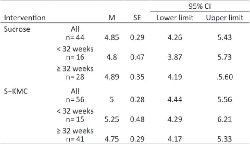
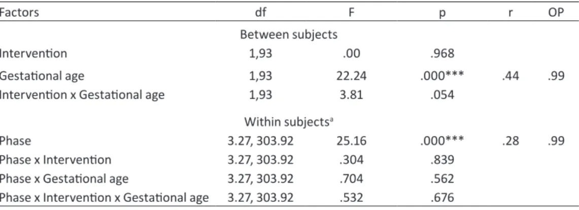
Em todos os testes foi utilizado como nível de significância α< .05. A ANOVA de medidas repetidas (fases do procedimento) a dois factores (intervenção e idade gestacional) revelou o efeito principal da intervenção sobre a percentagem de tempo em saliência inter-ciliar, F(1, 98)= 5.12, p= .026, e olhos apertados, F(1, 98)= 6.02, p= .015. A análise
post-hoc mostrou que no momento da punção, a saliência interciliar ocorria durante menos tem-po nos recém-nascidos do grutem-po S+CM (M= 15.89, EP= 4.58) do que no grupo Sacarose
(M= 29.22, EP= 4.75). O mesmo se verificou para o tempo em olhos apertados (M= 13.85,
A reactividade dos recém-nascidos durante o procedimento foi semelhante nos dois grupos de intervenção, observando-se o efeito principal da fase do procedimento sobre a PIPP, a frequência cardíaca, a saturação máxima de oxigénio, as expressões faciais e o índice baixa frequência da variabilidade da frequência cardíaca. Tal indica uma variação significa-tiva destes sinais de dor ao longo das fases do procedimento, com aumento dos sinais de dor desde o momento antes do procedimento até à punção, seguido de uma diminuição desses sinais até ao repouso.
O teste de Qui-Quadrado para cada fase do procedimento mostrou uma associa-ção significativa entre intervenassocia-ção e estado comportamental: em todas as fases, a propor-ção de bebés em estado de sono (versus estado de alerta) era significativamente mais eleva-da no grupo S+CM.
Apesar não ter havido uma diferença significativa no tempo médio de recuperação da frequência cardíaca de base após o procedimento, a probabilidade (odds-ratio) de ter re-cuperado aos 60 e 90 segundos após o procedimento foi cerca de 3 vezes mais elevada nos recém-nascidos do grupo S+CM com 32 ou mais semanas de gestação, do que nos do gru-po Sacarose.
Durante o procedimento não se verificaram efeitos adversos em qualquer dos gru-pos de intervenção.
A ansiedade materna foi baixa, sendo significativamente mais baixa nas mães do gru-po S+CM (M= 37.78, SD= 9.13) do que nas mães do grupo Sacarose (M= 43.48, SD=
9.82), t(87)= 2.65, p= .009.
Nas entrevistas, as mães salientaram a sensação de bem-estar em ter o bebé em con-tacto pele-a-pele, o contentamento em poder protegê-lo da dor e a importância que esse acontecimento havia tido para a realização do seu papel parental.
Estes resultados demonstram que a combinação sacarose, chupeta e canguru mater-no é eficaz e segura em recém-nascidos pretermo, permitindo reduzir a expressão facial e o tempo de recuperação quando comparada com a utilização de sacarose com chupeta; ní-veis baixos e moderados de ansiedade materna não interferem na redução das respostas de dor dos bebés; as mães apreciam o contacto pele-a-pele durante o procedimento doloroso e sentem o seu papel parental reforçado por poderem participar no alívio da dor do seu bebé.
Em conclusão, o canguru materno pode ser adicionado ao uso da sacarose com chu-peta para reduzir as respostas de dor de recém-nascidos pretermo acima das 28 semanas de gestação durante a colheita de sangue por venopunção.
Palavras-chave: dor, recém-nascido pretermo, sacarose, canguru materno, punção venosa.
Acronyms
ADHD - Attention deficit and hyperactivity disorder CNS - Central nervous system
CPAP - Continuous positive airway pressure DAN - Douleur-Aigue du Nouveau-Né
EDIN - Échelle Douleur et Inconfort du Nouveau-Né ELGA - Extremely low gestational age
FBW - Full birth weight HF - High frequency
HPA - Hypothalamic-pituitary-adrenocortical HRV - Heart rate variability
KC - Kangaroo Care
KMC - Kangaroo Mother Care LBW - Low birth weight LF - Low frequency MD - Mean difference
NEC - Necrotizing enterocolitis NFCS - Neonatal Facial Coding System
NIDCAP - Newborn Individualized Care and Assessment Program NIPS - Neonatal Infant Pain Scale
NNS - Non-nutritive sucking
PIPP - Premature Infant Pain Profile RSA - Respiratory sinus arrhythmia STAI - State-Trait Anxiety Inventory VAS - Visual analogue scale
VLBW - Very low birth weight WMD - Weighted mean difference
IINTRODUCTION——1
PART I
Theoretical background: The neonate and pain CHAPTER 1. The preterm neonate, a new paradigm——11
CHAPTER 2. Pain in the neonate
2.1 The pain experience——17
2.2 Animal models of infant pain——20
2.3 The capacity of preterm neonates to experience pain——25 2.4 Short term responses to painful stimulation——27
2.4.1 Behavioral cues——28
2.4.2 Physiological responses——30 2.5 Neonatal pain assessment tools——35
2.6 Long-term consequences of early pain exposure——38
CHAPTER 3. Pain exposure in the NICU environment
3.1 Epidemiology of pain in the NICU——48 3.2 Pain management in the NICU——52
CHAPTER 4. Non-pharmacological interventions to reduce procedural pain in the NICU——57
PART II
Empirical study: Kangaroo mother care, sucrose and pacifier vs sucrose and pacifier CHAPTER 6. Methods
6.1 Research design——99 6.2 Research setting——99 6.3 Sample——100
6.4 Variables and outcome measures——102 6.5 Research procedure——107
6.6 Ethical considerations——113
CHAPTER 7. Results
7.1 Participants’ characteristics——117 7.2 Responses to the interventions——123 7.2.1 Premature Infant Pain Profile——123 7.2.2 Heart rate——126
7.2.3 Oxygen saturation——133 7.2.4 Facial Behavior ——138 7.2.5 Behavioral state——147 7.2.6 Heart rate variability ——150 7. 2.7 Recovery time——153
7.2.8 Summary of pain responses to the interventions——156 7.3 Maternal anxiety and infants’ pain responses——156 7.4 Mothers’ perceptions of doing Kangaroo Care——160
CHAPTER 8. Discussion
8.1 Kangaroo care, sucrose and pacifier vs sucrose and pacifier ——175 8.2 Maternal anxiety and the pain responses of neonates in KMC——182 8.3 Mothers’ perceptions of doing KMC during venepuncture——183 8.4 Strengths and limitations ——185
8.5 Theoretical issues——188
8.6 Implications for clinical practice——189 8.7 Implications for research——191
CONCLUSION 197 Reference List——201
Appendix B - State-Trait Anxiety Inventory (STAI)——229
Appendix C - Authorization from the Administration Boards of the hospitals——231 Appendix D - Information sheet for parents——235
Appendix E - Parents’ consent form——237 Appendix F - Protocol——239
Appendix G - Data collection form——241
Appendix H - The Neonatal Facial Coding System——243
Appendix I - Comparison between final sample and lost cases——245 Appendix J - PIPP scores across phases of the procedure——247 Appendix K - Infants’ charcteristics ans PIPP scores——249 Appendix L - Hearth rate across the procedure——251
Appendix M – Oxygen saturation across the procedure——259 Appendix N - Facial actions across the procedure——263
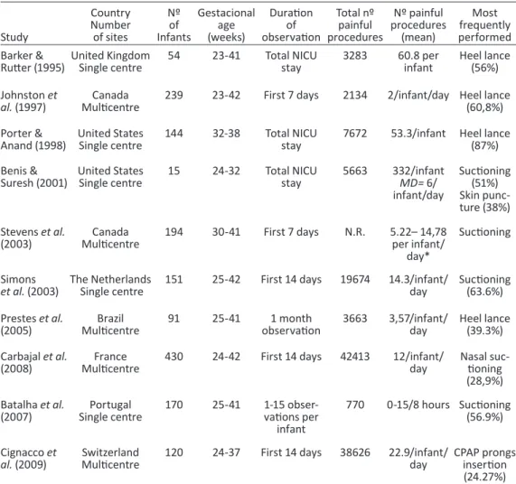
Table 1 - Studies examining the frequency of procedural pain in neonates——50
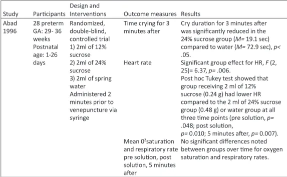
Table 2 - Studies examining the effects of sucrose to reduce pain from venepuncture in term and preterm infants——74
Table 3 - Studies examining the effect of Kangaroo Mother Care on pain responses of term and preterm infants during heel lance and venepuncture——86
Table 4 - Main health indicators related to birth in 2008 in the Centro region of Portugal100 Table 5 - Sample characteristics (categorical variables) by intervention group——120
Table 6 - Sample characteristics (numerical variables) by intervention group——121 Table 7 - Duration of the different phases of the procedure (seconds)——122 Table 8 - Need to repeat the procedure by intervention group——123 Table 9 - PIPP scores by intervention and gestational age group——124 Table 10 - Results of repeated-measures ANOVA for PIPP scores——124
Table 11 - Results of the t-test for maximum, average and minimum heart rate at Baseline126 Table 12 - Maximum heart rate for each intervention group and gestational age——127 Table 13 - Results of repeated-measures ANOVA for maximum heart rate——127 Table 14 - Average heart rate for each intervention group and gestational age——129 Table 15 - Results of repeated-measures ANOVA for average heart rate——129
Table 16 - Minimum heart rate for each intervention group and gestational age——130 Table 17 - Results of repeated-measures ANOVA for minimum heart rate——131 Table 18 - Pearson product-moment correlation coefficients (r) between maximum heart
rate values in different phases and infants’ characteristics by intervention, and corresponding coefficients of determination (r2)——132
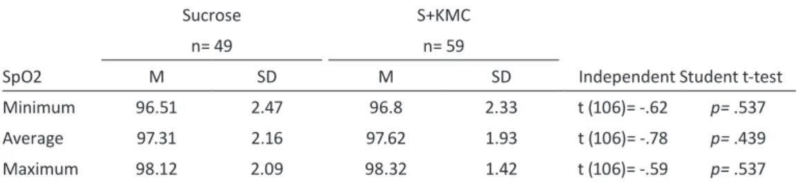
Table 19 - Baseline values for minimum, average and maximum oxygen saturation by intervention, and results of t-test——133
Table 20 - Minimum oxygen saturation (%) for each intervention group and gestational age——134
Table 21 - Results of repeated-measures ANOVA for minimum oxygen saturation——134 Table 22 - Average oxygen saturation (%) for each intervention group and gestational age 135 Table 23 - Results of repeated-measures ANOVA for average oxygen saturation——136 Table 24 - Average oxygen saturation (%) for each intervention group and gestational age 137 Table 25 - Results of repeated-measures ANOVA for maximum oxygen saturation——137 Table 26 - Percentage of time in brow bulge by intervention group and gestational age——139 Table 27 - Results of repeated-measures ANOVA for percentage of time in brow bulge—140
Table 28 - Percentage of time in eye squeeze by intervention group and gestational age——141 Table 29 - Results of repeated-measures ANOVA for percentage of time in eye squeeze 142 Table 30 - Percentage of time in nasolabial furrow by intervention group and gestational
age——143
Table 31 - Results of repeated-measures ANOVA for percentage of time in nasolabial furrow ——144
Table 32 - Significant Pearson product-moment correlation coefficients (r) between facial actions and corresponding coefficients of determination (r2)——145
Table 33 - Significant Pearson product-moment correlation (r) between brow bulge, eye squeeze, nasolabial furrow, and maximum heart rate during needle stick and compression phases and coefficients of determination (r2)——146
Table 34 - Number and percentage (below) of infants in each behavioral state, by phase of procedure and intervention group, and results of the Chi-square test——148 Table 35 - Mean low frequency peaks by intervention group and gestational age——150 Table 36 - Results of repeated-measures ANOVA for LF——151
Table 37 - High-frequency by intervention group and gestational age——151 Table 38 - Results of repeated-measures ANOVA for HF——152
Table 39 - LF/HF ratio by intervention group and gestational age——152 Table 40 - Results of repeated-measures ANOVA for LF/HF——153 Table 41 - Recovery time by intervention group and gestational age——154 Table 42 - Results of Two-way ANCOVA for recovery time——154
Table 43 - Association between recovery and intervention, by age group——155 Table 44 - Summary of significants results for pain responses——156
Table 45 - Maternal anxiety (STAI score) for each intervention group by gestational——157 Table 46 - Pearson product-moment correlation coefficients (r) between mothers’ anxiety and
maximum heart rate, in infants who had S+KMC, and corresponding coefficients of determination (r2)——158
Table 47 - Characteristics of mothers (n= 52) that were interviewed and their neonates 160 Table 48 - Maternal expectations at the beginning of the event——162
Table 49 - Maternal feelings during Kangaroo Care——164
Table 50 - Maternal perceptions of the baby’s feelings during Kangaroo Care——165 Table 51 - Maternal feelings during the blood draw——166
Table 52 - Maternal feelings about the blood draw——167
Figure 1. The preterm infant in the NICU and pain: problem statement 4 Figure 2. Study design——6
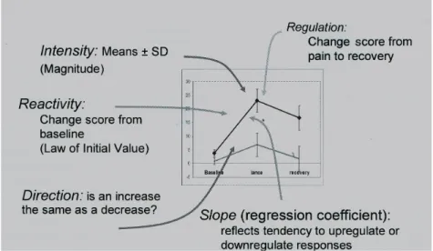
Figure 3. Hypothetical reactivity patterns comparing responses in Group A vs Group B— 38 Figure 4. Neonatal factors leading to long-term adverse neurologic outcomes——44
Figure 5. Sample flow——118
Figure 6. Evolution of PIPP scores across phases of the procedure, by intervention group 125 Figure 7. Maximum heart rate in beats per minute (bpm) across phases of the procedure, by
intervention group——128
Figure 8. Average heart rate in beats per minute (bpm) across phases of the procedure, by intervention group——130
Figure 9. Minimum heart rate in beats per minute (bpm) across phases of the procedure, by intervention group——131
Figure 10. Minimum oxygen saturation levels (%) across phases of the procedure, by intervention group——135
Figure 11. Average oxygen saturation levels (%) across phases of the procedure, by intervention group——136
Figure 12. Maximum oxygen saturation levels (%) across phases of the procedure, by intervention group——138
Figure 13. Percentage of time in brow bulge across phases of the procedure, by intervention group——140
Figure 14. Percentage of time in eye squeeze across phases of the procedure, by intervention group——142
Figure 15. Percentage of time in nasolabial furrow across phases of the procedure, by intervention group——144
Figure 16. Percentage of neonates in sleep states across phases of the procedure, by intervention group——147
Figure 17. Correlation between behavioral state and mean percentage of time in facial action, in each phase of the procedure——149
Diagrams
Diagram 1. Experimental protocol——108
It is currently well documented that infants who are born prematurely feel pain. Their pain was ignored for many years because of common beliefs that the immaturi-ty of the nervous system protected them from feeling pain and that there was no pain memory in infancy, and therefore no long-term consequences of suffering in early life. Difficulties in interpreting the infant’s expressions as being related to pain or to other stress conditions and lack of mastery over medications for pain relief for this age group have also contributed to poor pain-management in preterm infants (Schechter, Berde & Yaster, 1993).
In the past twenty-five years, however, research on pain in neonates has produced four major contributions to knowledge: the demonstration that even the smallest pterm neonates are equipped to and indeed experience pain; that neonates are able to re-spond to tissue-damaging stimuli through physiological and behavioral indicators that can be consistently measured; that repetitive and prolonged pain in the neonatal period has long-term consequences; and that a number of interventions, both pharmacological and non-pharmacological, can be effective and should be used to manage pain.
Studies of the neurobiology of pain development have put into evidence since the late eighties that innervation of the peripheral tissue and the basic connections be-tween primary sensory neurons and the cells in the dorsal horn of the spinal cord occur early in fetal development and that maturation of the afferents and chemical changes needed for pain processing at the spinal level are in place well before the third trimes-ter of gestation (Fitzgerald & Walker, 2009). However, the lack of inhibitory control at the spinal cord level, as well as the ineffectiveness of inhibitory pathways descend-ing from the brain stem, results in hypersensitivity to painful stimuli in preterm ne-onates. Knowledge about the supraspinal processing of pain is more recent and it has been shown that pain perception may occur in the absence of full cortical activity (Hall & Anand, 2005a). Yet, cortical activity is present in response to painful stimuli and has
2
now been measured through real-time near-infrared spectroscopy (Bartocci, Bergqvist, Lagercrantz, & Anand, 2006; Slater et al., 2006).
Infants born preterm usually need to be admitted to neonatal intensive care units (NICU). The environment of these units, as well the clinical condition of the infants, supplies multiple sources of stress and pain. These infants undergo a very high number of diagnostic and therapeutic procedures in order to improve their survival and most of these procedures, such as heel lance, venepuncture and suctioning, are invasive and cause acute pain (Carbajal et al., 2008; Cignacco et al., 2008). Neonatal diseases and
surgery, as well as prolonged ventilation, are sources of established and prolonged pain. Infants born at early gestational ages, with very low birth weight and sick infants are, by virtue of their clinical condition, more exposed to pain (Hall & Anand, 2005b).
Preterm infants respond to stress and painful events with physiological and be-havioral changes. The intensity of these responses is related to their gestational age, se-verity of illness and previous exposure to pain, younger and sicker infants’ responses being less robust than the responses of healthy term babies (Gibbins et al., 2008a;
Lucas-Thompson et al., 2008; Johnston & Stevens, 1996; Johnston, Stevens, Craig, & Grunau,
1993). Increase in heart rate, decrease in hemoglobin oxygen saturation levels, and corti-sol release are observed in the presence of painful stimulation. Facial grimacing, cry and body movement can be found in response to a painful procedure, facial grimacing be-ing a more specific response than others (Stevens et al., 2007).These indicators of pain
have been analyzed to build consistent assessment tools that facilitate the measurement of pain intensity and are valuable for clinical practice and research.
Early exposure to repetitive pain associated with maternal separation is not with-out consequence. Permanent changes in pain processing at the peripheral, spinal and supraspinal levels, in neuroendocrine function and in neurologic development may be manifested later by alteration in pain thresholds, in the response to stressful events, in cognitive functions, and by an array of long-term disabilities (Grunau & Tu, 2007; Anand & Scalzo, 2000; Gunnar & Barr, 1998).
The pain endured by neonates during their stay in the hospital is a major concern of parents (Gale, Franck, Kools, & Lynch, 2004). Being unable to protect their infant from pain and feeling dispossessed of their role as primary carers are referred by parents as important sources of distress (Franck, Cox, Allen, & Winter, 2004). Measures to en-hance maternal-infant interaction and empower parents in the care of their infants in neonatal intensive care units must therefore be considered part of a global approach of developmental, family-centered care.
3
The use of potent pharmacological agents like morphine and fentanyl for neo-natal surgery was demonstrated to successfully reduce mortality and morbidity more than two decades ago (Anand, Sippell, & Aynsley-Green, 1987). However, the use of these pharmacological agents for procedural pain such as related to heel lance, intra-ve-nous cannulation and endotracheal suctioning, is not an option given the high frequen-cy of these procedures and the potential of those agents for adverse effects. Morphine has not consistently been reported to be effective for acute procedural pain (Carbajal et al., 2005) and neither has Paracetamol (Shah, Taddio, & Ohlsson, 1998).
Lidocaine-prilocaine cream, known as EMLATM (Eutectic Mixture of Local Anesthetics), al-though safe in proper dosing, is not effective in preterm infants to reduce pain from heel lance (Stevens et al., 1999; Larsson, Jylli, Lagercrantz, & Olsson, 1995).
This obviously limited choice of pharmacological agents for common proce-dures has warranted research on Non-pharmacological interventions. Many studies have highlighted the positive effects of interventions like oral sweet solutions such as sucrose or glucose, non-nutritive sucking elicited through a pacifier, facilitated tuck-ing, breastfeeding and skin-to-skin contact between mother and infant, also known as kangaroo mother care, among others, in reducing the pain response of preterm infants during routine painful procedures. The mechanisms of action of some of these Non-pharmacological interventions are well known while others are still unclear and remain under research.
More important, when compared to placebo, the efficacy of these interventions in reducing the pain responses have been shown but more research is needed to devise combinations of interventions that will further decrease procedural pain levels.
The above considerations comprise the problem statement and can be represent-ed in Figure 1.
Pain as a human response is a focus of nursing practice, pain control is an expect-ed outcome and Non-pharmacological interventions for common procexpect-edural pain are within the scope of nursing practice (International Council of Nurses, 2010).
For this reason, with the aim of contributing to improve pain management prac-tices in neonatal care, we have considered the need to study interventions that, if effec-tive, will have a good potential to be integrated into clinical practice. Oral sucrose, with or without non-nutritive sucking, is currently considered standard care to manage pro-cedural pain in many neonatal units, and pacifiers are used frequently as a soothing in-tervention. Kangaroo Mother Care, on the other hand, is used more or less systematical-ly in these units to reduce parental stress and improve parent-child bonding but, as far
4
we know from published and unpublished reports, it is not currently used for pain man-agement (American Academy of Pediatrics, Committee on Fetus and Newborn and Section on Surgery, Canadian Paediatric Society, & Fetus and Newborn Committee, 2006). Adding kangaroo mother care to the standard use of sucrose and pacifier would therefore be feasible, since these interventions are known to neonatal staff, and might further reduce the pain responses of preterm infants during painful procedures. The question, however, was that the effect of this combination in reducing the pain respons-es of preterm infants during a painful procedure had not been studied before and was therefore unknown.
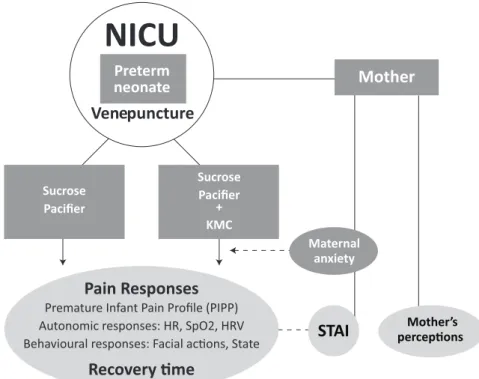
Figure 1. The preterm infant in the NICU and pain: problem statement. Preterm infants have the capacity to feel pain. During their stay in neonatal intensive care units, they endure a high num-ber of painful procedures and events. Their physiological and behavioral immediate responses to pain can be measured consistently. The long-term consequences of repetitive exposure to pain are reflected on alterations of pain pathways, behavior and cognition. Pain is a major concern for parents, who wish to be involved in the care of their infants. There are efficacious and safe avail-able interventions to reduce pain responses during procedures but more research is needed on the combination of these interventions to improve pain management.
Oral sucrose has been exhaustively studied for procedures such as heel lance and venepuncture and its efficacy has been demonstrated in very low doses, without more adverse events than with placebo (Stevens, Yamada, & Ohlsson, 2010). Infants given su-crose cry less and present lower pain scores. The stimulation of taste buds in the tongue by sweet-taste activates the release of endogenous opioids, responsible for cessation of cry in infants (Blass & Ciaramitaro, 1994). Pacifiers also reduce pain responses, either
5
alone or in combination with oral solutions (Cignacco et al., 2007). There are two
hy-potheses for the calming effect of sucking, none of them involving opioid mechanisms: it promotes self-regulation and it is a strong sensory stimulation mobilizing the limit-ed attentional resources of the neonate (Carbajal, Chauvet, Couderc, & Olivier-Martin, 1999). Kangaroo Mother Care is another well studied intervention, especially for heel lance. It promotes sleep states, reduces cry and pain scores and facilitates recovery of altered physiological parameters after procedural pain (Johnston, Campbell-Yeo, & Fernandes, 2009; Warnock et al., 2009). Opioid and non-opioid mechanisms may be at
stake, since during skin-to-skin contact between mother and infant, sensorimotor, ther-mal, olfactive and tactile interactions take place, which are hidden regulators of infant physiology and behavior (Hofer, 1994).
The main question of this study was therefore: are the pain responses of pre-term infants to a painful procedure reduced when kangaroo mother care is added to the standard care use of sucrose and pacifier? Given the known co-regulation of mother-infant physiology and behavior (Morelius, Theodorsson, & Nelson, 2005; Matthiesen, Ransjo-Arvidson, Nissen, & Uvnas-Moberg, 2001), a secondary question was raised: does maternal anxiety interfere with the potentially beneficial effects of kangaroo care? And finally, because mothers are an essential part of this intervention and their presence during painful procedures is often an issue for health professionals, a last question was raised: how do mothers perceive doing kangaroo care during a painful procedure, i.e., holding the baby skin-to-skin while the baby is enduring pain?
The painful procedure chosen to be examined was venepuncture because it is the most frequently performed needle-related procedure in our neonatal intensive care units.
In order to respond to the questions above, one main and two secondary objec-tives were defined: 1) to compare the efficacy of the combination of kangaroo mother care, sucrose and pacifier, with that of sucrose and pacifier, in reducing the pain respons-es of preterm infants undergoing venepuncture in the Neonatal Intensive Care Unit; 2) to examine the relation between maternal anxiety and the pain responses of preterm babies in KMC; and 3) to explore mothers’ perceptions of doing kangaroo care during venepuncture.
To attain the main objective of this study a single-blind randomized-control-led trial was conducted and, to respond to the secondary questions, mothers who performed kangaroo care were interviewed. The study took place in two level II/III Neonatal Intensive Care Units in Coimbra, Portugal. After authorization and consent
6
procedures, one-hundred and ten preterm babies (N= 110) with gestational ages be-tween 28 and 37 weeks, stratified into two gestational age groups (from 28 weeks to 31 weeks and six days and from 32 weeks to 36 weeks and 6 days) participated in this study. Infants were randomly assigned to receive one of two interventions to reduce pain during venepuncture for blood draw: 1) Oral sucrose with pacifier; or 2) Oral sucrose, pacifier and kangaroo mother care. Physiological indicators, namely heart rate and ox-ygen saturation, as well as facial behaviors were digitally recorded before, during and af-ter the venepuncture. The pain responses were examined using a composite pain scale, the Premature Infant Pain Profile (Stevens, Johnston, Petryshen, & Taddio, 1996) and analyzing its components separately: heart rate, oxygen saturation, facial actions and state. Heart rate variability and recovery time were also analyzed. The two intervention groups were compared across the procedure using repeated-measures ANOVA.
The second objective was attained undertaking a cross-sectional, correlational study. Mothers’ anxiety level was measured before the painful procedure using the State scale of the State-Trait Anxiety Inventory (Spielberger et al., 1983) and the relation with
infants’ pain responses was examined.
Finally, the third objective was reached through content analysis of the semi-structured interviews to mothers who performed kangaroo care.
The design of this study is represented in Figure 2.
Figure 2. Study design. Note: KMC - Kangaroo mother care; STAI - Stait-Trait Anxiety Inventory; NICU - Neonatal Intensive Care Unit.
7
This research report is organized in two parts — the background and the em-pirical study — and follows the recommendations contained in the Publication man-ual of the American Psychological Association, 5th edition (American Psychological Association, 2001).
In the first part, divided in five chapters, the theoretical and empirical back-ground for studying non-pharmacological pain interventions in the context of neona-tal care is presented. In the first chapter, the vision of the preterm neonate within the Model of the Synactive Organization of Behavioral Development (Als, Butler, Kosta & Anulty, 2005) offers a framework for integrating pain relief as an important part of the developmental care approach. Pain as a multidimensional experience and evidence from neurobiology of the capacity of preterm neonates to feel pain are examined next. Short term responses to pain and tools to assess pain intensity as well as long-term con-sequences of early exposure to pain are also addressed in the second chapter. The third chapter focuses the epidemiology of pain and pain management in neonatal intensive care units, confirming that pain occurs frequently and is often undertreated. The fourth chapter offers a comprehensive review of studies of non-pharmacological pain interven-tions for procedural pain in newborns. Parental concerns and the importance of a fam-ily-centered approach for pain management in the NICU are analyzed in the last chap-ter of the first part.
The second part presents the empirical study. The methods are described, name-ly the study design, the research settings, the sampling method, the outcome measures, the research procedure including the experimental protocol, the procedures for data ex-traction and statistical analysis, and the way in which ethical concerns were dealt with. The results are displayed in chapter seven, beginning with a presentation of participants’ characteristics, followed by the pain responses of infants to the interventions studied. In the third and fourth sections of the results chapter, the correlation between maternal anxiety and infants’ pain responses is presented as well as mothers’ perceptions of kan-garoo care during venepuncture. In the discussion, the results obtained in response to the research questions are summarized and interpreted according to the state of the art. Strengths and limitations of the study are disclosed, unresolved theoretical issues are put forward and implications are drawn for clinical practice and research.
The report concludes with an overall evaluation of the research process taking in-to consideration the objectives of the study, and opens the way for knowledge transla-tion of these results into practice as well as for near-future research.
Theoretical background:
The neonate and pain
CHAPTER 1.
The preterm neonate,
a new paradigm
CHAPTER 1. The preterm neonate, a new paradigm
For many years, preterm infants were considered immature, incomplete babies as mirrored in the way they were named: “premature babies”. Neonatal care was focused on compensating and treating the consequences of the immaturity of the different sys-tems – respiratory, gastro-intestinal, immune and nervous, among others. Infants born prematurely are abruptly deprived of the intrauterine environment, which provides the most favorable conditions for their development.
The conceptualization of development as proposed by the Model of the Synactive Organization of Behavioral Development (Als et al., 2005) offers a theoretical
back-ground for examining preterm neonates, their development and the aims of neonatal care in a different way. Under this model, preterm infants are not incomplete fullterms but rather, well-equipped, competently adapted fetuses that would function appropri-ately for their stage of development if they were in their natural environment: the ma-ternal uterus. They are seen as the initiators of the interaction with others, whether these are professionals or parents.
The infants’ functioning is viewed as continuous interactions between the in-traorganism subsystems (autonomic, motor, state, attentional/interactive and self-regu-lating) and the environment. Once exposed to the aggressive environment of a neonatal intensive care unit, a great amount of energy is consumed by infants in stabilizing their subsystems and little is left to pursue their development in the right direction unless the care provided helps them to do so (Als et al., 2004). A main goal of neonatal care, as
im-portant as to respond to physiological needs, is therefore to attend to the developmen-tal and emotional needs of preterm infants by adjusting the environment (Sparshott, 1997).
12
The first decades of development of neonatology, in the 1960s and 1970s, were devoted to the survival of preterm infants (Kennell, 1999) by maintaining the auto-nomic functioning: respiratory, cardiac, digestive and temperature control functions. The dawn of mechanical ventilation revolutionized neonatal care which became more and more intensive and invasive. Handling for procedures such as endotracheal suction-ing became a routine that had to be followed regularly.
It wasn’t until technology allowed non-invasive constant monitoring of oxygen blood levels through transcutaneous pO2 devices that the impact of care came to light.
Although some expressed concern for the lack of sleep and rest of these infants in the late 1970s (Lucey, 1977), state organization, motor system and sensory functioning were secondary concerns at that time (Als et al., 1986). It was in the 1980s, that
consid-ering the developmental and behavioral detrimental consequences of the aggressive en-vironment of neonatal intensive care, a number of interventions termed ‘Developmental Care’ were suggested to improve the neurodevelopmental outcome of preterm infants (Als et al., 1986). This broad group of interventions includes controlling external
stim-uli (vestibular, tactile, auditory, visual) during nursing routines, handling, feeding, pain management, adjusting these to the individual cues of the baby and involving parents in the care of their infant in the NICU in a family-centered approach (Aucott, Donohue, Atkins, & Allen, 2002). The idea of organizing care based on the individual behavio-ral cues of each baby, especially those who are very low birth weight (VLBW) was the basis of the Newborn Individualized Developmental Care and Assessment Program (NIDCAP). The impact of NIDCAP on neurodevelopment, maturation and morbid-ity remains controversial, some studies reporting clear positive effects (Kleberg et al.,
2008; Als et al., 2003; Kleberg, Westrup, Stjernqvist, & Lagercrantz, 2002; Kleberg,
Westrup, & Stjernqvist, 2000) and others not (Symington & Pinelli, 2006; Jacobs, Sokol, & Ohlsson, 2002; Ariagno et al., 1997). Yet, its foundational model, the Model
of the Synactive Organization of Behavioral Development, was innovative in stressing the competencies of preterm newborns, namely their capacity to interact with the envi-ronment, and the importance of modifying the physical and emotional environmental factors that can adversely affect the behavioral organization of these infants.
The refinement of neonatal care and the increasing use of high technology grad-ually pushed back the limits of viability of preterm infants in the past 40 years (Seri & Evans, 2008). While it was a common understanding in the 1970’s that infants born less than 28 weeks were not able to survive, today’s knowledge and technological re-sources in developed countries make it possible that infants born as early as 23 weeks of
13
gestational age are cared for in NICUs. This achievement however has not been with-out cost. Infants born very prematurely display an array of physical and psycho-behavio-ral consequences that are related to their early life events.
Studies about the effects of early experience on brain function and structure as well as on subsequent behavior (Als et al., 2004; Anand & Scalzo, 2000; Gunnar &
Barr, 1998) seem to confirm the need to reduce environmental stress factors in the neo-natal period, namely pain exposure and maternal deprivation.
Understanding pain in the neonate demands first of all that this concept is made clear. Knowledge of the peripheral, spinal and supraspinal processing of painful stim-uli owes a great deal to animal studies, which is why the most common animal models of infant pain will be briefly reviewed. The rate of development of the nervous system, both in growth and differentiation (birth and migration of neurons; growth of axons; formation of dendrites and synapses, myelination, pruning, to mention only a few proc-esses) is such, that it is not possible to discuss pain in the neonate without considering developmental issues. The capacity of preterm neonates to experience pain can be dem-onstrated by examining their immediate responses, measuring their intensity of pain and analyzing the long-term consequences of pain in early life.
2.1 The pain experience
The concept of pain as a multidimensional experience is fairly recent. Attempts to categorize pain as a sensation or as an emotion come from as far back as Aristotle (Melzack & Wall, 1987).
The International Association for the Study of Pain (IASP) defines pain as “An unpleasant sensory and emotional experience associated with actual or potential tissue damage or described in terms of such damage” (IASP Taskforce on Taxonomy, 1994) thus recognizing the multidimensional character of the pain experience.
The recognition that the pain experience is far more than the activity induced in the nociceptor and nociceptive pathways by a noxious stimulus, always involving a psy-chological state, finds support in the Gate-Control Theory (Melzack & Wall, 1987). Published in 1965 by Melzack and Wall, the Gate-Control Theory moved away from the Specificity Theory, which since Descartes postulated that a specific pain system transported pain messages from specific centers in the skin to a specific centre in the brain. The mechanism could be compared to pulling a string to ring a bell. This
mecha-18
nistic view was refined during the 19th and early 20th centuries by scientists like Müller and von Frey, under the new developments of physiology and histology but kept the idea of a fixed nervous system and direct pain pathways. The main contribution of this the-ory was the recognition of the specialized role of skin receptors. The existence of a di-rect and invariable relation between a particular quality of the physical stimulus and the psychological and sensory dimension of the experience defended by this theory, though, was not supported by empirical evidence and opened the way to other theories. Some of these theories like the Pattern theory and the Summation theory brought up important contributions to the understanding of pain but none of them alone could consistent-ly offer a comprehensive explanation for the complexity and variety of pain syndromes (Melzack & Wall, 1987).
The conceptual mode that underlies the Gate-Control Theory is based on the fol-lowing propositions:
1. The transmission of nerve impulses from afferent fibers to spinal cord transmission (T) cells is modulated by a spinal gating (SG) mechanism in the dorsal horns.
2. The spinal gating mechanism is influenced by the relative amount of ac-tivity in large-diameter (L) and small-diameter (S) fibers: Acac-tivity in large fibers tends to inhibit transmission (close the gate) and small-fiber activity tends to facilitate transmission (open the gate).
3. The spinal gating mechanism is also influenced by nerve impulses that descend from the brain.
4. A specialized system of large-diameter, rapidly conducting fibers (the central control trigger) activate higher cognitive processes that then influ-ence, by way of descending fibers, the modulating properties of the spinal gating mechanism.
5. When the output of the spinal cord transmission (T) cells exceed a crit-ical level, it activates the action system – those neural areas that underlie the complex, sequential patterns of behavior and experience characteristic of pain. (Jeans & Melzack, 1992, p. 22)
This model acknowledges the fundamental role of the central nervous system (CNS) in filtering, selecting and modulating pain inputs from high-threshold small fib-ers (Melzack, 1999) and definitely breaks the Cartesian dichotomy between mind and body. Brain activities related to attention, emotion and memory exert control over the sensory input.
19
However, how the brain functions to produce the qualities of the experience in the absence of sensory peripheral inputs has not been explained by this theory. Analyzing the phantom limb phenomena, Melzack developed the Neuromatrix Theory of Pain (Melzack, 2001; Melzack, 1999). This new conceptual model of the nervous sys-tem proposes that a widespread network of neurons, called the neuromatrix, is the ana-tomical substrate of our experience of the body-self and the somatosensory qualities we feel. The neuromatrix consists of loops between the thalamus and cortex and between the cortex and the limbic system, the spatial distribution and synaptic links of the neu-romatrix being initially determined genetically and later “sculpted” by sensory inputs. These loops are responsible for the sensory-discriminative, affective-motivational and evaluative-cognitive components of the pain experience. They diverge to permit paral-lel processing in different components of the neuromatrix and converge to permit in-teraction between the outputs of this processing. This cyclical processing and synthesis of nerve impulses has a characteristic output pattern named “neurosignature”. The neu-rosignature for the pain experience is, again, determined by the synaptic architecture of the neuromatrix resulting from the genetic and sensory influences and is modulated by sensory and cognitive inputs, such as psychological stress, to produce the particular qualities and properties of the pain experience. Multiple inputs act on the neuromatrix and contribute to produce the output signature. Painful stimuli might then trigger the neurosignature output but do not produce it. The neurosignature pattern, a continu-ous outflow of nerve impulses from the body-self neuromatrix, is projected into areas in the brain, the sentient neural hub, where it is modulated by ongoing inputs producing a continually changing stream of awareness. In the same way, the activation of neural net-works responsible for movement produces the movement itself while the projection to the sentient neural hub produces the experience of movement. The inputs to body-self neuromatrix include sensory (e.g. cutaneous, visceral, visual, vestibular inputs) as well as motivational-affective (e.g. hypothalamic-pituitary-adrenal system, noradrenalin-sym-pathetic system, immune system, cytokines, endogenous opiates) and cognitive-evalua-tive inputs (e.g. learning, past experience, personality, attention, anxiety). The outputs from body-self neuromatrix involve the pain perception in its sensory-discriminative, motivational-affective and cognitive-evaluative dimensions, patterns of action, commu-nication and coping, and stress-regulation programs.
This theory proposes a model of brain functioning that reinforces the concept of pain as a multidimensional experience integrating the role of higher psychoneural proc-esses in addition to the previous modulation and descending control of sensory nerve
20
inputs caused by injury, and therefore offers an explanation for the experience of pain in the absence of injury or peripheral sensory pathways as is the case in most chronic pain syndromes.
In neonates, the Gate-Control Theory is a useful framework for acute procedur-al pain and the interventions that can be used as pain gating mechanisms. Sensory and motivational-affective inputs are certainly present in preterm infants. While cognitive-evaluative inputs such as learning and culture might have a minor role, other inputs such as past experience, attention (state) and anxiety (distress), may play an important role in the experience of pain. The long-term consequences of pain in early life, however, may come to find some explanation under the Neuromatrix Theory of Pain.
The capacity of neonates to feel pain was challenged for a long time, given that the experience of acute pain requires the structures in the CNS to be connected in order that the sensory inputs reach the brain cortex. The full development of pain pathways and the activity of the cortex have been considered critical issues in recognizing that ne-onates feel pain. Research with animal models has been extremely useful in elucidat-ing about the development of neuronal structures and functionelucidat-ing of sensory pathways.
2.2 Animal models of infant pain
Much of what we know about pain and development is inferred from animal studies. For ethical reasons, certain kinds of experiments are not justifiable in humans unless strong evidence suggests that the results may be beneficial in clinical practice. Given the amount of animal studies that have generated hypotheses about pain reliev-ing interventions and their mechanisms in neonates, a brief overview of common out-comes of experiments in animal models of infant pain may help understand the ration-ale of such studies.
Rodents represent a useful model for the investigation of human neonatal pain for three main reasons: it is possible to parallel the stages of neurosensory development of rat pups and human infants; the quick rate of maturation allows the study of long-term consequences of neonatal pain in a short time; and the control of extraneous vari-ables is easier than in human research. Although the developmental timetvari-ables are dif-ferent in rats and in humans, the basic sequence of events in the maturation of sensory systems is the same in both species (Fitzgerald & Anand, 1993). Rat pups are born fair-ly immature compared to fullterm infants and their neurological maturation stage at birth, in terms of somatosensory and motor development, is comparable to the human infant development around 24 weeks of gestational age. Studies of the developmental neuroanatomy and neurophysiology of pain as well as studies of pain behaviors relate
21
data obtained from newborn rats in the first week of life to preterm infants at the sec-ond trimester of gestation. By postnatal day 10 (P10) the stage of development is relat-ed to that of fullterm infants; data from 2-3 week-old rats corresponds to infants during the first years of life (Sternberg & Al-Chaer, 2007; Johnston, Walker, & Boyer, 2002; Fitzgerald & Anand, 1993). In addition, laboratory rodents have a short gestation (ap-proximately 3 weeks) yielding large litters of pups. These have a rapid rate of postnatal maturation: they are weaned at around 20 days, reach sexual maturation around 6-7 weeks of age and are adults near the 10th week (P60). It is therefore possible to study the long-term consequences of neonatal pain in only a few months (Sternberg & Al-Chaer, 2007). Animal models offer the possibility to control the timing, frequency and intensity of the pain stimulation in a way that is not possible in the clinical environ-ment where pain and the outcomes studied are related to clinical care. Genetic factors responsible for individual variability can also be controlled in animal studies, by using selected strains of rats. Smaller samples can be big enough to show small differences in effect sizes (Johnston et al., 2002a). The clinical relevance of animal studies though, has
some limitations and Johnston and colleagues articulate the questions that can be val-idly answered by animal studies, considering the asynchronous development of the vari-ous brain regions and the higher complexity of human behavior compared to rodent be-havior: “The specific questions relate to the effects of peripheral injury of differing types and magnitude on the central nervous system (CNS), how long the effects last, how widespread the changes are (peripheral, spinal, supraspinal), and what mechanisms can block the change” (Johnston et al., 2002a, p. 397).
Studies that have examined the immediate responses and long-term consequenc-es of early exposure to pain use the paradigm of acute needle pain (Johnston & Walker, 2003; Anand, Coskun, Thrivikraman, Nemeroff, & Plotsky, 1999), persistent inflam-matory pain caused by chemical agents or cutaneous tissue injury (Ririe, Bremner, & Fitzgerald, 2008; Ruda, Ling, Hohmann, Peng, & Tachibana, 2000; De Lima, Alvares, Hatch, & Fitzgerald, 1999; Reynolds & Fitzgerald, 1995) and nerve injury (Lee & Chung, 1996).
Acute pain can be elicited by single or repeated needle stick in the dorsum or plantar surface of the hindpaw (Johnston et al., 2002a; Anand et al., 1999) or by
repeat-ed footshock (Sternberg & Al-Chaer, 2007). Inflammatory pain is frequently obtainrepeat-ed through injections into paws of formalin, a mild inflammatory agent producing short-lasting local inflammation (30 to 60 minutes) while carrageenan, capsaicin, bee-venom or complete Freund’s adjuvant (CFA) are stronger inflammatory agents causing
long-22
lasting pain and in the case of CFA1, long-term activation of immune responses more suitable to mimic chronic pain (Johnston et al., 2002a).
Outcome measures used to examine the effect of single or repetitive pain and the modulating effect of interventions on different types of pain include pain thresholds to thermal or mechanical stimuli as well as pain behaviors, stress responsiveness, changes in tissue innervation and pain circuitry, and peripheral, spinal and supraspinal activity of neurons and neurotransmitters.
Pain thresholds
Thermal sensitivity threshold is measured using the Hargreaves test, the hot plate test or the tail flick test. In the Hargreaves test the rat is placed in an acrylic box and a beam of light is directed to the footpad of one paw. The temperature of the beam ris-es rapidly and latency to nociceptive behavior i.e., the time elapsed until the rat displays behaviors such as paw lifting, licking, shaking or flicking, is considered to be the pain threshold (Johnston et al., 2002a). The hot plate test is similar but the surface of the box
in which the rat is placed is at a constant temperature of 50 to 60 degrees Celsius. In the tail flick test a heated beam of light set at a certain temperature is directed onto the tail of the animal placed in a narrow acrylic box not allowing him to move, and the latency to flick the tail out of the heat source is the pain threshold.
Mechanical sensitivity is most commonly measured through stimulation with von Frey hairs, nylon filaments of graded calibrated diameters. Von Frey hairs are ap-plied sequentially in increasing diameters on the animal’s paw, until the cutaneous flex-or reflex is elicited. The end of the filament is pressed against the skin requiring a precise force to form a buckle, the caliber of the filament in grams being considered the me-chanical threshold. The cutaneous flexor reflex is a protective response and depends up-on the development of spinal sensory processes (Johnstup-on et al., 2002a).
Variations in thermal and mechanical sensitivity in inflamed versus non-inflamed animals allow the study of analgesic agents as well as and the modulating effect of cer-tain Non-pharmacological interventions such as non-nutritive suckling (Anseloni, Ren, Dubner, & Ennis, 2004) or sucrose.
The mediating role of maternal rearing on adult pain thresholds as a consequence of neonatal repetitive pain has recently been explored testing thermal sensitivity (de Medeiros, Fleming, Johnston, & Walker, 2009).
1Complete Freund’s Adjuvant (CFA) is a mineral oil emulsion containing heat-killed
23
Pain behaviors
Specific pain behaviors of the animal when inflammatory pain is inflicted in-clude ultrasonic vocalizations, licking or shaking the paw, lifting the paw and protect-ing the paw. The formalin test, consistprotect-ing of a formalin injection in the paw, is a well validated method of testing used as a model of inflammatory pain (Johnston & Walker, 2003; Teng & Abbott, 1998; Abbott & Guy, 1995). It involves supraspinal mechanisms and allows the understanding of maturation processes and consequences of repeated, long-lasting or severe pain in higher structures of the central nervous system. Recently, this model has been used to examine maternal behavior (grooming) in the presence of repeated neonatal pain in offspring (Walker, Kudreikis, Sherrard, & Johnston, 2003).
Stress responses
Behavioral responses such as rats’ exploratory activity in an open field or in a new environment are used as a measure of discomfort associated with ongoing pain and to study the long-term consequences of neonatal pain on the responses to distress, anxie-ty and agoraphobia under the assumption that early pain experiences will interfere with adult stress responsiveness (Sternberg & Al-Chaer, 2007; Anand et al., 1999). Social
discrimination, i.e. time spent investigating a novel juvenile has been used as a meas-ure of chemosensory memory (Anand et al., 1999). Since pain is a stressor, hormones
such as ACTH, cortisol or corticosterone are used to measure the activation of the hy-pothalamic-pituitary-adrenal axis (Walker et al., 2003). Alcohol preferences of adult
rats have also been studied as a consequence of repetitive neonatal pain by measuring the intake of solutions of sucrose and sucrose with alcohol and comparing rats exposed to repetitive neonatal pain to rats exposed to non-noxious touch stimulation (Anand et al., 1999), although this was not replicated in a subsequent study (Bhutta et al., 2001).
Structural and functional changes in nervous tissue and neurons
Inflammatory pain elicited by inflammatory agents such as formalin or CFA or by skin wound is used as a stimulus to identify the structural and physiological chang-es that occur during tissue insult and in the long-term. Skin injury causchang-es inflammato-ry pain as a result of sensitization of peripheral nociceptors and central neuronal path-ways followed by sprouting of sensory nerve terminals and hypersensitivity (Sternberg & Al-Chaer, 2007).
Innervation of the skin, dorsal horns and root ganglia as well as nociceptive pathways, are studied through immunocytochemistry and immunohistochemistry techniques. These methods use antibodies to target components of cells or tissues,
re-24
spectively. Components identified include neurotransmitters such as glutamate or γ-aminobutyric acid (GABA), neuropeptides such as substance P and enkephalins, neu-rotransmitters and neuropeptides receptors such as N-methyl-D-aspartate (NMDA), GABA and opiate receptors, immediate early genes such as c-fos expression (Johnston
et al., 2002a).
Examining the responses to tissue injury at different ages, it is possible to identi-fy which populations of sensory fibers are more sensitive to nerve sprouting, playing a more significant role in skin hyperinnervation, and which are the critical stages of devel-opment (Reynolds & Fitzgerald, 1995). The effect of interventions such as nerve blocks before skin wound in young animals can be studied using cutaneous hyperinnervation and sensory thresholds as outcomes (De Lima, Alvares, Hatch, & Fitzgerald, 1999).
Measurements of electrophysiological activity in the dorsal horn of rats of dif-ferent ages elucidate the postnatal development of spinal cord mechanisms of inflam-matory pain, changes in receptive fields at the dorsal horn as well as the disruption of structural and functional organization of nerve connections at the spinal level as con-sequence of inflammatory pain and the role of neurotransmitters in fiber connectivi-ty (Peng, Ling, Ruda, & Kenshalo, 2003; Torsney & Fitzgerald, 2002; Beggs, Torsney, Drew, & Fitzgerald, 2002).
To summarize, several models of neonatal pain are used in animal studies to un-derstand basic pain mechanisms related to development, long-term consequences of ear-ly exposure to pain and factors that can mediate or block those effects. Acute needle pain and inflammatory pain by chemical agents or tissue injury are the most common ones. Outcomes of studies using these paradigms alone or combined include thermal and mechanical sensitivity, pain and stress behaviors, structural and functional chang-es in nervous tissue, neurons and nociceptive pathways, measured through a variety of methods. The number of possible combinations of model, studied outcomes and meas-urement techniques is such that nearly each study reaches findings that are difficult to compare with others.
Furthermore, the parallel between pain inflicted in these experimental condi-tions and pain experienced by human neonates under clinical care is hard to establish, requiring caution when drawing clinically useful conclusions from animal studies. The findings in animal studies about the development of pain circuitry, widening receptive fields and decreased threshold following injury have led to studies showing similar re-sults in humans (Andrews, Desai, Dhillon, Wilcox, & Fitzgerald, 2002; Fitzgerald & De Lima, 2001; Andrews & Fitzgerald, 1999; Andrews & Fitzgerald, 1994; Fitzgerald, Millard, & McIntosh, 1989; Fitzgerald, Shaw, & MacIntosh, 1988)
25
Understanding the immediate and long-term effects of different types of pain on the developing peripheral and central nervous system of animals and the factors that can mediate theses consequences increases clinicians’ awareness of the detrimental ef-fects of pain in human infants and generates hypotheses regarding interventions to be tested in the clinical environment. As Johnston and colleagues point out: “Interaction between clinicians and basic scientists, with an understanding of the domain in which each group is working, is critical to the meshing of efforts from these domains. With collaboration between these groups, more relevant research can be conducted that can lead to the decrease in pain and its consequences in neonates.” (Johnston et al., 2002a,
pp. 411-412).
2.3 The capacity of preterm neonates to experience pain
The requirements for the occurrence of pain are the existence of functioning pe-ripheral, spinal and supraspinal anatomic structures related to the pain/tactile system as well as the neurochemical system associated with pain transmission and modulation. Some have argued that the development of the mind to allow consciousness of pain is also needed for the pain experience (Derbyshire, 2006). While it is clear that fetuses in the second trimester of gestation have endocrine and reflex responses to noxious stim-ulation (Glover & Fisk, 1999), it is controversial whether this can be considered pain or just nociception. It is accepted, however, that the interaction with the outside world that occurs at birth marks the beginning of consciousness and is the key to consider that very preterm neonates are able to feel pain. Consciousness may be defined by sensory aware-ness of the body, the self and the world (Lagercrantz & Changeux, 2009). Early preterm neonates exhibit sensory awareness when they react to sound, smell, touch and taste. In responding to painful stimuli through both behavioral and physiological signs, they ex-press emotions, differentiate self and non-self touch and show signs of shared feelings (Lagercrantz & Changeux, 2009). Yet, these authors argue, they are present-oriented and self-awareness is limited, which is why they can be considered in a minimal level of consciousness that will increase with age.
Regarding the peripheral anatomical and functional requirements for the pain ex-perience, it is known that nociceptive neurons are specified in early fetal life (Fitzgerald, 2005). Sensory neurons in the dorsal root ganglia begin to grow towards the skin and towards the spinal cord by 6 weeks of gestation. Specialized sub-populations of these sensory neurons reach all the cutaneous and mucosal surfaces by 20 weeks. The final density of nociceptive nerve endings in the skin of newborns is at least the same as in adults (Anand & Hickey, 1987) and is a result of the balance between cell growth and cell death (Fitzgerald, 2005).