DOI: http://dx.doi.org/10.18363/rbo.v75.2018.e1164 Original Article/Operative Dentistry
Influence of buccal-lingual width on the survival
rate of proximal posterior Atraumatic Restorative
Treatment (ART) restorations
Carolina Guedes Barquete,1 Antônio Fernando Monnerat,2 Cesar dos Reis Perez31Department of Dentistry, School of Dentistry, Federal Fluminense University (UFF), Rio de Janeiro, RJ, Brazil
2Department of Integrated Clinical Procedures, School of Dentistry, State University of Rio de Janeiro (UERJ), Rio de Janeiro, RJ, Brazil 3Department of Prosthesis, School of Dentistry, State University of Rio de Janeiro (UERJ), Rio de Janeiro, RJ, Brazil
• Conflicts of interest: none declared. AbstrAct
Objective: this study examined the relationship between the buccal-lingual width and the survival rate of multiple surface restorations, after simulated mechanical
cycling. Material and Methods: three cavity’s widths were evaluated: Regular (R = 3.21mm), Large (L = 4.81mm), and Very Large (VL - 6.42mm). Thirty extracted teeth were selected, prepared, restored with high viscosity glass ionomer cement through the ART technique and subject to simulated mechanical cycling. In each group, the samples were classified according to four different scores: 0 – no fracture, 1 – fracture with no repair needed, 2 - fracture with possible repair, and 3 – complete resto-ration loss/impossible to repair. Kruskal-Wallis test was used for the analysis of categorical outcomes. Results: there was a significant statistical difference between all groups (p<0.05), where the variable “bucco-lingual width” directly affected the results. Conclusion: cavities with controlled dimensions, closer to what is academically indicated, have a more favorable success rate, compatible with those found in occlusal cavities.
Keywords: Dental atraumatic restorative treatment; Dental stress analysis; Glass ionomer cement; Dental caries.
Introduction
T
he Atraumatic Restorative Treatment (ART) is an ap-proach, created by J. Frencken, to manage dental car-ies. The ART has been performing breakthroughs for using a simple technique, involving only hand instruments, without the need of rotatory instruments or anesthesia. The infected tooth tissue is removed with an excavator based on the concept of partial caries removal and the cavity is re-stored with a high viscosity glass ionomer cement (GIC) or a resin modified glass ionomer cement (RMGIC).1The GIC was chosen for this technique because it ensures a true adhesion to the tooth, in addition to biocompatibility, low coefficient of thermal expansion, and easy insertion and repair. However, some low mechanical properties of this ce-ment contribute to some of the ART’s lack of success due to reduced wear, resistance, tensile, and fatigue, in relation to other materials.2 This cement would not be appropriated for areas subjected to severe tensions.
Seeking to achieve longevity and clinical success in this type of restoration, some factors influence are considered crucial, such as cavities dimensions and number of involved surfaces. Clinical studies demonstrate lower survival rates for proximal posterior restorations in relation to single oc-clusal restorations. Single surface ART restorations have 93% to 96% success rates in permanent teeth3-5 and 82% to 95% in deciduous teeth.6,7 However, multiple surface cavities within the same period have about 67% success rates in per-manent teeth and 55% to 75% in primary teeth.8-10
In addition to considering the number of involved sur-faces, the ART restoration should take into account its size. Longitudinal studies about multiple surface cavities demon-strate that the medium size cavities have best results when compared to very small or enlarged cavities.11 However, it’s important to consider the rate of fracture failure is directly influenced by the fact that it usually is a heroic treatment, and in this way, approach much larger cavities than conven-tionally indicated.12,13
Because of this reduced longevity of the ART in posterior teeth, there is some resistance within the dental and scien-tific community to recommend this technique for everyday use.14 However, it has been shown that the ART restorations have a longevity similar to conventional amalgam, compos-ite resin, and compomer restorations when restoring single occlusal cavities. And according to one meta-analysis, class II ART restorations presented a similar success rate in pri-mary molars when compared to those with amalgam.15 But after one year, the survival rate was 75 percent.16
It is suggested that the real problem might be related to the type/extent of the cavity and not the restorative materi-al.17 The main factor that motivated this work was, in fact, the direct clinical observation that most of the failures were related to cavities with great opening in the proximal box. Thus, although it is known that there is no standard prepa-ration in this type of treatment, standardized dimensions were established, based on previous articles, in order to eval-uate the box-width variable and its failure rate.
The purpose of this study is to mechanically test the behavior of proximal ART posterior restorations restored with a high viscosity GIC, using the cavity’s buccal-lingual width as the variable. The null hypothesis is that the resto-rationswith three different cavity’s widths behave similar-ly after mechanical cycling.
Material and Methods
Specimens
Impressions with polyvinylsiloxane (Futura AD, DFL, Rio de Janeiro, RJ, Brazil) of the right upper second bicus-pid, first molar, and second molar were made on a den-tal arch mannequin (Prodens, Rio de Janeiro, RJ, Brazil), where the right upper first molar was removed. This mold was filled with transparent acrylic resin (Clássico, São Pau-lo, SP, Brazil) to make thirty acrylic dental arch models.
Then, thirty teeth (all right upper third molars) were obtained for the study through the Teeth’s Bank of UERJ School of Dentistry. The took into account the similarity in shape and size. Adjustments were made in each third mo-lar’s roots, so that it would becorrectly placed in the “arti-ficial socket” (that corresponendedto the right upper first molar) with a contact point on both sides. When an ade-quate contact point was not obtained, a correction of the contour with acrylic resin (over the bicuspid distal face) was done. Finally, all acrylic models were fixed in PVC cyl-inders, measuring 2.5cm in width and 1.5cm in height, in order to standardize the samples according to the test ma-chine. This step was very important because the area of the restoration that receives the action of the machine’s active point, about 2mm of the marginal restoration crest, should be well-centered in the apparatus.
Tooth Preparation
The teeth were then randomly selected and prepared with one of the three types of cavities described below:
1. Regular Cavity (R): cervical-occlusal (C-O), mesi-al-distal (M-D), and buccal-lingual (B-L) dimensions of 3.21mm.
2. Large Cavity (L): C-O and M-D dimensions of 3.21mm and B-L of 4.81mm.
3. Very Large Cavity (VL): C-O and M-D dimensions of 3.21mm and B-L of 6.42mm.
These parameters were measured with a caliper (Elec-tronic Digital Caliper-MTX, Toolsworld, Guarulhos, SP, Brazil) and the variable between the groups was the buc-cal-lingual dimension. The cavities were prepared with a cylindrical diamond point (KG Sorensen FG 1090, São Paulo, SP, Brazil). The active part of this point presents a width of 3,21mm. So, this dimension was used to obtain the different cavities’ buccal-lingual measures (1X, 1,5X and 2X the active part of de diamond point). Ten teeth were
prepared for each type of cavity. After preparation, poly-ether impression material (Impregum, 3M ESPE, Seefeld, Germany) was applied around the tooth root with the aid of a brush in order to simulate a periodontal ligament. The teeth were fixed in position and the sockets were filled with acrylic resin.
The restorative material chosen in this study was Ketac Molar (3M ESPE, Seefeld, Germany), a high viscosity GIC used for the ART. The material was then manipulated fol-lowing the manufacturer’s instructions and the clinical steps of the conventional technique for the ART Class II cavity restoration were followed, as described below:
1. The GIC was inserted into the cavity with ART In-struments (SSWhite Duflex, Rio de Janeiro, RJ, Brazil).
2. A digital pressure was held with a finger smeared with pretoleum jelly (Rioquimica, São José do Rio Preto, SP, Brazil) for 1minute. After that, dental floss (Hillo, Aperibé, RJ, Brazil) was inserted in the interproximal region be-tween the restoration and the adjacent tooth.
3. The digital pressure was held again for a further 2 minutes.
4. A layer of pretoleum jelly was applied over the resto-ration in order to obtain surface protection.
The models were stored in distilled water (Asfer chem-ical factory, São Caetano do Sul, SP, Brazil) for 24 hours.
Mechanical Cycling
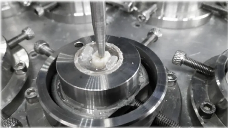
The specimens were carried to the thermo-mechanical cycler Erios ER- 37000NG (Erios, São Paulo, SP, Brazil), with a previously stipulated force and cycles (Figure 1). The active point of the cycler was put in position in contact to the chosen GIC restoration region (2mm inboard from the point of contact with the neighbor) and the machine was configured to do only axial force without sliding, with 50Kgf and 100,000 cycles (18 hours 37 minutes 5 seconds), at a frequency of 2 Hz. This protocol wasbased on previous studies.18 It was necessary to do a total of 3 cycles, with 10 specimens per test, obtaining a total of 30 specimens.
Figure 1. Specimen in position on the cycler ready for the start of the
Specimens’ Analysis
After each cycle, the specimens were submerged in dis-tilled water for 24 hours. Then, they were inspected and clas-sified according to the criteria previously defined (Figure 2): score 0 (no fracture), score 1 (presence of fracture but with-out need for repair), score 2 (presence of reparable fracture), and score 3 (presence of irreparable fracture or complete loss of restoration).
Statistical Analysis
Kruskal-Wallis test was used for statistical analysis with a 0.05 significance level and was performed using SPSS soft-ware (IBM SPSS Statistics 22, IBM Corp., Armonk, NY, USA).
Results
The results obtained after mechanical cycling are shown in Figure 3. Within group R, five specimens did not show fracture (score 0) and five specimens were classified as “no need for repair” (score 1). Considering Group L, five spec-imens needed repair (score 2), four of them needed no re-pair (score 1), and only one specimen did not fracture (score 0). In Group VL, nine specimens were classified as score 3 (complete loss of GIC) and one as score 2. There was a statis-tically significant difference between all groups, where the variable buccal-lingual width affected the results (p <0.05).
Figure 2. Examples of specimens with different scores: 0, 1, 2 and 3
Discussion
Dental caries in children and adults represents an im-portant issue of public health, affecting predominantly the poorest and low-income population. The ART reduces costs, using manual excavation of dental caries, eliminating the need for anesthesia and expensive equipment, and restores the cavity with a high viscosity GIC.19 However the longev-ity of ART restorations appears to be associated with lower success rates for occlusoproximal restorations in permanent and primary teeth.20
Certainly, one of the variables that most strongly affects the mechanical strength and the restoration survival is the opening of the proximal isthmus. Several studies reported reasons for the reduced longevity and sought solutions to improve the technique. However, the size of the cavities was not described in most of these studies about the ART fail-ure.7,21-23 Thus, the rate of failures in proximal posterior ART restorations may be overestimated since no specific criteria was adopted to characterize very large cavity restorations and consider them separately.
The aim of this study is to help to understand ifthe open-ing of the isthmus can influence the mechanical resistance and longevity of proximal posterior ART restorations. Based on the results, the null hypothesis was rejected because a sig-nificant statistical difference was found between all groups. Group VL had the worst results, with 90% of score 3 and 10% of score 2. Group L had 50% of score 2, 40% of score 1, and 10% with score 0. Group R presented 50% of score 1 and 50% with score 0.
This study tested three buccal-lingual openings: R would correspond to a proximal posterior cavity with a conven-tional buccal-lingual width for a molar; L would correspond to a more open buccal-lingual width but carried out within the limits of the proximal contact with the neighbor; and VL would correspond to a cavity with a buccal-lingual opening beyond the proximal contact with the neighbor.
Figure 3. Graph demonstrating the score results among the three cavity
In Kemoli’s study,24 the cavities with a 3.60mm buccal lingual dimension where considered medium size and showed the best clinical results. This size is similar to the R cavity described in the present study (3.21mm). It is believed that in small isthmus situations, buccal-lingual walls are better preserved, and the cavity is more retentive. This characteristic contributes to the preservation of the restoration. Also, the exposure of the GIC is lower, mak-ing it less subject to tensions. Buccal-lmak-ingual dimensions of approximately 3.21mm have a high probability of success.
The L Group showed lower results than the R Group, but there was no specimen with a complete failure. All the GIC restorations were capable of being repaired. Finally, the VL group (cavities with the largest isthmus – 6.42mm) showed very poor results. As GIC has low tensile and com-pressive strengths, the restorations tend to fracture more frequently in this type of width. In very large restorations, with or without involvement of the cusp, there is only a small amount of remaining structure for adherence to the GIC.8,25 This size is not suitable for the use of the TRA tech-nique in order to avoid premature loss of the restoration. In Gurunathan and Tandon’s study,26 the authors useda cavity’s classification in which small proximal posterior cavities were defined as less than 4mm, (similar to R) and medium cavities as larger than 4mm and less than 8mm. This study reported highersurvival rate to the restorations in regular size cavities’, in comparison to cavities of higher dimensions.
The cervical-incisal height and mesiodistal distance were also predefined in this work. However, these two mea-sures were not considered as decisive factors for longevity. All specimens had the same size of 3.21mm in height and depth, differing only in the buccolingual width that is con-sidered a crucial factor in determining the survival of the restoration.
Obtaining the contact point with the adjacent tooth is an important factor for maintaining the GIC in the tooth. For this, metal strips are usually utilized as a form of in-sulation and making the contact point.2,22,27 However, the methodology used in this study was the flossing technique. The floss should be used after the GIC starts hardening: the floss is compressed against the adjacent tooth to avoid the GIC adhering to the adjacent proximal surface but allow-ing the contact point. This is a simple and practical tech-nique that does not lead to any additional damage.
The mechanical cycling was used for its ability to simu-late chewing movements. This kind of test is used to mea-sure fracture resistance of materials in a simulated stress situation.28 Some studies have already submitted the reg-ular GIC or RMGIC to mechanical cycling. Most of these studies have sought to evaluate the presence of
microleak-age after the restorations cycling.29-31
Studies that analyze the high viscosity GIC restorations with approximate involvement subjected to mechanical cycles were not found in the literature. The strength of 50N was chosen as a moderately low strength present during mastication,32 which, when used by a high number of cy-cles (100,000 in this study), can promote fatigue at the teeth-restoration complex.18
After performing mechanical cycling, the restoration had a visual inspection by three experienced operators in the ART. In this study, failure was considered when there was a large or total loss of material, which could conse-quently cause accumulation of food, secondary caries, or a loss in clinical function. When the restoration presented cracks or little loss of material, which would not clinically damage the restoration, it was considered as “without need of repair” or “with possible repair”.
In literature, the restorations involving two or more surfaces represent a great dilemma for ART technique and, along with other reasons, is attributed to its failure. Two main reasons include:1) inadequate GIC resistance in areas subject to masticatory stress and 2) poor fracture strength33 which implies surface wear and fractures in both margins and in the restoration’s body, causing partial or total loss.34 The gradual dissolution of the proximal surface, changes in anatomical shape and marginal collapse of restorations were also noticed.9,33
Conclusion
Within the limitations of an in vitro study, this work’s findings suggest that the buccal-lingual width is a relevant variable in the prognosis of a proximal posterior ART res-torations. In this work a width larger than 4.81mm pre-sented a significant performance drop. Further studies are needed, especially clinical follow-up studies, in order to understand more precisely the behavior of this type of restoration. However, it should be emphasized that the treatment is a valuable option in public health, especially in poor countries, and that the restoration of cavities wider than academically accepted should be considered separate-ly in epidemiological and in follow-up studies, so as not to underestimate the clinical success of proximal posterior restorations with conventional size.
Acknowledgements
We would like to thank the extension project of the State University of Rio de Janeiro (Brazil) Atraumatic Restor-ative Treatment for the support to research in this area. We would like also to express our gratitude to Dentistry Post- Graduation from UFF for their availability during the tests.
Dent. 2004;6(3):221-6.
19. Mickenautsch S, Munshi I, Grossman ES. Comparative cost of ART and con-ventional treatment within a dental school clinic. Sadj. [Comparative Study]. 2002;57(2):52-8.
20. Bonifácio CC, Hesse D, Raggio DP, Bönecker M, van Loveren C, van Amer-ongen WE. The effect of GIC-brand on the survival rate of proximal-ART resto-rations. Int J Paediatr Dent. 2013;23(4):251-8.
21. Bonifácio CC, Hesse D, Bönecker M, Van Loveren C, Van Amerongen WE, Raggio DP. A preliminary clinical trial using flowable glass-ionomer cement as a liner in proximal-ART restorations: the operator effect. Med Oral Patol Oral Cir Bucal. 2013;18(3):529-32.
22. Cefaly DF, Barata TJ, Bresciani E, Fagundes TC, Lauris JR, Navarro MF. Clin-ical evaluation of multiple-surface ART restorations: 12- month follow-up. J Dent Child (Chic). 2007;74(3):203-8.
23. Hilgert LA, de Amorim RG, Leal SC, Mulder J, Creugers NH, Frencken JE. Is high-viscosity glass-ionomer-cement a successor to amalgam for treating prima-ry molars? Dent Mater. 2014;30(10):1172-8.
24. Kemoli AM, van Amerongen WE, Opinya G. Influence of the experience of operator and assistant on the survival rate of proximal ART restorations: two-year results. Eur Arch Paediatr Dent. 2009;10(4):227-32.
25. Hickel R, Manhart J. Longevity of restoration in posterior teeth and reasons for failure. J Adhes Dent. 2001;3(1):45-64.
26. Deepa G, Shobha T. A clinical evaluation of two glass ionomer cements in primary molars using atraumatic restorative treatment technique in India: 1 year follow up. Int J Paediatr Dent. 2010;20(6):410-8.
27. Hesse D, Bonifácio CC, Guglielmi CA, Bönecker M, van Amerongen WE, Raggio DP. Bilayer technique and nano-filled coating increase success of approx-imal ART restorations: a randomized clinical trial. Int J Paediatr Dent. 2015 Sep 15. doi:10.1111/ipd.12194.
28. Bapna MS, Gadia CM, Drummond JL. Effects of aging and cyclic load-ing on the mechanical properties of glass ionomer cements. Eur J Oral Sci. 2002;110(4):330-4.
29. Parveen N, Ahmed B, Butt AM, Bari A. The effects of mechanical stresses on the comparative microleakage of two different restorative materials. J Univ Med Dent Col. 2012;3(1):48-53.
30. De Mattos Pimenta Vidal C, Pavan S, Briso AL, Bedran-Russo AK. Effects of three restorative techniques in the bond strength and nanoleakage at gingi-val wall of Class II restorations subjected to simulated aging. Clin Oral Inves-tig.2013;17(2):627-33.
31. Mirzaie M, Yasini E, Kermanshah H, Omidi BR. The effect of mechanical load cycling and polishing time on microleakage of class V glass-ionomer and composite restorations: A scanning electron microscopy evaluation. Dent Res J (Isfahan).2014;11(1):100-8.
32. Anderson DJ. Measurement of stress in mastication. I. J Dent Res. 1956;35(5):664-70.
33. Anusavice KJ. Does ART have a place in preservative dentistry? Community Dent Oral Epidemiol. 1999;27(6):442-8.
34. Frankenberger R, Sindel J, Krämer N. Viscous glass-ionomer cements: a new alternative to amalgam in the primary dentition? Quintessence Int. 1997;28(10):667-7.
References
1. Frencken JE. Atraumatic restorative treatment and minimal intervention den-tistry. Br Dent J. 2017;223(3):183-9.
2. Scholtanus JD, Huysmans MC. Clinical failure of class-II restorations of a highly viscous glass-ionomer material over a 6-year period: a retrospective study. J Dent. 2007;35(2):156-62.
3. Frencken JE, Songpaisan Y, Phantumvanit P, Pilot T. An atraumatic restorative treatment (ART) technique: evaluation after one year. Int Dent J. 1994;44(5):460-4. 4. Frencken JE, Makoni F, Sithole WD. Atraumatic restorative treatment and glass-ionomer sealants in a school oral health programme in Zimbabwe: evalua-tion after 1 year. Caries Res. 1996;30(6):428-33.
5. Taifour D, Frencken JE, Beiruti N, van’t Hof MA, Truin GJ, van Palenstein Helderman WH. Comparison between restorations in the permanent dentition produced by hand and rotary instrumentation-- survival after 3 years. Commu-nity Dent Oral Epidemiol. 2003;31(2):122-8.
6. Menezes JP, Rosenblatt A, Medeiros E. Clinical evaluation of atraumatic res-torations in primary molars: a comparison between 2 glass ionomer cements. J Dent Child (Chic). 2006;73(2):91-7.
7. Yip HK, Smales RJ, Yu C, Gao XJ, Deng DM. Comparison of atraumatic re-storative treatment and conventional cavity preparations for glass-ionomer res-torations in primary molars: one-year results. Quintessence Int. 2002;33(1):17-21. 8. Lo EC, Holmgren CJ. Provision of Atraumatic Restorative Treatment (ART) restorations to Chinese pre-school children--a 30-month evaluation. Int J Paedi-atr Dent. 2001;11(1):3-10.
9. Taifour D, Frencken JE, Beiruti N, van’t Hof MA, Truin GJ. Effectiveness of glass-ionomer (ART) and amalgam restorations in the deciduous dentition: re-sults after 3 years. Caries Res. 2002;36(6):437-44.
10. Yip KH, Smales RJ, Gao W, Peng D. The effects of two cavity preparation methods on the longevity of glass ionomer cement restorations: an evaluation after 12 months. J Am Dent Assoc. 2002;133(6):744-51.
11. Kemoli AM, van Amerongen WE. Influence of the cavity-size on the sur-vival rate of proximal ART restorations in primary molars. Int J Paediatr Dent. 2009;19(6):423-30.
12. Frencken JE, Holmgren CJ. Atraumatic Restorative Treatment (ART) for Dental Caries, ed 1. Nljmegen, The Netherlands: STl Books. 1999.
13. Mickenautsch S, Grossman E. Atraumatic Restorative Treatment (ART): fac-tors affecting success. J Appl Oral Sci. 2006;14Suppl:34-6.
14. Frencken JE. The state-of-the-art of ART restorations. Dent Update. 2014;41(3):218-20. 222-4.
15. Raggio DP, Hesse D, Lenzi TL, AB Guglielmi C, Braga MM. Is atraumat-ic restorative treatment an option for restoring occlusoproximal caries lesions in primary teeth? A systematic review and meta-analysis. Int J Paediatr Dent 2013;23(6):435-43.
16. Lo E, Holmgren C. Provision of atraumatic restorative treatment (ART) res-torations to Chinese preschool children: a 30-month evaluation. Int J Paediatr Dent 2001;11(1):3-10.
17. Raggio DP, Hesse D, Lenzi TL, Guglielmi CAB, Braga MM. Is atraumatic restorative treatment an option for restoring occlusoproximal caries lesions in primary teeth? A systematic review and meta-analysis. Int J Paediatr Dent. 2013;23(6):435-43.
18. Bedran-de-Castro AK, Pereira PN, Pimenta LA, Thompson JY. Effect of ter-mal and mechanical load cycling on nanoleakage of Class II restorations. J Adhes
Submitted: 07/06/2018 / Accepted for publication: 09/10/2018
Corresponding Author Cesar dos Reis Perez
E-mail: [email protected]
Mini Curriculum and Author’s Contribution
1. Carolina Guedes Barquete – MS. Contribution: collaboration of the methodology; preparation of the manuscript; writing the manuscript. ORCID: 0000-0002-6757-4207
2. Antônio Fernando Monnerat – DDS. Contribution: conception and design of the study. ORCID: 0000-0002-5298-0157
3. Cesar dos Reis Perez – DDS. Contribution: conception and design of the study; standardization of the methodology; data interpretation; preparation of the manus-cript; critical review and final approval. ORCID: 0000-0001-9640-1578