cells
CommunicationIdentification of Novel Hemangioblast Genes in the
Early Chick Embryo
José Serrado Marques1, Vera Teixeira2ID, António Jacinto1and Ana Teresa Tavares1,*ID 1 CEDOC, Chronic Diseases Research Centre, NOVA Medical School, Universidade NOVA de Lisboa,
Campo dos Mártires da Pátria, 130, 1169-056 Lisbon, Portugal; [email protected] (J.S.M.); [email protected] (A.J.)
2 Instituto Gulbenkian de Ciência, Rua da Quinta Grande, 6, 2780-156 Oeiras, Portugal;
* Correspondence: [email protected]; Tel.: +351-218-803-101
Received: 10 December 2017; Accepted: 27 January 2018; Published: 31 January 2018
Abstract: During early vertebrate embryogenesis, both hematopoietic and endothelial lineages derive from a common progenitor known as the hemangioblast. Hemangioblasts derive from mesodermal cells that migrate from the posterior primitive streak into the extraembryonic yolk sac. In addition to primitive hematopoietic cells, recent evidence revealed that yolk sac hemangioblasts also give rise to tissue-resident macrophages and to definitive hematopoietic stem/progenitor cells. In our previous work, we used a novel hemangioblast-specific reporter to isolate the population of chick yolk sac hemangioblasts and characterize its gene expression profile using microarrays. Here we report the microarray profile analysis and the identification of upregulated genes not yet described in hemangioblasts. These include the solute carrier transporters SLC15A1 and SCL32A1, the cytoskeletal protein RhoGap6, the serine protease CTSG, the transmembrane receptor MRC1, the transcription factors LHX8, CITED4 and PITX1, and the previously uncharacterized gene DIA1R. Expression analysis by in situ hybridization showed that chick DIA1R is expressed not only in yolk sac hemangioblasts but also in particular intraembryonic populations of hemogenic endothelial cells, suggesting a potential role in the hemangioblast-derived hemogenic lineage. Future research into the function of these newly identified genes may reveal novel important regulators of hemangioblast development.
Keywords:chicken embryo; yolk sac; hemangioblast; microarray analysis; novel genes
1. Introduction
During vertebrate embryogenesis, there is a close developmental relationship between hematopoiesis and vasculogenesis. In the early embryo, the first hematopoietic and endothelial cells arise in the extraembryonic yolk sac blood islands from a common precursor known as the hemangioblast [1,2]. Recent evidence suggests that hemangioblasts give rise to hematopoietic cells through two types of intermediate progenitors, hemogenic angioblasts and hemogenic endothelial cells [3,4]. In the yolk sac, a first wave of hematopoiesis arises from hemogenic angioblasts that give rise to primitive hematopoietic cells, such as primitive erythrocytes, embryonic macrophages and megakaryocytes [4–6]. Also in the yolk sac, a second wave of hematopoiesis originates from hemogenic endothelial cells that give rise to definitive erythrocytes and to most myeloid lineages, including tissue-resident macrophages and microglial cells that persist into adulthood [7,8]. Finally, a third wave of hematopoiesis arises from intraembryonic hemogenic endothelial cells and produces definitive hematopoietic stem/progenitor cells (HSPC) that will colonize the fetal hematopoietic organs [9]. Cell-tracing studies have shown that these intraembryonic precursors also have an extraembryonic origin, as they migrate from the yolk sac prior to the onset of circulation [10,11].
Cells 2018, 7, 9 2 of 12
Together, these evidences suggest that most (if not all) hematopoietic cells in the embryo derive from yolk sac hemangioblasts.
The identification of novel hemangioblast markers and regulators has great clinical potential in regenerative medicine, for it may contribute to the implementation of new hemangioblast-based therapies for the treatment of various hematologic and vascular disorders. Although several factors have been shown to play a role in hemangioblast formation, such as Lmo2 [12,13], Tal1/Scl [14], Runx1 [15] and Sox7 [16], our knowledge on the molecular players involved in hemangioblast specification and differentiation remains largely incomplete. We therefore sought to identify novel potential hemangioblast regulators by analyzing the gene expression profile of yolk sac hemangioblasts isolated from the early chick embryo, as previously attempted in other model systems [17,18]. In the past, we identified and characterized a novel hemangioblast-specific enhancer (Hb) that is able to specifically drive the expression of a reporter gene (enhanced green fluorescent protein, eGFP) in yolk sac hemangioblasts of the chicken embryo [19,20]. This work introduced the Hb-eGFP reporter as a powerful tool for labeling the hemangioblast population and studying the dynamics of blood island morphogenesis in live imaging assays. Moreover, this reporter was used to describe the transcriptional profile of the hemangioblast [19]. In this communication, we report the pathway and gene network analysis of the hemangioblast transcriptome and the identification of novel genes expressed in yolk sac hemangioblasts. In addition to genes known to have a role in other cell types and developmental processes, we introduce a previously uncharacterized gene, DIA1R, and describe its expression pattern in the chick embryo at different stages of development.
2. Materials and Methods
2.1. Embryo Ex Ovo Electroporation
Fertilized chicken eggs were purchased from Quinta da Freiria (Bombarral, Portugal) and incubated for the appropriate period at 37.5◦C in a humidified incubator. Embryos were staged according to Hamburger and Hamilton (HH; [21]) and processed as previously described [19]. In brief, HH3 chicken embryos were injected with Hb-eGFP and pCAGGS-RFP reporter plasmids and electroporated using 2-mm square electrodes (CY700-1Y electrode; Nepa Gene, Chiba, Japan) and a square wave electroporator (ECM830; BTX, Holliston, MA, USA). Electroporated embryos were grown until stages HH5-6 in New culture [22] and imaged using a Zeiss SteREO Lumar.V12 fluorescence stereomicroscope equipped with a Zeiss MRc.Rev3 camera and ZEN 2 Pro software (Carl Zeiss, Oberkochen, Germany).
2.2. Immunohistochemistry
Hb-eGFP-electroporated embryos were fixed in 4% paraformaldehyde, cryoprotected in 15% sucrose, embedded in 7.5% gelatine/15% sucrose and cryosectioned at 20 µm. Immunostaining was performed with a primary antibody against the extracellular domain of avian VEGFR2 (gift from Anne Eichmann) [23] and a secondary antibody labeled with Alexa Fluor 568 (A11004; Thermo Fisher Scientific, Waltham, MA, USA). Images were acquired on a Leica DMRA2 upright microscope (Leica Microsystems, Wetzlar, Germany) with a CoolSNAP HQ CCD camera (Photometrics, Tucson, AZ, USA) and MetaMorph V7.5.1 software (Molecular Devices, Sunnyvale, CA, USA).
2.3. Microarray Data Analysis
Microarray expression profiling of the yolk-sac hemangioblast transcriptome is described in detail in our previous work [19]. In brief, embryos were electroporated with the Hb-eGFP and pCAGGS-RFP reporter constructs, harvested at stage HH5-6 and dissociated into a single cell suspension. The eGFP+/RFP+ and eGFP-/RFP+ cell populations were sorted on a Mo-Flo high-speed fluorescence-activated cell sorter (Beckman Coulter, Brea, CA, USA). Total RNA was isolated from triplicates of each population and processed for RNA integrity evaluation, reverse transcription
Cells 2018, 7, 9 3 of 12
and amplification. cRNA samples were hybridized against six Affymetrix GeneChip Chicken Genome arrays and scanned on an Affymetrix GeneChip scanner 3000 7G (Thermo Fisher Scientific). The microarray dataset was deposited in NCBI’s Gene Expression Omnibus (GEO) under the accession number GSE32494.
Differentially expressed genes with a fold change greater than 1.2 were analyzed using Ingenuity Pathway Analysis (IPA) software (Ingenuity Systems, Redwood City, CA, USA;www.ingenuity.com). The Functional Analysis was used to identify the biological processes and/or diseases, whereas the Canonical Pathways Analysis was used to identify the signaling pathways that were most significant to the dataset. The significance of the association between the dataset and the functional class or canonical pathway was expressed as negative log p-value using Fisher’s exact test. The molecular relationships between gene products were represented in a network generated from information contained in the Ingenuity Pathways Knowledge Base. This analysis was restricted to four functional classes associated with early embryonic development: Cellular Development, Cardiovascular System Development and Function, Organismal Development, Organ Development and Cell Signaling.
2.4. In Situ Hybridization
The chick DIA1R riboprobe was generated from a fragment of the cDNA clone ChEST746d11 (nucleotides 1–585; GenBank accession number BX931741). For whole-mount in situ hybridization, chicken embryos were collected at stages HH3 to HH18 and processed as previously described [24]. Selected embryos were dehydrated in 30% sucrose, embedded in gelatin, frozen and cryosectioned. Embryos at embryonic day 10 (E10) were cryosectioned before being processed for in situ hybridization on tissue sections, as described [25]. Whole-mount embryos were imaged on Zeiss SteREO Lumar.V12, whereas tissue sections were imaged on a Leica DMLB2 upright microscope, equipped with a Leica DFC250 color CCD camera (Leica Microsystems), using IrfanView software (Irfan Skiljan, Wiener Neustadt, Austria;www.irfanview.com).
3. Results and Discussion
During the study of chick Cerberus transcriptional regulation [26], we isolated a cis-regulatory region that drives reporter gene expression specifically in yolk sac hemangioblasts [19]. The specificity of this hemangioblast reporter (Hb-eGFP) is highlighted in Figure1. In chick embryos co-electroporated with Hb-eGFP and the ubiquitous reporter pCAGGS-RFP, eGFP fluorescence is restricted to a population of cells in the posterior extraembryonic region (Figure1A) and co-localizes with cVEGFR2 (Flk1), a marker of early hemangioblasts (Figure1B; [23]).
In our previous work, we used the Hb-eGFP reporter to isolate the hemangioblast population and characterize its gene expression profile by microarray analysis ([19]; GSE32494). At the time, this analysis was used to confirm the specificity of the hemangioblast reporter. Here we have taken a deeper look at our microarray data in order to uncover the pathways most active in the hemangioblast and identify novel genes expressed in this cell population.
Cells 2018, 7, 9 4 of 12
Cells 2018, 7, x FOR PEER REVIEW 3 of 12
Differentially expressed genes with a fold change greater than 1.2 were analyzed using Ingenuity
Pathway Analysis (IPA) software (Ingenuity Systems, Redwood City, CA, USA; www.ingenuity.com).
The Functional Analysis was used to identify the biological processes and/or diseases, whereas the
Canonical Pathways Analysis was used to identify the signaling pathways that were most significant
to the dataset. The significance of the association between the dataset and the functional class or
canonical pathway was expressed as negative log p-value using Fisher’s exact test. The molecular
relationships between gene products were represented in a network generated from information
contained in the Ingenuity Pathways Knowledge Base. This analysis was restricted to four functional
classes associated with early embryonic development: Cellular Development, Cardiovascular System
Development and Function, Organismal Development, Organ Development and Cell Signaling.
2.4. In Situ Hybridization
The chick DIA1R riboprobe was generated from a fragment of the cDNA clone ChEST746d11
(nucleotides 1–585; GenBank accession number BX931741). For whole-mount in situ hybridization,
chicken embryos were collected at stages HH3 to HH18 and processed as previously described [24].
Selected embryos were dehydrated in 30% sucrose, embedded in gelatin, frozen and cryosectioned.
Embryos at embryonic day 10 (E10) were cryosectioned before being processed for in situ hybridization
on tissue sections, as described [25]. Whole-mount embryos were imaged on Zeiss SteREO Lumar.V12,
whereas tissue sections were imaged on a Leica DMLB2 upright microscope, equipped with a Leica
DFC250 color CCD camera (Leica Microsystems), using IrfanView software (Irfan Skiljan, Wiener
Neustadt, Austria; www.irfanview.com).
3. Results and Discussion
During the study of chick Cerberus transcriptional regulation [26], we isolated a cis-regulatory
region that drives reporter gene expression specifically in yolk sac hemangioblasts [19]. The
specificity of this hemangioblast reporter (Hb-eGFP) is highlighted in Figure 1. In chick embryos
co-electroporated with Hb-eGFP and the ubiquitous reporter pCAGGS-RFP, eGFP fluorescence is
restricted to a population of cells in the posterior extraembryonic region (Figure 1A) and co-localizes
with cVEGFR2 (Flk1), a marker of early hemangioblasts (Figure 1B; [23]).
Figure 1. Expression of the yolk sac hemangioblast reporter in the early chick embryo (HH5). (A)
Chick embryo co-electroporated with the pCAGGS-RFP ubiquitous reporter (red) and the Hb-eGFP hemangioblast reporter (green); (B) Transverse section of an Hb-eGFP-electroporated embryo
Figure 1. Expression of the yolk sac hemangioblast reporter in the early chick embryo (HH5). (A) Chick embryo co-electroporated with the pCAGGS-RFP ubiquitous reporter (red) and the Hb-eGFP hemangioblast reporter (green); (B) Transverse section of an Hb-eGFP-electroporated embryo immunolabeled for cVEGFR2 (magenta). At this early stage, the Hb-eGFP reporter specifically labels the yolk sac population of hemangioblasts, which can be identified by the expression of cVEGFR2 (B). This membrane receptor is detected at the surface of the eGFP-expressing cells. BF, bright field. Scale bar: 100 µm.
3.1. Gene Expression Analysis of the Hemangioblast Transcriptome
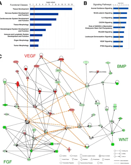
For the microarray analysis of the yolk-sac hemangioblast, we electroporated chick embryos with Hb-eGFP and pCAGGS-RFP reporter constructs, isolated the Hb-eGFP+/RFP+ and Hb-eGFP-/RFP+ cell populations and compared their gene expression profiles [19]. We analyzed the microarray dataset using Ingenuity Pathway Analysis (IPA; Ingenuity Systems,http://www.ingenuity.com) in order to identify the functional classes and signaling pathways that were most significantly represented in the hemangioblast transcriptome (Figure2).
The functional pathway analysis identified 65 classes of biological functions that are significantly enriched in the dataset from hemangioblasts, eight of which are displayed in Figure2A. As expected, hemangioblast genes were assigned to functional classes related to embryonic development, such as Tissue Development and Organ Morphology. In addition, the high representation of the classes Cardiovascular System Development and Function, Hematological System Development and Function and Immune and Lymphatic System Development and Function suggests that hemangioblasts express genes associated with both vascular and hematopoietic lineages, as previously shown [27].
Cells 2018, 7, 9 5 of 12
Cells 2018, 7, x FOR PEER REVIEW 5 of 12
Figure 2. Ingenuity pathway analysis of genes differentially expressed in yolk sac hemangioblasts. (A) Top classes of biological functions and (B) canonical signaling pathways most significantly represented in the Hb-eGFP+ microarray dataset (Ingenuity Pathways Analysis (IPA) library; Ingenuity Systems, www.ingenuity.com). Genes that met the fold change cutoff of 1.2 were considered for the analysis. Bars indicate the minus log of the p-value of each functional class/canonical pathway. The threshold line (orange) corresponds to a p-value of 0.05. The yellow line in (B) represents the ratio between the number of genes from the dataset in a given pathway that meet the cutoff criteria and the total number of genes of that pathway; (C) Network diagram representing the molecular relationships between genes differentially expressed in hemangioblasts. This graphical representation generated by IPA includes gene products of four functional classes: Cellular Development, Cardiovascular System Development and Function, Organismal Development, Organ Development and Cell Signaling. Gene products are represented as nodes (shapes) and the biological
Figure 2.Ingenuity pathway analysis of genes differentially expressed in yolk sac hemangioblasts. (A) Top classes of biological functions and (B) canonical signaling pathways most significantly represented in the Hb-eGFP+ microarray dataset (Ingenuity Pathways Analysis (IPA) library; Ingenuity Systems,www.ingenuity.com). Genes that met the fold change cutoff of 1.2 were considered for the analysis. Bars indicate the minus log of the p-value of each functional class/canonical pathway. The threshold line (orange) corresponds to a p-value of 0.05. The yellow line in (B) represents the ratio between the number of genes from the dataset in a given pathway that meet the cutoff criteria and the total number of genes of that pathway; (C) Network diagram representing the molecular relationships between genes differentially expressed in hemangioblasts. This graphical representation generated by IPA includes gene products of four functional classes: Cellular Development, Cardiovascular System Development and Function, Organismal Development, Organ Development and Cell Signaling. Gene products are represented as nodes (shapes) and the biological relationship between two nodes is represented as an edge (line). Orange lines represent interactions between gene products from different canonical pathways. All edges are supported by at least one reference from the literature, from a textbook, or from canonical information stored in the Ingenuity Pathways Knowledge Base. The intensity of the node color indicates the degree of upregulation (red) or downregulation (green). Nodes are displayed using various shapes that represent the functional class of the gene product, while edges are displayed with various labels that describe the nature of the biological relationship between the nodes (see legend in the figure).
Cells 2018, 7, 9 6 of 12
The canonical pathway analysis identified 78 pathways that are significantly enriched in the dataset from hemangioblasts, nine of which are displayed in Figure2B. Two of these pathways are the VEGF signaling pathway, which play a well-established role in vasculogenesis [28], and the CXCR4 signaling pathway, which regulates HSPC homing and engraftment in the bone marrow [29] and may be involved in the migration of yolk sac hemangioblast-derived angioblasts into intraembryonic regions [7,11]. Alternatively, CXCR4 signaling may modulate the hemogenic potential of the yolk sac hemangioblast, as recently shown in embryonic stem cell cultures [30]. We also identified several hemangioblast genes that are involved in axon guidance. Indeed, the vascular and neural networks are known to have several common morphogenetic signals [31]. Interestingly, another pathway over-represented in hemangioblasts is the leukocyte extravasation signaling pathway, which includes molecules responsible for the interactions between blood and endothelial cells [32]. These molecules are likely to play a similar role in the blood-endothelial interactions that take place during the differentiation of hemangioblasts in the blood islands.
We then designed and analyzed the network of molecular interactions of the major signaling pathways differentially expressed in hemangioblasts (Figure2C). As expected, many upregulated genes belong to the VEGF signaling pathway, such as FLT1 (VEGFR1) and FLT4 (VEGFR3), or interact with it, such as CDH5 (VE-cadherin), an endothelium adhesion molecule that is expressed in hemogenic endothelial cells prior to their differentiation [33], and LMO2, a transcription factor essential for hemangioblast development and hematopoiesis [12,13,34]. On the other hand, several downregulated genes belong to or interact with the BMP, FGF and WNT signaling pathways. These three pathways include genes that are highly expressed in cell types other than the hemangioblast, such as the paraxial mesoderm (e.g., FST—BMP signaling pathway; [35]), primitive streak (e.g., FGF19—FGF signaling pathway; [36]) and axial mesendoderm (e.g., DKK1—NT signaling pathway; [37]).
3.2. Identification of Novel Hemangioblast Genes
In addition to genes that are known to play a role in hemangioblast development, such as LMO2 [12,13] and TAL1/SCL [14], our differential screening of hemangioblast transcripts led to the identification of several genes unknown to have a function in hematopoiesis or vasculogenesis, as well as some previously uncharacterized genes (Table1). These include SLC15A1 (+5.1) and SCL32A1 (+4.19), RhoGap6 (+3.07), CTSG (+3.7), MRC1 (+3.2), LHX8 (+2.77), CITED4 (+2.23), PITX1 (+2.2), and the novel gene DIA1R (+4.2).
SLC15A1 and SLC32A1 are members of the solute carrier family. The transmembrane transporter SLC15A1 is involved in amino acid uptake in the intestinal epithelium [38], whereas the vesicular transporter SLC32A1 acts as a carrier of inhibitory amino acid neurotransmitters in the central nervous system [39]. Their expression in hemangioblasts may indicate that these cells have a particular amino acid requirement. The cytoskeletal protein RhoGap6 was shown to promote the formation of filopodia-like processes in mammalian cell cultures [40], and it may have a similar role in hemangioblasts as they actively migrate in the extraembryonic region [19].
The serine protease CTSG (Cathepsin G) is expressed at the promyelocytic stage of myeloid development [41]. In addition, CTSG participates in tissue remodeling at sites of inflammation [42] and in the degradation of endothelial VE-cadherin during neutrophils transmigration [43]. In hemangioblasts, CTSG may play an active role in extracellular matrix remodeling during blood island formation and endothelial-to-hematopoietic transition. MRC1 is a transmembrane mannose receptor that mediates the phagocytosis of microorganisms by antigen-presenting cells [44]. During development, MRC1 transcripts are found in the zebrafish caudal hematopoietic tissue and endothelial cell precursors [45] and in the mouse yolk sac blood islands [46]. Taken together, the presence of both CTSG and MRC1 in yolk sac hemangioblasts indicates that myeloid lineage genes are already expressed in these progenitors.
Cells 2018, 7, 9 7 of 12
Table 1.List of selected genes upregulated in yolk sac hemangioblasts. Genes without a known function in hematopoiesis or vasculogenesis are highlighted in graya.
FCb Gene Symbol Gene Name Gene ID Molecular Function Biological Function Expression in Early Embryos
+5.1 SLC15A1 Solute carrier family 15, member 1 378789 Membrane transporter Oligopeptide transport -+4.2 C1HXorf36
(DIA1R)
Chromosome 1 open reading frame, human
CXorf36 (Deleted in Autism 1 Related) 418555 - - (this study)
+4.19 SLC32A1 Solute carrier family 32, member 1 419167 Vesicular transporter GABA vesicular transporter -+4.15 SOX7 SRY (sex determining region Y)-box 7 771337 Transcription factor Vasculogenesis and
hematopoiesis Angioblasts
+3.73 LMO2 LIM domain only 2 374129 Transcription factor Hematopoiesis Hematopoietic progenitors
+3.7 CTSG Cathepsin G 426049 Serine protease Tissue remodeling Myeloid progenitors
+3.62 TAL1 (SCL) T-cell acute lymphocytic leukemia 1 (stem
cell leukemia) 396298 Transcription factor Hematopoiesis Hematopoietic progenitors +3.51 RUNX1 Runt-related transcription factor 1 396152 Transcription factor Hematopoiesis Blood islands
+3.44 EGR1 Early growth response 1 373931 Transcription factor HSPC proliferation Vasculogenic mesoderm
+3.2 MRC1 Mannose receptor C-type 1 420516 Membrane receptor Endocytosis Blood islands
+3.17 KLHL6 Kelch-like 6 424762 Transcription factor Lymphocyte differentiation
-+3.3 SPI1 (PU.1) Spleen focus forming virus (SFFV) proviral
integration oncogene spi1 395879 Transcription factor Hematopoiesis Hematopoietic progenitors +3.07 RhoGap6 Similar to Rho-GTPase-activating protein 6
(LOC422284 locus) 422284 Cytoskeleton regulator Actin remodeling
-+2.77 LHX8 LIM homeobox 8 424721 Transcription factor Neurogenesis Blood islands
+2.71 FLT1 (VEGFR1) Fms-related tyrosine kinase 1 (vascular
endothelial growth factor receptor 1) 374100 Receptor tyrosine kinase Vasculogenesis/Angiogenesis
Hemangioblasts and endothelial cells
+2.63 SOX18 SRY (sex determining region Y)-box 18 374200 Transcription factor Vasculogenesis Blood islands +2.49 CDH5 Cadherin 5, type 2, VE-cadherin (vascular
epithelium) 374068 Cell adhesion molecule Vasculogenesis/Angiogenesis Endothelial cells +2.39 CD34 Hematopoietic progenitor cell antigen CD34 419856 Cell surface antigen - Hematopoietic progenitors +2.29 FLT4 (VEGFR3) Fms-related tyrosine kinase 4 (vascular
endothelial growth factor receptor 3) 395742 Receptor tyrosine kinase Angiogenesis Blood islands and endothelial cells +2.23 CITED4 Cbp/p300-interacting transactivator, with
Glu/Asp-rich carboxy-terminal domain, 4 395465 Transcription regulator in vitro cardiogenesis Blood islands +2.2 PITX1 Paired-like homeodomain 1 374201 Transcription factor Pituitary and hindlimb
development Posterior extraembryonic mesoderm +2.09 Fli1 Friend leukemia virus integration 1 gene 419723 Transcription factor Vasculogenesis and
hematopoiesis
Endothelial and erythroid progenitors +1.96 HHEX Hematopoietically expressed homeobox 396182 Transcription factor Vasculogenesis and
hematopoiesis Blood islands
a Gene function and expression patterns were obtained from the literature and from ZFIN (http://zfin.org), GEISHA (http://geisha.arizona.edu/geisha) and EMAGE
Cells 2018, 7, 9 8 of 12
LHX8 is involved in the differentiation of cholinergic neurons in the mouse telencephalon [47], CITED4 regulates the proliferation of embryonic stem cell-derived cardiac progenitor cells [48], and PITX1 plays a role in pituitary and hindlimb development [49]. These transcription factors may also regulate the differentiation of HSPCs, as do their respective family members LHX2 [50], CITED2 [51] and PITX2 [52]. In the future, the expression of these newly identified genes in yolk sac hemangioblasts should be validated in the early embryo. In addition, further investigation will be required to resolve their potential roles in the hemangioblast.
3.3. Expression Pattern of DIA1R in the Chick Embryo
The second most highly expressed gene in the hemangioblast transcriptome was the chick ortholog of human cXorf36 or DIA1R (deleted in autism 1 related; +4.2 fold change; Table1), a gene implicated in autism spectrum disorders and X-linked mental retardation [53,54]. The chick DIA1R gene (cDIA1R or C1HXorf36) encodes a protein of 430 amino acids that is 65% identical and 87% similar to the human protein [54]. DIA1R genes are found exclusively in vertebrates and their function is largely unknown. Based on sequence analysis, DIA1R proteins were predicted to contain a signal peptide and a highly conserved PIP49_C protein-kinase domain, characteristic of the FAM69 family of kinase-like proteins [53,55]. These features suggest that DIA1R proteins may regulate molecular traffic or interfere with the function of secreted factors [53].
The analysis of cDIA1R expression during chick development revealed that this gene is expressed in yolk sac hemangioblasts, blood islands and endothelial cells of the dorsal aorta, endocardium and head vasculature (Figure3), which are regions known to have hemogenic capability [56]. In particular, cDIA1R expression appears to be higher in cells that are morphologically similar to hemogenic endothelial cells (Figure3D’,E’,F”). In the brain neuroepithelium, cDIA1R is expressed in the vascular endothelium and in isolated cells that resemble microglial cells (Figure3H; [57]). Consistently with our observations, transcripts of DIA1R orthologs have been detected in hematopoietic progenitors and endothelial cells in zebrafish embryos ([17]; cc058 gene), in endothelial and microglial cells from embryonic and adult mouse brains ([58]; 4930578C19Rik gene) and in human endothelial cells [59]. Taken together, these findings suggest that DIA1R may be a good marker and potential regulator of the intraembryonic hemogenic lineages that derive from yolk sac hemangioblasts.
Cells 2018, 7, 9 9 of 12
Cells 2018, 7, x FOR PEER REVIEW 8 of 12
3.3. Expression Pattern of DIA1R in the Chick Embryo
The second most highly expressed gene in the hemangioblast transcriptome was the chick
ortholog of human cXorf36 or DIA1R (deleted in autism 1 related; +4.2 fold change; Table 1), a gene
implicated in autism spectrum disorders and X-linked mental retardation [53,54]. The chick DIA1R
gene (cDIA1R or C1HXorf36) encodes a protein of 430 amino acids that is 65% identical and 87%
similar to the human protein [54]. DIA1R genes are found exclusively in vertebrates and their function
is largely unknown. Based on sequence analysis, DIA1R proteins were predicted to contain a signal
peptide and a highly conserved PIP49_C protein-kinase domain, characteristic of the FAM69 family
of kinase-like proteins [53,55]. These features suggest that DIA1R proteins may regulate molecular
traffic or interfere with the function of secreted factors [53].
The analysis of cDIA1R expression during chick development revealed that this gene is
expressed in yolk sac hemangioblasts, blood islands and endothelial cells of the dorsal aorta,
endocardium and head vasculature (Figure 3), which are regions known to have hemogenic
capability [56]. In particular, cDIA1R expression appears to be higher in cells that are morphologically
similar to hemogenic endothelial cells (Figure 3D’,E’,F”). In the brain neuroepithelium, cDIA1R is
expressed in the vascular endothelium and in isolated cells that resemble microglial cells (Figure 3H;
[57]). Consistently with our observations, transcripts of DIA1R orthologs have been detected in
hematopoietic progenitors and endothelial cells in zebrafish embryos ([17]; cc058 gene), in endothelial
and microglial cells from embryonic and adult mouse brains ([58]; 4930578C19Rik gene) and in
human endothelial cells [59]. Taken together, these findings suggest that DIA1R may be a good
marker and potential regulator of the intraembryonic hemogenic lineages that derive from yolk sac
hemangioblasts.
Figure 3. cDIA1R expression in the chick embryo. cDIA1R in situ hybridization was performed on
whole-mount embryos at HH5 (A), 8 (B), 11 (C), 13 (D), 17 (E), 18 (F,G) and on a E10 brain cryosection (H). (B’,D’,E’,F’,F”) Sections of correspondent whole-mount embryos. (C1–4) Regions of embryo in (C) at high magnification. cDIA1R expression starts to be detected at HH5 (A) in the extraembryonic mesoderm that will form the yolk sac blood islands at later stages (B,B’,C4). In HH11 embryos (C), cDIA1R is also expressed in the endocardium (arrow in C1), in the developing head vasculature (arrowheads in C1 and C2), in cells associated with the dorsal mid- and hindbrain (asterisks in C2) and in the dorsal aorta region (DA; C3). At later stages (D–H), cDIA1R expression is detected in most blood vessels of the embryo, such as the dorsal aorta (DA in D,D’), intersomitic vessels (E), head vasculature (E’,F,F’,F”) and allantois (G). Higher intensity is found in particular blood vessel cells that resemble hemogenic endothelial cells (D’,E’,F”; arrowheads). In the brain neuroepithelium (H), cDIA1R is detected both in the neurovasculature (arrows) and in isolated cells that may be microglial cells (arrowheads). Scale bars: 50 μm in B’,E’,F”; 100 μm in D’,F’.
Over the past decade, increasing evidence supports a key role for microglia in the pathogenesis
of neurodevelopmental disorders such as autism [60–62]. In addition, reduced rates of angiogenesis
Figure 3. cDIA1Rexpression in the chick embryo. cDIA1R in situ hybridization was performed on whole-mount embryos at HH5 (A), 8 (B), 11 (C), 13 (D), 17 (E), 18 (F,G) and on a E10 brain cryosection (H). (B’,D’,E’,F’,F”) Sections of correspondent whole-mount embryos. (C1–C4) Regions of embryo in (C) at high magnification. cDIA1R expression starts to be detected at HH5 (A) in the extraembryonic mesoderm that will form the yolk sac blood islands at later stages (B,B’,C4). In HH11 embryos (C), cDIA1R is also expressed in the endocardium (arrow in C1), in the developing head vasculature (arrowheads in C1 and C2), in cells associated with the dorsal mid- and hindbrain (asterisks in C2) and in the dorsal aorta region (DA; C3). At later stages (D–H), cDIA1R expression is detected in most blood vessels of the embryo, such as the dorsal aorta (DA in D,D’), intersomitic vessels (E), head vasculature (E’,F,F’,F”) and allantois (G). Higher intensity is found in particular blood vessel cells that resemble hemogenic endothelial cells (D’,E’,F”; arrowheads). In the brain neuroepithelium (H), cDIA1R is detected both in the neurovasculature (arrows) and in isolated cells that may be microglial cells (arrowheads). Scale bars: 50 µm in B’,E’,F”; 100 µm in D’,F’.
Over the past decade, increasing evidence supports a key role for microglia in the pathogenesis of neurodevelopmental disorders such as autism [60–62]. In addition, reduced rates of angiogenesis and perfusion abnormalities have been found in autistic brains [63]. Therefore, DIA1R may play a role in microglia and/or in neurovascular development, which can underlie its implication in autism and mental retardation disorders. We are currently investigating DIA1R potential function using overexpression and loss-of-function approaches in different vertebrate models.
Acknowledgments:The authors would like to thank Rui Gardner, Jörg Becker, Isabel Marques, Gabriel Martins and Telmo Pereira for technical assistance, Domingos Henrique for the pCAGGS-RFP construct, Anne Eichmann for the avian VEGFR2 antibody, Lara Carvalho for critically revising the manuscript and Fundação para a Ciência e a Tecnologia for financial support.
Author Contributions:A.T.T. conceived and designed the experiments; V.T. and A.T.T. performed the experiments; J.S.M. and A.T.T. analyzed the data; A.J. contributed reagents and materials; J.S.M. and A.T.T. wrote the paper.
Conflicts of Interest:The authors declare no conflict of interest. The founding sponsors had no role in the design of the study; in the collection, analyses, or interpretation of data; in the writing of the manuscript, and in the decision to publish the results.
References
1. Sabin, F.R. Studies on the origin of blood vessels and of red corpuscles as seen in the living blastoderm of the chick during the second day of incubation. Contrib. Embryol. 1920, 9, 213–262.
2. Murray, P.D.F. The development in vitro of the blood of the early chick embryo. Proc. R. Soc. Lond. B 1932, 111, 497–521. [CrossRef]
3. Gritz, E.; Hirschi, K.K. Specification and function of hemogenic endothelium during embryogenesis. Cell. Mol. Life Sci. 2016, 73, 1547–1567. [CrossRef] [PubMed]
Cells 2018, 7, 9 10 of 12
4. Lacaud, G.; Kouskoff, V. Hemangioblast, hemogenic endothelium, and primitive versus definitive hematopoiesis. Exp. Hematol. 2017, 49, 19–24. [CrossRef] [PubMed]
5. Palis, J.; Robertson, S.; Kennedy, M.; Wall, C.; Keller, G. Development of erythroid and myeloid progenitors in the yolk sac and embryo proper of the mouse. Development 1999, 126, 5073–5084. [PubMed]
6. Tober, J.; Koniski, A.; McGrath, K.E.; Vemishetti, R.; Emerson, R.; de Mesy-Bentley, K.K.; Waugh, R.; Palis, J. The megakaryocyte lineage originates from hemangioblast precursors and is an integral component both of primitive and of definitive hematopoiesis. Blood 2007, 109, 1433–1441. [CrossRef] [PubMed]
7. Ginhoux, F.; Guilliams, M. Tissue-Resident Macrophage Ontogeny and Homeostasis. Immunity 2016, 44, 439–449. [CrossRef] [PubMed]
8. Tay, T.L.; Hagemeyer, N.; Prinz, M. The force awakens: Insights into the origin and formation of microglia. Curr. Opin. Neurobiol. 2016, 39, 30–37. [CrossRef] [PubMed]
9. De Bruijn, M.F.; Speck, N.A.; Peeters, M.C.; Dzierzak, E. Definitive hematopoietic stem cells first develop within the major arterial regions of the mouse embryo. EMBO J. 2000, 19, 2465–2474. [CrossRef] [PubMed] 10. Samokhvalov, I.M.; Samokhvalova, N.I.; Nishikawa, S. Cell tracing shows the contribution of the yolk sac to
adult haematopoiesis. Nature 2007, 446, 1056–1061. [CrossRef] [PubMed]
11. Tanaka, Y.; Sanchez, V.; Takata, N.; Yokomizo, T.; Yamanaka, Y.; Kataoka, H.; Hoppe, P.S.; Schroeder, T.; Nishikawa, S. Circulation-independent differentiation pathway from extraembryonic mesoderm toward hematopoietic stem cells via hemogenic angioblasts. Cell Rep. 2014, 8, 31–39. [CrossRef] [PubMed]
12. Patterson, L.J.; Gering, M.; Eckfeldt, C.E.; Green, A.R.; Verfaillie, C.M.; Ekker, S.C.; Patient, R. The transcription factors Scl and Lmo2 act together during development of the hemangioblast in zebrafish. Blood 2007, 109, 2389–2398. [CrossRef] [PubMed]
13. Stanulovic, V.S.; Cauchy, P.; Assi, S.A.; Hoogenkamp, M. LMO2 is required for TAL1 DNA binding activity and initiation of definitive haematopoiesis at the haemangioblast stage. Nucleic Acids Res. 2017, 45, 9874–9888. [CrossRef] [PubMed]
14. Gering, M.; Rodaway, A.R.; Göttgens, B.; Patient, R.K.; Green, A.R. The SCL gene specifies haemangioblast development from early mesoderm. EMBO J. 1998, 17, 4029–4045. [CrossRef] [PubMed]
15. Lacaud, G.; Gore, L.; Kennedy, M.; Kouskoff, V.; Kingsley, P.; Hogan, C.; Carlsson, L.; Speck, N.; Palis, J.; Keller, G. Runx1 is essential for hematopoietic commitment at the hemangioblast stage of development in vitro. Blood 2002, 100, 458–466. [CrossRef] [PubMed]
16. Lilly, A.J.; Costa, G.; Largeot, A.; Fadlullah, M.Z.; Lie-A-Ling, M.; Lacaud, G.; Kouskoff, V. Interplay between SOX7 and RUNX1 regulates hemogenic endothelial fate in the yolk sac. Development 2016, 143, 4341–4351. [CrossRef] [PubMed]
17. Cannon, J.E.; Place, E.S.; Eve, A.M.; Bradshaw, C.R.; Sesay, A.; Morrell, N.W.; Smith, J.C. Global analysis of the haematopoietic and endothelial transcriptome during zebrafish development. Mech. Dev. 2013, 130, 122–131. [CrossRef] [PubMed]
18. Nasrallah, R.; Fast, E.M.; Solaimani, P.; Knezevic, K.; Eliades, A.; Patel, R.; Thambyrajah, R.; Unnikrishnan, A.; Thoms, J.; Beck, D.; et al. Identification of novel regulators of developmental hematopoiesis using Endoglin regulatory elements as molecular probes. Blood 2016, 128, 1928–1939. [CrossRef] [PubMed]
19. Teixeira, V.; Arede, N.; Gardner, R.; Rodríguez-León, J.; Tavares, A.T. Targeting the hemangioblast with a novel cell type-specific enhancer. BMC Dev. Biol. 2011, 11, 76. [CrossRef] [PubMed]
20. Gordon-Keylock, S.; Medvinsky, A. Endothelio-hematopoietic relationship: Getting closer to the beginnings. BMC Biol. 2011, 9, 88. [CrossRef] [PubMed]
21. Hamburger, V.; Hamilton, H.L. A series of normal stages in the development of the chick embryo. J. Morphol.
1951, 88, 49–92. [CrossRef] [PubMed]
22. New, D.A.T. A New Technique for the Cultivation of the Chick Embryo in vitro. Development 1955, 3, 326–331. 23. Eichmann, A.; Corbel, C.; Nataf, V.; Vaigot, P.; Bréant, C.; Le Douarin, N.M. Ligand-dependent development of the endothelial and hemopoietic lineages from embryonic mesodermal cells expressing vascular endothelial growth factor receptor 2. Proc. Natl. Acad. Sci. USA 1997, 94, 5141–5146. [CrossRef] [PubMed] 24. Wilkinson, D.G. Whole mount in situ hybridisation of vertebrate embryos. In In Situ Hybridization;
Wilkinson, D.G., Ed.; Oxford University Press: Oxford, UK, 1992; pp. 75–83.
25. Braissant, O.; Wahli, W. A simplified in situ hybridization protocol using non-radioactively labelled probes to detect abundant and rare mRNAs on tissue sections. Biochemica 1998, 1, 10–16.
Cells 2018, 7, 9 11 of 12
26. Tavares, A.T.; Andrade, S.; Silva, A.C.; Belo, J.A. Cerberus is a feedback inhibitor of Nodal asymmetric signaling in the chick embryo. Development 2007, 134, 2051–2060. [CrossRef] [PubMed]
27. Minko, K.; Bollerot, K.; Drevon, C.; Hallais, M.F.; Jaffredo, T. From mesoderm to blood islands: Patterns of key molecules during yolk sac erythropoiesis. Gene Expr. Patterns 2003, 3, 261–272. [CrossRef]
28. Olsson, A.K.; Dimberg, A.; Kreuger, J.; Claesson-Welsh, L. VEGF receptor signaling—In control of vascular function. Nat. Rev. Mol. Cell Biol. 2006, 7, 359–371. [CrossRef] [PubMed]
29. Sharma, M.; Afrin, F.; Satija, N.; Tripathi, R.P.; Gangenahalli, G.U. Stromal-derived factor-1/CXCR4 signaling: Indispensable role in homing and engraftment of hematopoietic stem cells in bone marrow. Stem Cells Dev.
2011, 20, 933–946. [CrossRef] [PubMed]
30. Ahmed, T.; Tsuji-Tamura, K.; Ogawa, M. CXCR4 Signaling Negatively Modulates the Bipotential State of Hemogenic Endothelial Cells Derived from Embryonic Stem Cells by Attenuating the Endothelial Potential. Stem Cells 2016, 34, 2814–2824. [CrossRef] [PubMed]
31. Carmeliet, P.; Tessier-Lavigne, M. Common mechanisms of nerve and blood vessel wiring. Nature 2005, 436, 193–200. [CrossRef] [PubMed]
32. Barreiro, O.; Sánchez-Madrid, F. Molecular Basis of Leukocyte–Endothelium Interactions during the Inflammatory Response. Rev. Española Cardiol. 2009, 62, 552–562. [CrossRef]
33. Costa, G.; Mazan, A.; Gandillet, A.; Pearson, S.; Lacaud, G.; Kouskoff, V. SOX7 regulates the expression of VE-cadherin in the haemogenic endothelium at the onset of haematopoietic development. Development 2012, 139, 1587–1598. [CrossRef] [PubMed]
34. Yamada, Y.; Warren, A.J.; Dobson, C.; Forster, A.; Pannell, R.; Rabbitts, T.H. The T cell leukemia LIM protein Lmo2 is necessary for adult mouse hematopoiesis. Proc. Natl. Acad. Sci. USA 1998, 95, 3890–3895. [CrossRef]
[PubMed]
35. Connolly, D.J.; Patel, K.; Seleiro, E.A.; Wilkinson, D.G.; Cooke, J. Cloning, sequencing, and expressional analysis of the chick homologue of follistatin. Dev. Genet. 1995, 17, 65–77. [CrossRef] [PubMed]
36. Kurose, H.; Bito, T.; Adachi, T.; Shimizu, M.; Noji, S.; Ohuchi, H. Expression of Fibroblast growth factor 19 (Fgf19) during chicken embryogenesis and eye development, compared with Fgf15 expression in the mouse. Gene Expr. Patterns 2004, 4, 687–693. [CrossRef] [PubMed]
37. Chapman, S.C.; Schubert, F.R.; Schoenwolf, G.C.; Lumsden, A. Analysis of spatial and temporal gene expression patterns in blastula and gastrula stage chick embryos. Dev. Biol. 2002, 245, 187–199. [CrossRef]
[PubMed]
38. Smith, D.E.; Clémençon, B.; Hediger, M.A. Proton-coupled oligopeptide transporter family SLC15: Physiological, pharmacological and pathological implications. Mol. Asp. Med. 2013, 34, 323–336. [CrossRef]
[PubMed]
39. Schiöth, H.B.; Roshanbin, S.; Hägglund, M.G.; Fredriksson, R. Evolutionary origin of amino acid transporter families SLC32, SLC36 and SLC38 and physiological, pathological and therapeutic aspects. Mol. Asp. Med.
2013, 34, 571–585. [CrossRef] [PubMed]
40. Prakash, S.K.; Paylor, R.; Jenna, S.; Lamarche-Vane, N.; Armstrong, D.L.; Xu, B.; Mancini, M.A.; Zoghbi, H.Y. Functional analysis of ARHGAP6, a novel GTPase-activating protein for RhoA. Hum. Mol. Genet. 2000, 9, 477–488. [CrossRef] [PubMed]
41. Hanson, R.D.; Connolly, N.L.; Burnett, D.; Campbell, E.J.; Senior, R.M.; Ley, T.J. Developmental regulation of the human cathepsin G gene in myelomonocytic cells. J. Biol. Chem. 1990, 265, 1524–1530. [PubMed] 42. Heutinck, K.M.; ten Berge, I.J.M.; Hack, C.E.; Hamann, J.; Rowshani, A.T. Serine proteases of the human
immune system in health and disease. Mol. Immunol. 2010, 47, 1943–1955. [CrossRef] [PubMed]
43. Hermant, B.; Bibert, S.; Concord, E.; Dublet, B.; Weidenhaupt, M.; Vernet, T.; Gulino-Debrac, D. Identification of Proteases Involved in the Proteolysis of Vascular Endothelium Cadherin during Neutrophil Transmigration. J. Biol. Chem. 2003, 278, 14002–14012. [CrossRef] [PubMed]
44. East, L.; Isacke, C.M. The mannose receptor family. Biochim. Biophys. Acta 2002, 1572, 364–386. [CrossRef] 45. Wong, K.S.; Proulx, K.; Rost, M.S.; Sumanas, S. Identification of vasculature-specific genes by microarray
analysis of etsrp/etv2 overexpressing zebrafish embryos. Dev. Dyn. 2009, 238, 1836–1850. [CrossRef]
[PubMed]
46. Takahashi, K.; Donovan, M.J.; Rogers, R.A.; Ezekowitz, R.A. Distribution of murine mannose receptor expression from early embryogenesis through to adulthood. Cell Tissue Res. 1998, 292, 311–323. [CrossRef]
Cells 2018, 7, 9 12 of 12
47. Zhao, Y.; Marin, O.; Hermesz, E.; Powell, A.; Flames, N.; Palkovits, M.; Rubenstein, J.L.R.; Westphal, H. The LIM-homeobox gene Lhx8 is required for the development of many cholinergic neurons in the mouse forebrain. Proc. Natl. Acad. Sci. USA 2003, 100, 9005–9010. [CrossRef] [PubMed]
48. Miake, J.; Notsu, T.; Higaki, K.; Hidaka, K.; Morisaki, T.; Yamamoto, K.; Hisatome, I. Cited4 is related to cardiogenic induction and maintenance of proliferation capacity of embryonic stem cell-derived cardiomyocytes during in vitro cardiogenesis. PLoS ONE 2017, 12, e0183225. [CrossRef] [PubMed]
49. Szeto, D.P.; Rodriguez-Esteban, C.; Ryan, A.K.; O’Connell, S.M.; Liu, F.; Kioussi, C.; Gleiberman, A.S.; Izpisúa-Belmonte, J.C.; Rosenfeld, M.G. Role of the Bicoid-related homeodomain factor Pitx1 in specifying hindlimb morphogenesis and pituitary development. Genes Dev. 1999, 13, 484–494. [CrossRef] [PubMed] 50. Pinto do O, P.; Richter, K.; Carlsson, L. Hematopoietic progenitor/stem cells immortalized by Lhx2 generate
functional hematopoietic cells in vivo. Blood 2002, 99, 3939–3946. [CrossRef] [PubMed]
51. Du, J.; Yang, Y.-C. Cited2 in hematopoietic stem cell function. Curr. Opin. Hematol. 2013, 20, 301–307. [CrossRef] [PubMed]
52. Kieusseian, A.; Chagraoui, J.; Kerdudo, C.; Mangeot, P.E.; Gage, P.J.; Navarro, N.; Izac, B.; Uzan, G.; Forget, B.G.; Dubart-Kupperschmitt, A. Expression of Pitx2 in stromal cells is required for normal hematopoiesis. Blood 2006, 107, 492–500. [CrossRef] [PubMed]
53. Aziz, A.; Harrop, S.P.; Bishop, N.E. DIA1R is an X-linked gene related to Deleted in Autism-1. PLoS ONE
2011, 6, e14534. [CrossRef] [PubMed]
54. Aziz, A.; Harrop, S.P.; Bishop, N.E. Characterization of the deleted in autism 1 protein family: Implications for studying cognitive disorders. PLoS ONE 2011, 6, e14547. [CrossRef] [PubMed]
55. Dudkiewicz, M.; Lenart, A.; Pawłowski, K. A novel predicted calcium-regulated kinase family implicated in neurological disorders. PLoS ONE 2013, 8, e66427. [CrossRef] [PubMed]
56. Swiers, G.; Rode, C.; Azzoni, E.; de Bruijn, M.F. A short history of hemogenic endothelium. Blood Cells Mol. Dis. 2013, 51, 206–212. [CrossRef] [PubMed]
57. Ignacio, A.R.; Muller, Y.M.; Carvalho, M.S.; Nazari, E.M. Distribution of microglial cells in the cerebral hemispheres of embryonic and neonatal chicks. Braz. J. Med. Biol. Res. 2005, 38, 1615–1621. [CrossRef]
[PubMed]
58. Gay, L.; Miller, M.R.; Ventura, P.B.; Devasthali, V.; Vue, Z.; Thompson, H.L.; Temple, S.; Zong, H.; Cleary, M.D.; Stankunas, K.; et al. Mouse TU tagging: A chemical/genetic intersectional method for purifying cell type-specific nascent RNA. Genes Dev. 2013, 27, 98–115. [CrossRef] [PubMed]
59. Bhasin, M.; Yuan, L.; Keskin, D.B.; Otu, H.H.; Libermann, T.A.; Oettgen, P. Bioinformatic identification and characterization of human endothelial cell-restricted genes. BMC Genom. 2010, 11, 342. [CrossRef] [PubMed] 60. Prinz, M.; Priller, J. Microglia and brain macrophages in the molecular age: From origin to neuropsychiatric
disease. Nat. Rev. Neurosci. 2014, 15, 300–312. [CrossRef] [PubMed]
61. Edmonson, C.A.; Ziats, M.N.; Rennert, O.M. A Non-inflammatory Role for Microglia in Autism Spectrum Disorders. Front. Neurol. 2016, 7, 9. [CrossRef] [PubMed]
62. Mosser, C.A.; Baptista, S.; Arnoux, I.; Audinat, E. Microglia in CNS development: Shaping the brain for the future. Prog. Neurobiol. 2017, 149–150, 1–20. [CrossRef] [PubMed]
63. Emanuele, E.; Orsi, P.; Barale, F.; di Nemi, S.U.; Bertona, M.; Politi, P. Serum levels of vascular endothelial growth factor and its receptors in patients with severe autism. Clin. Biochem. 2010, 43, 317–319. [CrossRef]
[PubMed]
© 2018 by the authors. Licensee MDPI, Basel, Switzerland. This article is an open access article distributed under the terms and conditions of the Creative Commons Attribution (CC BY) license (http://creativecommons.org/licenses/by/4.0/).