C-ERB-2, P53, Ki67 Proteins and Receptors of Estrogen
and Progesterone on the Prognosis of Epithelial
Ovarian Cancer
Tomé A
1, Leal I
2, Palmeiras C
3, Matos E
4, Amado J
5*, Abreu M
6, Lopes C
71Centre of Gynecological Oncology and Breast, Hospital Santo Antonio (CHP), Porto, Portugal 2Department of Pathology, Hospital Santo Antonio, Porto, Portugal
3Department of Immunology and Pathology, Portuguese Institute of Oncology (IPO), Porto, Portugal 4Department of Community Medicine, ICBAS, Porto University, Porto, Portugal
5Universidade Católica Portuguesa, Centre of Interdisciplinary Health Investigation - Health Sciences Institute
(CIIS-ICS), Portugal
6Department of Oncologic Medicine, Portuguese Institute of Oncology (IPO), Porto, Portugal 7Department of Molecular Biology and Immunology, ICBAS, Porto University, Porto, Portugal
*Correspondence: João Amado, Universidade Católica Portuguesa, Centre of Interdisciplinary Health Investigation
- Health Sciences Institute (CIIS-ICS), Portugal, E-mail: pbatista@porto.ucp.pt Received: February 08, 2019; Accepted: March 18, 2019; Published: March 23, 2019
Abstract
Ovarian cancer is the seventh most common cancer diagnosed in women worldwide. To date, many studies in epithelial ovarian cancer (EOC) have reported on the association HER-2/neu, p53 proteins and steroid hormones and their respective receptors with prognosis and/or the carcinogenesis process, but no definitive conclusion has been reached.
Objectives: To assess the proteins c-erbB-2, p53, Ki67 and receptors of estrogen (ER) and progesterone (PR) of EOC, with regard to clinical stage findings and its effect on survival.
Methods: 125 patients with a diagnosis of EOC treated by primary surgery and chemotherapy have participated. A surgical stage was noted and analyzed the correlation with c-erbB-2, p53, Ki67, ER and PR. Immunohistochemical analysis, using the anti-c-erbB-2, p53, Ki67 monoclonal antibodies, the antibody cod PR clone PgR and code ER-6-F11 Anti human estrogen. The c-erbB-2 study was complemented by genetic amplification and was reported univariate and multivariate analysis.
Results: Age 55.7 ± 16; 50.2% with residual disease (< 2 cm); initial (54.6%) and advanced (45.4%) stage. Univariate analysis showed positive staining for c-erbB-2, p-53, Ki67, PR and ER. The patients with negative receptors had a significantly shortened survival time (p = 0.01) than patients with positive receptors. Multivariable analysis revealed only clinical FIGO stage as an independent prognostic of overall survival (p = 0.002). Other variables like c-erbB-2, p53, Ki67, and ER were not significantly related to survival.
Conclusions: We concluded that patients with negative PR had a significantly shortened survival time than patients with positive receptors. The overexpression of markers c-erbB-2, p53, Ki67, and ER, were not significantly related to survival in EOC. Only the FIGO stage was achieved to be an independent predictor of overall survival. They should be evaluated together with the patient’s clinical status and other prognostic factors.
Keywords: Epithelial ovarian carcinoma, Immunohistochemistry and CISH, Proteins: c-erbB-2, p53, Ki67, Steroid
receptors, Prognosis
Research Article
ISSN: 2637-4617
Introduction
Ovarian cancer is the seventh most common cancer diagnosed in women worldwide [1]. Approximately one-fourth of all gynecologic malignancies are of ovarian origin, and 47% of all gynecologic cancer-related deaths are due to ovarian cancer. Ovarian cancer carries the highest mortality among all gynecological malignancies. The high mortality is due mostly to the fact that the tumor is frequently diagnosed late, in advanced stages (III, or even IV), because the early stages are often asymptomatic, and no effective screening methods are available [2]. The average 5-year survival of patients in all stages of the disease is only 40%, in patients with the advanced disease only 10-20% [3]. Some studies reported a decreasing incidence in women, attributed to the use of an oral contraceptive pill [4,5].
It seems that there has been no significant decrease in incidence or mortality from ovarian cancer since the early 1980s, although the imagiologic exams had permitted diagnosed more and more the disease in early stages. In the absence of preventable etiologic factors or effective tools for screening, the only possible means of improving survival currently lies with the optimal management of patients after initial diagnosis. The prognosis of epithelial ovarian cancer can be correlated with biological (age), social (performance status) and clinical factors (tumor stage, histological grade, histological type, presence or absence of ascites; size and number of residual lesions after primary cytoreduction surgery, and chemotherapy) [6-8]. Identification of new prognostic factors might be useful in directing the therapy and intensifying the follow-up of a selected group of patients. A variety of prognostic factors have been reported, but their independent prognostic significance remains unclear. Immunohistochemistry has been widely used in the biomarkers search.
The oncoprotein c-erbB-2 (HER-2/neu; Neu), encodes a transmembrane glycoprotein that is member of the class I receptor tyrosine kinase family, which includes the epidermal growth factor, HER-2/neu, HER-3 and HER-4 [9]. Proto-oncogene HER-2/neu is located on chromosome 17 and is not activated by a point mutation but through amplification and overexpression of the wild-type gene. Amplification of the HER-2/neu oncogene may be observed in 20-30% of cases in a wide spectrum of neoplastic disorders (e.g., breast, lung carcinomas, others), and HER-2/neu overexpression has been associated with a poor prognosis of patients with cancer arising from other primary sites, but studies of ovarian cancer have
produced conflicting results [10-12]. Patients with breast carcinomas with amplified or overexpressed HER-2/neu can benefit from anthracycline-based regimens as well as Trastuzumab (Herceptin), a recombinant humanized monoclonal antibody against the Her-2/neu protein [13-15]. HER-2/neu is generally assessed as protein overexpression by using immunohistochemistry (IHC), and patients with tumors that either have 2+ or 3+ results with this method become good candidates for treatment with Trastuzumab.
However, studies indicate that HER-2/neu determined as gene amplification provides better prognostic information and is associated with a better response to Trastuzumab. HER-2/neu gene amplification is primarily detected by in situ hybridization and uses fluorescence (FISH) to detect the signals [16]. This method is expensive, it requires a fluorescence microscope, appropriate filters, and a sophisticated camera; so it is not practical as a screening tool. Chromogenic in situ hybridization (CISH) is a recently introduced method, and although it makes use of the in situ hybridization technology of FISH, it also takes advantage of the chromogenic signal detection of IHC that can be detected with the ordinary light microscope and has fewer costs [17,18]. CISH is potentially able to detect HER-2/neu gene amplification and to minimize, if not eliminate, the false positive fraction with the IHC procedure.
To date, many studies in epithelial ovarian cancer have reported on the association between HER-2/ neu expression and outcome; some earlier studies reported that HER-2/neu overexpression was a poor prognostic factor, but later studies reported that HER-2/neu expression had no relationship with prognosis. Thus, no definitive conclusion has been reached as to the relationship between HER-2/neu expression and prognosis [14,19]. Just like of studies in breast also for the ovary, more studies should be conducted for studies in order to enhance the HER2 prognostic value and advantage therapy with monoclonal antibodies (Trastuzumab or other) [9,20].
P53 is a tumor suppressor gene (inhibit cell division and/or promote cell death/apoptosis) located on the short arm of chromosome17 [21]. It suppresses cell growth by controlling entry into the S-phase of the cell cycle. Mutation or deletion of the p53 gene is believed to result in uncontrolled cell proliferation. Most p53 gene mutations result in stabilization of the protein. In contrast to the short half-life of the wild type p53 protein,
increased the stability of the mutant forms allows their detection by immunohistochemical techniques. Mutations of the p53 gene are the most common genetic abnormalities described in human cancers and have been implicated in the pathogenesis of several human tumors [22]. Mutations of the p53 have been found in approximately 40-80% of epithelial ovarian cancer cases. Studies have demonstrated an association between p53 protein overexpression and poor prognosis in patients with several tumor types. In epithelial ovarian carcinoma, the role of p53 protein is contentious, and there are a number of studies with contradictory results [23]. There are several studies that identified the p53 protein as an adverse prognostic factor for survival in ovarian cancer; others studies suggested that alterations in p53 expression in ovarian cancer can to affect the sensibility to chemotherapy [24]. In contrast, there are a number of studies that suggest that p53 expression has no prognostic value in epithelial ovarian cancer [25].
The proliferation activity of the tumor cell can be determined using a variety of methods, but many of these methods have significant technical limitations (DNA flow cytometry, DNA image cytometry, immunohistochemistry and others) [26]. Immunohistochemistry allows evaluating the Ki67 a nuclear non-histone protein expressed in cells in G1, S, G2, and M cell cycle phases, but absenting from quiescent cells in G0. High cellular proliferative activity was associated with poor outcome. On the other hand, other studies did not confirm the relationship between proliferation activity and epithelial ovarian cancer prognosis [27].
Steroid hormones (estrogen and progesterone) are important hormones secreted by the ovary and acting through specific receptors. The interaction between steroid hormones and their respective receptors (estrogen receptor (ER) and progesterone receptor (PR)) are thought to play an important role in the process of carcinogenesis in gynecologic cancers as well as other primary tumors. Since ER and PR were first recognized as prognostic factors for breast cancer, much interest has been focused on steroid receptors in tumors thought to be related to gonadal hormones (endometrium, prostatic, ovarian cancer). ER and PR have been found in about 50% of ovarian tumors. Although the significance of their presence in the pathogenesis of epithelial ovarian tumors has not yet been defined [28], a role similar to that in breast cancer has been claimed, in that their presence seems to be inversely related to tumor differentiation but has not yet been confirmed the relationship. Tumor
expression of ER and/or PR, as well as their pattern of combinations (ER+/PR+, ER+/PR-, ER-/PR-), has been identified as predictive factors for response to endocrine treatment. Some studies found expression of PR to be an independent indicator of favorable prognosis in epithelial ovarian cancer [29,30]. However, other studies did not confirm these results.
Thus, we studied c-erbB-2 (ERBB2, HER-2/neu, neu), p53, Ki67 and steroid receptors (ER and PR) tumor expression and their possible prognostic value in epithelial ovarian cancer.
Objective
The objective was to evaluate the value of proteins c-erbB-2, p53, Ki67 and steroids receptors (ER and PR) in predicting long-term survival of patients with epithelial ovarian cancer.
Material and Methods
This retrospective study comprised one hundred and twenty-five patients with an epithelial ovarian cancer diagnosis and treated, at the Gynecologic Oncology Centre, Hospital Geral Santo Antonio, Porto, Portugal.
All patients were treated by multidisciplinary medical-surgical teams and by international protocols. All patients were staged according to the criteria of the International Federation of Gynecology and Obstetrics FIGO) staging system I-IV, and after surgery received six courses of chemotherapy basis platinum.
In this series, all were invasive tumors. All histological sections were reviewed by reference pathologist and histological classifications were performed using the criteria defined by the World Health Organization (WHO). The tumors were graded according to the WHO histologic grading system as grade 1, 2 or 3. Clinical information was available for all patients (age, date of initial diagnosis, surgical stage, histological type, tumor grade, initial tumor volume, residual tumor volume, treatment, follow-up), and the date of death confirmed.
Immunohistochemical staining
The expression of markers was detected using the three-step streptavidin-biotin immunoperoxidase method. The monoclonal antibodies to human c-erbB-2 (1:100, CB11, Zymed), p53 (1:30, D07, Novocastra), Ki67 (1:20, MM1, Novocastra), ER (1:20, ER-6-F11, Novocastra), and PR (1:20, PGR, Novocastra) were used to identify the immune staining. Tissue sections from
paraffin-embedded tissue were deparaffinized in xylene, rehydrated through a downgraded alcohol series and washed with phosphate-buffered saline (PBS).
To improve antigen detection, the sections were pre-treated in a microwave oven (900 W) for 20 min in a 10 mM citrate buffer pH 6.0 or EDTA buffer pH 8.0, for the deferent’s markers. After cooling, the sections were immersed in 3% hydrogen peroxide (H2O2) and distilled water for 30 min to block endogenous peroxidase activity. Nonspecific staining was eliminated by 60 min incubation. Excess normal serum was removed and replaced by primary antibodies used and incubated overnight (4°C) in a humidified chamber. After washing the slides, the sections were incubated in streptavidin-biotin-complex (HRP, Labvision Corporation) for 20 min at room temperature. Subsequently, the color was developed with 3, 3 diaminobenzidine tetrahydrochloride with H2O2 in PBS buffer for 5min. Slides were counterstained with Gill’s hematoxylin, were dehydrated and mounted. Primary antibodies and biotinylated secondary antibodies were diluted in PBS. Negative controls were carried out by replacing the primary antibody with PBS. Paraffin sections from ovarian cancer with known immunoreactivity to c-erbB-2, p53, Ki67, ER, and PR antigens were used as positive controls. For each case, positively-stained tumor cells within five microscopic fields with the highest immunoreactivity (“hot spot” areas) were counted at high magnification (400x) using a 10×10 grid.
For the determination of c-erbB-2 protein overexpression, only the membrane staining intensity was evaluated. In samples in which CB11 were used as a primary antibody, intensity was graded as 0 (staining not greater than the negative control), 1+ (light staining - < 10%), 2+ (moderate staining -10 to 30%), or 3+ (intense staining - > 30%). The samples were considered to present c-erbB-2 overexpression when staining was intense (3+).
The study of c-erbB-2 was complemented by genetic amplification through the chromogenic in
situ hybridization technique (CISH), with the following
procedure: tissues 4-5 μm thick were mounted on Histogrip-treated microscope slides, dried at 37°C, and baked for 2-4 hours at 60°C. The slides were deparaffinized for 15 min three times in xylene at room temperature (22-27°C) and washed for 2 min three times in 100% ethanol at room temperature. The slides were microwaved in SPOT-Light Tissue Heat Pretreatment Buffer for 10 min at 92°C and washed for 3 min twice in PBS. They were covered with 100 μL SPOT-Light Tissue Pretreatment
Enzyme for 10 min at 37°C and washed for 2 min three times in PBS at room temperature. The slides were then dehydrated in 70%, 85%, 95%, and 100% ethanol for 2 min each, then air-dried. Denatured probe (15 μL) was added to the center of each sample and covered with a 24 mm × 32 mm coverslip, the edges of which were sealed with a thin layer of rubber cement to prevent the evaporation of probe solution during incubation. The slides were denatured at 94°C for 3 min and placed in a dark humidity box for 16-24 hours at 37°C. After removal of the rubber cement and coverslip, the slides were immersed in 0.5 × SCC buffer in a Coplin jar for 5 min at 75°C. They were then washed for 2 min three times in PBS-Tween 20 buffer at RT. The slides were submerged in peroxidase quenching solution and then washed for 2 min three times with PBS, after which endogenous biotin blocking was performed with Reagent A (100 μL of CAS Block). Using Zymed’s SPOT-Light Detection Kit, 100 μL each of fluorescein isothiocyanate-labeled sheep anti-digoxigenin, horseradish peroxidase-labeled goat anti-fluorescein isothiocyanate, and diaminobenzidine chromogen were sequentially added to the slides, with three 2 min rinses with PBS-Tween between the additions of reagents. The slides were counterstained with 150 μL of Gill-2 hematoxylin and incubated for 3 min. They were then dehydrated with a graded series of alcohol, cleared in xylene, and mounted with a coverslip. The results of amplification were recorded as follows: two to four signs - lack of amplification; four to six signs - result misunderstanding; more than six signs - the presence of amplification. In all cases where there was amplified, the minimum number of signs was always clearly exceeding 6. The signs were or clearly distinct from each other or confluent, among themselves, in the stain.
Positive staining for p53 was nuclear. The reaction for p53 was considered positive when more than 25% of the tumors cells exhibited strong diffuse immunostaining. The Ki67 labeling index (LI) was calculated as the percentage of positive nuclei divided by the total number of cells examined. At least 1000 cells per tumor were examined. Staining in more than 10% (LI) of the tumor cells was considered positive. For the ER and PR, the percentage of tumor cells that exhibited nuclear staining for a particular receptor regardless of intensity was considered positive when more than 10% of cells showed stained.
Statistical analysis
The statistical analysis was done using the SPSS statistical package for Windows, version 22 (IBM SPSS, Chicago, IL). For univariate analysis, survival time was
analyzed by the Kaplan-Meier method, and the log-rank test was used to assess differences among groups. For multivariate analysis, Cox proportional hazard regression model was used to examine all factors found to be predictive of survival in univariate analysis simultaneously. Associations between tested parameters were studied by Spearman rank correlation. Differences were considered statistically significant at p = 0.20.
Results
The mean age of 86 patients was 55.7 ± 16 years (range 23-85 years). The median follow-up of patients was 70.5 months. In this series, the FIGO’s stage I and II were 47 cases and stage III and IV were 39 cases. Concerning the survival rate, the group I and II were grouped and groups III and IV as well. Overall survival was defined as the time from diagnosis until death or the date of the last follow-up.
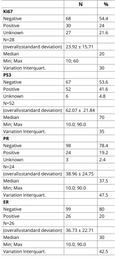
The immunohistochemical results (N and %) for Ki67, p53, PR, and ER are presented in table 1.
The study of HER-2/neu in our series was some difficulties arising largely because the older cases have had histologic, stages processing more or less time-consuming Bouin fixing, which is an excellent fixative for certain tissues (e.g., for lymphomas), but it has the disadvantage of hamper studies immunocytochemistry, as occurs with the HER-2/neu. This limitation is felt in studies of immunocytochemistry and much more obvious becomes when we perform molecular genetic studies, such as CISH. Therefore, only 42 cases to obtain a material with an acceptable quality of fixing to perform immunocytochemistry and CISH. The results are shown in table 2, which underline the following data: of the 42 cases included 7 had intensity of positivity 2; 8 with the intensity was 3. In the other, the intensity 1 was 22 cases and 0 presented 5 cases.
In all cases, where the intensity of reaction was 0 or 1, gene amplification was through CISH. In 7 cases in which the intensity of positivity was 2, there were 4 amplification and absence in the other 3. In 8 cases, where the intensity was 3, all gene amplification showed to CISH. So, the concordance between 3+ IHC and CISH-amplified cases was 100%, denoting all gene CISH-amplified cases to be overexpressing the HER-2/neu protein.
The entire cases with gene amplification and thus positive, for HER-2/neu, was 12 in 42, that is 28.57% of cases. We have not found an association between
overexpression/amplification (IHC/CISH) and prognosis. We noted that IHC/positive cases, as well as CISH-positive-only cases, had the same prognosis regarding survival, whereas IHC-positive-only cases had a prognosis similar to that of IHC/CISH-negative tumors.
Table 1: Immunohistochemical: Ki67, p53, PR and ER.
N % Ki67 Negative 68 54.4 Positive 30 24 Unknown 27 21.6 N=28 (overall±standard deviation) 23.92 ± 15.71 Median 20 Min; Max 10; 60 Variation Interquart. 30 P53 Negative 67 53.6 Positive 52 41.6 Unknown 6 4.8 N=52 (overall±standard deviation) 62.07 ± 21.84 Median 70 Min; Max 10.0; 90.0 Variation Interquart. 35 PR Negative 98 78.4 Positive 24 19.2 Unknown 3 2.4 N=24 (overall±standard deviation) 38.96 ± 24.75 Median 37.5 Min; Max 10.0; 90.0 Variation Interquart. 47.5 ER Negative 99 80 Positive 26 20 N=26 (overall±standard deviation) 36.73 ± 22.71 Median 30 Min; Max 10.0; 90.0 Variation Interquart. 42.5
The amplification evidenced by CISH was the second two patterns: or present themselves as signals and points individually and independently; or existing between
themselves, presenting itself as a stain (Figures 1 and 2).
HER 2 - ICQ
2 + 3 +
Figure 1: HER-2/neu - Immunocytochemistry (IHC)-(400x).
Figure 2: HER-2/neu– Chromogenic in situ hybridization (CISH).
Results for FIGO stage showed that this variable correlated significantly with survival (P = 0.002) (Table 3 and Figure 3).
In univariate analysis, the survival time was longer in patients with stage I-II disease than in those with stages III-IV disease (Cox p = 0.002). In the first group (I-II) the overall survival was 179.7 months (IC = 143.2-216.2) and in the second group (III-IV), the overall survival was 73.4 months (IC = 39.4-107.0).
The univariate analysis did not show an association of HER-2/neu protein expression with overall survival. Also, in the multivariate analysis the overexpression/ amplification protein was not proven as an independent prognostic indicator.
Figure 3: Overall survival of patients with epithelial ovarian carcinoma related to the FIGO stage. The survival in relation to markers HER-2/neu, p53, Ki67, and steroids receptors (ER and PR), the univariate analysis of clinical follow-up data revealed that patients with negative PR, had a significantly shortened survival time (p = 0.01) than patients with positive (Table 3 and Figure 4).
Figure 4: Overall survivals of patients with epithelial ovarian carcinoma relative to progesterone receptor. Table 2: HER-2/neu: Immunohistochemistry and CISH.
Nº of cases HER-2/neu (ICQ) CISH (AMPLIFICATION)
0 1+ 2+ 3+ 0 1+ 2+ 3+ Total
Other variables like HER-2/neu, P53, Ki67 and ER were not significantly related to survival (Table 3).
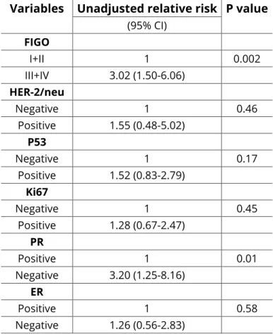
Table 3: Univariate survival analysis (Kaplan-Meier) Variables Unadjusted relative risk P value
(95% CI) FIGO I+II 1 0.002 III+IV 3.02 (1.50-6.06) HER-2/neu Negative 1 0.46 Positive 1.55 (0.48-5.02) P53 Negative 1 0.17 Positive 1.52 (0.83-2.79) Ki67 Negative 1 0.45 Positive 1.28 (0.67-2.47) PR Positive 1 0.01 Negative 3.20 (1.25-8.16) ER Positive 1 0.58 Negative 1.26 (0.56-2.83)
In the multivariable statistical analysis only FIGO stage (P = 0.002) was achieved to be independent predictor of overall survival (Table 4). None of the other variables showed any independent predictive value for patient prognosis.
In this study, the p53 suppressor gene showed a positive percentage (41.6%) and the univariate analysis did not show an association of p53 overexpression and prognosis (with overall survival) (Table 1). Also, the Multivariate analysis not showed that the p53 overexpression protein was not proven as an independent prognostic indicator.
Other market studded by immunohistochemical, the proliferative market Ki67 (with positive percentage, 24.0%) did not show association Ki67 expression with overall survival by the univariate analysis, as well as in multivariate analysis, the Ki67 was not proven as an independent prognostic indicator (Table 1).
The percentage of PR positive was 19.2% and ER positive 20.0%. We found that PR was associated with a better prognosis. Univariate analysis of clinical follow-up data revealed that patients with negative PR, had a significantly shortened survival time (p = 0.01) than
patients with positive (Table 3 and Figure 4). The same was not observed with the ER.
Table 4: Multivariable analysis of survival time using cox’s regression proportional hazards model for identification of independent prognostic factors for
patients with ovarian carcinoma.
Variables Adjusted Relative risk P value
(95% CI) FIGO I+II 1 0.02 III+IV 2.52 (1.13-5.61) PR Positive 1 0.16 Negative 2.02 (0.75-5.44) P53 Negative 1 0.94 Positive 1.03 (0.48-2.20)
Discussion
Wide substantial progress has been made and although more and more patients are living longer with their disease, the majority of patients with advanced ovarian cancer are not cured. The prognosis of ovarian cancer is discouraging compared to other malignancies of the female genital tract. Despite aggressive surgery and intensified chemotherapy, the outcome of patients with stage III and IV is poor. The importance of staging with regard to survival also stems from the influence it has on subsequent patient management. The prognostic value of stage according to the FIGO has been well established [8]. In a surgical staging procedure, there is some controversy over some aspects, especially in the early stages (I-II), regarding the status of retroperitoneal lymph nodes. When is presumed FIGO I and II stage, the extensive surgical staging showed that tumors were a more advanced stage, in general stage III. The “aggressive surgical staging” cannot significantly change the survival of the patients, but it can increase the operation risk [3,31]. In our study, unlike the great majority of the series, the percentage of early stages (I-II) cancers was higher compared to the advanced stage cancers. This is due to many of these cases been referred to the hospital. Most of the patients were diagnosed in routine gynecologic exams or by other specialties after the realization of imagiologic exams, for different clinical indications. In fact, nowadays, it seems like patients with ovarian cancer may be diagnosed at early stages, as a result of better primary care by general practitioners. Nevertheless, we still diagnose a great number of advanced stage cancers,
although this number may be decreasing.
In this study, only the FIGO stage was achieved to be an independent predictor of overall survival by the multivariable statistical analysis. None of the other variables showed any independent predictive value for patient prognosis. The stage is reorganized as the most important prognostic factor in epithelial ovarian cancer [8,32,33].
Valid prognostic factors are necessary to estimate the course of the disease and to define biologically similar subgroups for the analysis of therapeutic efficacy [32,34]. Many studies have been devoted to finding “Prognostic Factors”, and numerous features have been described that can help predict the prognosis of early and advanced ovarian cancer with varying degrees of accuracy and may provide a better understanding of the biological behavior of ovarian tumors. Immunohistochemistry has been widely used in the search of such marchers [35-37].
The HER-2/neu expression can be determined through IHC, FISH, CISH, and ELISA among other tests. This oncogene has been studied mainly in breast cancer by IHC and FISH where it has a prognostic, predictive and therapeutic target value. The HER-2/neu expressionin epithelial ovarian cancer has been less studied and we studied the overexpression by IHC and the amplification by CISH, a promising practical alternative to FISH. After the first CISH study by Tanner and colleagues [38] other reports favorably validated CISH results and the concordance between CISH and FISH [17,39-41]. Some reports noted the advantages of CISH over FISH; because CISH is a specific, sensitive, and easily applicable method for the detection of HER-2/neu gene amplification, and it can be used together with IHC for the evaluation of patients with breast carcinoma [17,18]. In this study, different series reported positivity frequencies of overexpression, it may be attributable to the type of material analyzed (fresh or embedded in paraffin) and to differences in specificity among antibodies used. Enzyme and microwave treatment of the tissue during the staining process may greatly affect the staining results, and tissue fixation procedures may also influence immunostaining. Different scoring methods and subjective interpretation of immunohistochemical analysis may also be reasons for different results obtained by different studies.
In our study, the percentage positive of HER-2/neu has not found an association between overexpression/ amplification (IHC/CISH) and it was not proven as an
independent prognostic indicator. Researchers have been trying to assess the real prognostic significance of HER-2/neu in ovarian cancer, but the results of their studies have been controversial. Some earlier studies reported that HER-2/neu overexpression was a poor prognostic factor [42,43], but later studies reported that HER-2/neu expression had not any relationship with prognosis [11,12,44,45]. Thus, no definitive conclusion has been reached as to the relationship between HER-2/ neu expression and prognosis.
The same happens with the role of the p53 protein in epithelial ovarian cancer, several studies have identified the p53 protein as an adverse prognostic factor for survival in epithelial ovarian cancer [46]. However, other studies suggest that p53 expression has not prognostic value in epithelial ovarian cancer [47]. De Graeff and collaborators showed in meta-analysis that the outcome of analysis was influenced by FIGO stage, for example: in some studies the p53 overexpression was associated with shorter survival of patients in stage I and II, but not in advanced stage III and IV; in other studies, found contradictory results, with shorter survival in an advanced stage but not in the early stage of the tumors with p53 overexpression [47]. In our study, we don’t find any association of the p53 overexpression with the predictive value of overall survival in early stage or advanced stage.
As for the other markers studied by immunohistochemical, we used the proliferative marker Ki67 that did not show association Ki67 expression with overall survival, as well as the Ki67 was not proven as an independent prognostic indicator. Studies on DNA content and proliferation in epithelial ovarian cancer have contradictory results regarding the prognostic significance of these parameters. Authors showed that ovarian cancer had a higher median percentage of Ki67 staining than borderline and benign tumors, and they found a significant relationship between the proliferative market Ki67 and disease-free survival that was independent the others parameters as histologic type, histologic grade, and stage [48]. Others authors also observed that Ki67 is a marker that differs in expression significantly between the short and long-term survivors [14,48]. Poor outcome was associated with the high proliferative activity, however series of studies did not confirm the relationship between proliferative activity and prognosis in epithelial ovarian cancer.
In our study, the percentage of PR was associated with a better prognosis, the univariate analysis of clinical
follow-up data revealed that patients with negative PR, had a significantly shortened survival time than patients with positive. The same was not observed with ER. Therefore, some of the latest studies of the analysis of ER and PR status showed that steroids receptors have significant favorable prognostic value in ovarian cancer. Especially progesterone positive tumor receptor status is proved to be an independent prognostic variable of improved progression for free-survival among patients with ovarian carcinoma. This anti-tumor effect of PR (significant survival benefit) may be due to two hypotheses that have been proposed. Estrogen-responsive cells efficiently repair DNA and avoid apoptosis; progesterone promotes cell differentiation and apoptosis, and stimulation of PR inhibits DNA synthesis and cell division. What justified the theory, proposed to explain the causal mechanism of carcinogenesis, the hypothesis “incessant ovulation”, that argues that repeated cycles of ovulation-induced trauma and repair of the ovarian surface epithelium (OSE) at the site of ovulation. According to with this hypothesis, the protective effect of progesterone will be: decreasing the exposure of the OSE to high levels of estrogens; antagonizing the growth-promoting effect of estrogens on OSE; inducing the apoptosis of tumor cells [49]; loss chromosome 11q23.3-24.3 decreasing PR expression and elevated risk for ovarian cancer and poorer prognosis. So, this may explain why patients with ER + and PR- tumors have the worst and patients with ER- and PR+ tumors the best prognosis. It is important to identify reliable prognostic markers, such as PR. So, maybe possible targets of therapy.
Conclusion
Although this study, with a relatively small number of cases, we conclude that patients with negative PR had a significantly shortened survival time (p = 0.01) than patients with positive. The overexpression of markers HER-2/neu, p53, Ki67 and ER, were not significantly related to survival in ovarian carcinoma. CISH is a promising, practical alternative to FISH that can be used in conjunction with IHC, which remains the first screening procedure of choice. IHC is easy to perform, relatively inexpensive, and able to detect a majority of ovarian cancer patients whose tumors have negative (0 or 1+) or positive (3+) HER-2/neu status, all three of which have complete concordance with CISH.
Only FIGO stage was achieved to be an independent predictor of overall survival. They should be evaluated together with the patient’s clinical status and other prognostic factors.
References
1. Stordal BK, Kalachand R, Hall N. Taxane monotherapy regimens for the treatment of recurrent epithelial ovarian cancer. Cochrane Database Syst Rev. 2018. Doi: https://doi.org/10.1002/14651858.CD008766.pub2 2. Akahira JI, Yoshikawa H, Shimizu Y, et al. Prognostic
factors of stage IV epithelial ovarian cancer: A multicenter retrospective study. Gynecol Oncol. 2001;81(3):398-403. Doi: https://doi.org/10.1006/ gyno.2001.6172
3. Benedet JL, Bender H, Jones H, et al. FIGO staging classifications and clinical practice guidelines in the management of gynecologic cancers. FIGO Committee on Gynecologic Oncology. Int J Gynaecol
Obstet. 2000;70(2):209-262. https://www.ncbi.nlm.
nih.gov/pubmed/11041682
4. Hannaford PC, Selvaraj S, Elliott AM, et al. Cancer risk among users of oral contraceptives: Cohort data from the Royal College of General Practitioner’s oral contraception study. BMJ. 2007;335(7621):651. Doi: https://doi.org/10.1136/bmj.39289.649410.55
5. Menczer J, Liphshitz I, Barchana M. A decreasing incidence oaf ovarian carcinoma in Israel. Int J
Gynecol Cancer. 2006;16(1):41-44. Doi: https://doi.
org/10.1111/j.1525-1438.2006.00275.x
6. Berek JS, Friedlander ML, Bast RC. Epithelial ovarian, fallopian tube, and peritoneal cancer. In: Holland-Frei Cancer Medicine. American Cancer Society; 2017:1-27. Doi: https://doi.org/10.1002/9781119000822. hfcm105
7. Clark TG, Stewart ME, Altman DG, et al. A prognostic model for ovarian cancer. Br J Cancer. 2001;85(7):944-952. Doi: https://doi.org/10.1054/bjoc.2001.2030 8. DiSilvestro P, Peipert JF, Hogan JW, et al. Prognostic
value of clinical variables in ovarian cancer. J Clin
Epidemiol. 1997;50(5):501-505. Doi: https://doi.
org/10.1016/S0895-4356(97)00002-4
9. Wang Y, Wang D, Ren M. Prognostic value of HER-2/ neu expression in epithelial ovarian cancer: A meta-analysis. Tumour Biol. 2014;35(1):33-38. Doi: https:// doi.org/10.1007/s13277-013-1003-9
10. Goff B, Muntz H, Greer B, et al. Oncogene expression: Long-term compared with short-term survival in patients with advanced epithelial ovarian cancer.
Obstet Gynecol. 1998;92(1):88-93. Doi: https://doi.
org/10.1016/S0029-7844(98)00121-5
11. Høgdall EVS, Christensen L, Kjaer SK, et al. Distribution of HER-2 overexpression in ovarian carcinoma tissue
and its prognostic value in patients with ovarian carcinoma: from the Danish MALOVA Ovarian Cancer Study. Cancer. 2003;98(1):66-73. Doi: https://doi. org/10.1002/cncr.11476
12. Rubin SC, Finstad CL, Wong GY, et al. Prognostic significance of HER-2/neu expression in advanced epithelial ovarian cancer: A multivariate analysis. Am J
Obstet Gynecol. 1993;168(1):162-169. Doi: https://doi.
org/10.1016/S0002-9378(12)90907-2
13. Emens LA. Trastuzumab: Targeted therapy for the management of HER-2/neu-overexpressing metastatic breast cancer. Am J Ther. 2005;12(3):243-253. https:// www.ncbi.nlm.nih.gov/pubmed/15891269
14. Meng S, Tripathy D, Shete S, et al. HER-2 gene amplification can be acquired as breast cancer progresses. Proc Natl Acad Sci. 2004;101(25):9393-9398. Doi: https://doi.org/10.1073/pnas.0402993101 15. Mabuchi S, Morishige K, Kimura T. Use of monoclonal
antibodies in the treatment of ovarian cancer. Curr
Opin Obstet Gynecol. 2010;22(1):3-8. Doi: https://doi.
org/10.1097/GCO.0b013e3283324114
16. Odicino FE, Bignotti E, Rossi E, et al. HER-2/neu overexpression and amplification in uterine serous papillary carcinoma: Comparative analysis of immunohistochemistry, real-time reverse transcription-polymerase chain reaction, and fluorescence in situ hybridization. Int J Gynecol Cancer. 2008;18(1):14-21. Doi: https://doi.org/10.1111/j.1525-1438.2007.00946.x
17. Kumamoto H, Sasano H, Taniguchi T, et al. Chromogenic in situ hybridization analysis of HER-2/neu status in breast carcinoma: application in screening of patients for trastuzumab (Herceptin) therapy. Pathol Int. 2001;51(8):579-584. https://www. ncbi.nlm.nih.gov/pubmed/11564211
18. Yan B, Choo SN, Mulyadi P, et al. Dual-colour HER2/chromosome 17 chromogenic in situ hybridisation enables accurate assessment of HER2 genomic status in ovarian tumours. J Clin Pathol. 2011;64(12):1097-1101. Doi: https://doi.org/10.1136/ jclinpath-2011-200082
19. Ferretti G, Felici A, Papaldo P, et al. HER2/neu role in breast cancer: From a prognostic foe to a predictive friend: Curr Opin Obstet Gynecol. 2007;19(1):56-62. Doi: https://doi.org/10.1097/GCO.0b013e328012980a 20. Wu S, Ma C, Yang Y. The prognostic value of HER-2/neu
overexpression in colorectal cancer: Evidence from 16 studies. Tumour Biol. 2014;35(11):10799-10804. Doi: https://doi.org/10.1007/s13277-014-2376-0
21. Ozaki T, Nakagawara A. p53: The attractive tumor suppressor in the cancer research field.
Biomed Res Int. 2011;2011:1-13. Doi: https://doi.
org/10.1155/2011/603925
22. Leel W, Park E, Kim T. Expression of p53, p27 and Jab 1 protein in epithelial ovarian tumors. Eur J
Gynaec Oncol. 2012. https://www.ncbi.nlm.nih.gov/
pubmed/23091890
23. Shahin MS, Hughes JH, Sood AK, et al. The prognostic significance of p53 tumor suppressor gene alterations in ovarian carcinoma. Cancer. 2000;89(9):2006-2017. Doi: https://doi.org/10.1002/1097-0142(20001101)89:9<2006::AID-CNCR18>3.3.CO;2-Z 24. Agarwal R, Kaye SB. Ovarian cancer: Strategies
for overcoming resistance to chemotherapy. Nat
Rev Cancer. 2003;3(7):502-516. Doi: https://doi.
org/10.1038/nrc1123
25. Fallows S, Price J, Atkinson RJ, et al. p53 mutation does not affect prognosis in ovarian epithelial malignancies. J Pathol. 2001;194(1):68-75. Doi: https:// doi.org/10.1002/path.857
26. El-Naggar AK, Vielh P. Solid tumor DNA content analysis. Methods Mol Biol. 2004;263:355-370. Doi: https://doi.org/10.1385/1-59259-773-4:355
27. Garzetti GG, Ciavattini A, Goteri G, et al. Ki67 Antigen immunostaining (MIB 1 monoclonal antibody) in serous ovarian tumors: Index of proliferative activity with prognostic significance. Gynecol Onco. 1995;56(2):169-174. Doi: https://doi.org/10.1006/ gyno.1995.1026
28. Kommoss F, Pfisterer J, Thome M, et al. Steroid receptors in ovarian carcinoma: Immunohistochemical determination may lead to new aspects. Gynecol Oncol. 1992;47(3):317-322. Doi: https://doi.org/10.1016/0090-8258(92)90133-4
29. Ayadi L, Chaabouni S, Khabir A, et al. Correlation between immunohistochemical biomarkers expression and prognosis of ovarian carcinomas in tunisian patients. World J Oncol. 2010;1:118. Doi: https://doi.org/10.4021/wjon2010.06.213w
30. Lee P, Rosen DG, Zhu C, et al. Expression of progesterone receptor is a favorable prognostic marker in ovarian cancer. Gynecol Oncol. 2005;96(3):671-677. Doi: https://doi.org/10.1016/j. ygyno.2004.11.010
31. Sharp F, Mason W, Creasman W. Ovarian cancer. In: Biology, Diagnosis and Management. 1st ed. London; 1992.
32. Friedlander M. Prognostic factors. Ovarian cancer. 1998;5:143-152.
33. Holschneider CH, Berek JS. Ovarian cancer: Epidemiology, biology, and prognostic factors.
Semin Surg Oncol. 2000;19(1):3-10. Doi: https://doi.
org/10.1002/1098-2388(200007/08)19:1<3::AID-SSU2>3.0.CO;2-S
34. Geisler JP, Geisler HE. Tumor markers and molecular biological markers in gynecologic malignancies. Curr
Opin Obstet Gynecol. 2001;13(1):31-39. Doi: https://
doi.org/10.1097/00001703-200102000-00005
35. Akahira J, Inoue T, Suzuki T, et al. Progesterone receptor isoforms A and B in human epithelial ovarian carcinoma: Immunohistochemical and RT-PCR studies. Br J Cancer. 2000;83(11):1488-1494. Doi: https://doi.org/10.1054/bjoc.2000.1463
36. Tomsová M, Melichar B, Sedláková I, et al. Prognostic markers in ovarian carcinoma - retrospective study.
Cesk Patol. 2005;41(2):51-59. https://www.ncbi.nlm.
nih.gov/pubmed/15966333
37. Yemelyanova A, Vang R, Kshirsagar M, et al. Immunohistochemical staining patterns of p53 can serve as a surrogate marker for TP53 mutations in ovarian carcinoma: an immunohistochemical and nucleotide sequencing analysis. Mod Pathol. 2011;24(9):1248-1253. Doi: https://doi.org/10.1038/ modpathol.2011.85
38. Tanner M, Gancberg D, Di Leo A, et al. Chromogenic in situ hybridization: a practical alternative for fluorescence in situ hybridization to detect HER-2/ neu oncogene amplification in archival breast cancer samples. Am J Pathol. 2000;157(5):1467-1472. Doi: https://doi.org/10.1016/S0002-9440(10)64785-2 39. Chi DS, Liao JB, Leon LF, et al. Identification of
prognostic factors in advanced epithelial ovarian carcinoma. Gynecol Oncol. 2001;82(3):532-537. Doi: https://doi.org/10.1006/gyno.2001.6328
40. Ozalp SS, Yalcin OT, Basaran GN, et al. Prognostic significance of deletion and over-expression of the p53 gene in epithelial ovarian cancer. Eur J Gynaecol
Oncol. 2000;21(3):282-286. https://www.ncbi.nlm.nih.
gov/pubmed/10949395
41. Röhlke P, Milde-Langosch K, Weyland C, et al. p53
is a persistent and predictive marker in advanced ovarian carcinomas: Multivariate analysis including comparison with Ki67 immunoreactivity. J Cancer
Res Clin Oncol. 1997;123(9):496-501. Doi: https://doi.
org/10.1007/BF01192204
42. Ananiev J, Gulubova M, Manolova I, et al. Prognostic significance of HER2/neu expression in gastric cancer.
Wiener klinische Wochenschrift.
2011;123(13-14):450-454. Doi: https://doi.org/10.1007/s00508-011-0025-9 43. Camilleri-Broët S, Hardy-Bessard AC, Le Tourneau A,
et al. HER-2 overexpression is an independent marker of poor prognosis of advanced primary ovarian carcinoma: A multicenter study of the GINECO group.
Ann Oncol. 2004;15(1):104-112. https://www.ncbi.nlm.
nih.gov/pubmed/14679128
44. Halon A, Donizy P, Biecek P, et al. HER-2 Expression in immunohistochemistry has no prognostic significance in gastric cancer patients. Sci World J. 2012;2012:1-6. Doi: https://doi.org/10.1100/2012/941259
45. Frutuoso C, Silva MR, Amaral N, et al. Prognosis value of p53, C-erB-2 and Ki67 proteins in ovarian carcinoma. Acta Med Port. 2001;14(3):277-283. https:// www.ncbi.nlm.nih.gov/pubmed/11552325
46. Bali A, O’Brien PM, Edwards LS, et al. Cyclin D1, p53, and p21Waf1/Cip1 expression is predictive of poor clinical outcome in serous epithelial ovarian cancer.
Clin Cancer Res. 2004;10(15):5168-5177. Doi: https://
doi.org/10.1158/1078-0432.CCR-03-0751
47. de Graeff P, Crijns APG, de Jong S, et al. Modest effect of p53, EGFR and HER-2/neu on prognosis in epithelial ovarian cancer: A meta-analysis. Br J Cancer. 2009;101(1):149-159. Doi: https://doi.org/10.1038/ sj.bjc.6605112
48. Gursan N, Sipal S, Calik M, et al. P53, bcl-2, Ki67 Li (labeling index) status in benign, proliferative, and malignant ovarian surface epithelial neoplasms.
Eurasian J Med. 2009;41(1):10-14. https://www.ncbi.
nlm.nih.gov/pmc/articles/PMC4261648/
49. Syed V, Ho S-M. Progesterone-induced apoptosis in immortalized normal and malignant human ovarian surface epithelial cells involves enhanced expression of FasL. Oncogene. 2003;22(44):6883-6890. Doi: https://doi.org/10.1038/sj.onc.1206828
Copyright: © Tomé et al. This is an Open Access article distributed under the terms of the Creative Commons Attribution License, which permits unrestricted use, distribution, and reproduction in any medium, provided the original work is properly cited.